. 2013 Sep;51(9):667-75.
doi: 10.1002/dvg.22407. Epub 2013 Jul 9.

Troponin T3 expression in skeletal and smooth muscle is required for growth and postnatal survival: characterization of Tnnt3(tm2a(KOMP)Wtsi) mice

Affiliations

Troponin T3 expression in skeletal and smooth muscle is required for growth and postnatal survival: characterization of Tnnt3(tm2a(KOMP)Wtsi) mice

Yawen Ju et al. Genesis. 2013 Sep.

Abstract

The troponin complex, which consists of three regulatory proteins (troponin C, troponin I, and troponin T), is known to regulate muscle contraction in skeletal and cardiac muscle, but its role in smooth muscle remains controversial. Troponin T3 (TnnT3) is a fast skeletal muscle troponin believed to be expressed only in skeletal muscle cells. To determine the in vivo function and tissue-specific expression of Tnnt3, we obtained the heterozygous Tnnt3+/flox/lacZ mice from Knockout Mouse Project (KOMP) Repository. Tnnt3(lacZ/+) mice are smaller than their WT littermates throughout development but do not display any gross phenotypes. Tnnt3(lacZ/lacZ) embryos are smaller than heterozygotes and die shortly after birth. Histology revealed hemorrhagic tissue in Tnnt3(lacZ/lacZ) liver and kidney, which was not present in Tnnt3(lacZ/+) or WT, but no other gross tissue abnormalities. X-gal staining for Tnnt3 promoter-driven lacZ transgene expression revealed positive staining in skeletal muscle and diaphragm and smooth muscle cells located in the aorta, bladder, and bronchus. Collectively, these findings suggest that troponins are expressed in smooth muscle and are required for normal growth and breathing for postnatal survival. Moreover, future studies with this mouse model can explore TnnT3 function in adult muscle function using the conditional-inducible gene deletion approach

Keywords: development; knockout mice; muscle; troponin.


Figures

Fig 1
Fig 1. Genomic structure of Tnnt3tm2a(KOMP)Wtsi mice
(A) The Tnnt3/lacZ knock-in allele (Tnnt3tm2a(KOMP)Wtsi) is shown to illustrate the mutated Tnnt3 gene in which the lacZ and neomycin expression cassette was inserted between Exon 9 and Exon 10, resulting in a Tnnt3 null gene, and is flanked by FRT (Flippase Recognition Target) sites. This cassette contains the splice acceptor of mouse engrailed 2 exon 2 (En2 SA), an internal ribosome entry sequence (IRES) to initiate lacZ translation, and polyadenylation (pA) to terminate transcription after the lacZ gene. The neo gene is driven by human beta actin promoter (hBactP) and contains it own pA. Additionally, Exon 10 is flanked by loxP sites. (B) The resulting Tnnt3 conditional knockout gene structure that is generated by crossing Tnnt3tm2a(KOMP)Wtsi mice with mice that express the Flp recombinase is shown to illustrate removal of the lacZ and neomycin cassette. (C) The resulting Tnnt3 knockout allele generated after crossing with Cre expression mice is shown.
Fig 2
Fig 2. Tnnt3 is required for postnatal survival, normal growth, and tnnc2 and tnni2 expression
(A) The gross phenotype of a representative (n>4) Tnnt3lacZ/+ mouse at 2 month of age is shown next to its WT (+/+) littermate to illustrate the smaller sized of the mutant animals. Photographs of homozygous (lacZ/lacZ) mutant mice could not be obtained since these mice die shortly after birth. (B) The body weights of Tnnt3 lacZ/+ and their WT littermates were measured weekly from 3 to 9 weeks (wk) after birth, and the data are presented as the mean +/− SD (n=4, *p<0.05). A photograph (C) and radiograph (D) of representative Tnnt3lacZ/lacZ, Tnnt3lacZ/+ and Tnnt3+/+ embryos harvested at E18.5 (n≥3) is presented to illustrate the gene dose effect on growth. QRT-PCR was performed on RNA obtained from muscle of WT, Tnnt3lacZ/+ or Tnnt3lacZ/lacZ adult mice (E), or from E18.5 embryos (F), and the data are presented as the mean +/− SD (n=3, *p<0.05).
Fig. 3
Fig. 3. The tissue specific expression pattern of Tnnt3
(A) Whole mount was performed on 1-day old Tnnt3lacZ/+mice and their WT littermates (n=2). The photograph illustrates the broad X-gal staining in skeletal muscle throughout the Tnnt3lacZ/+ animal except in brown fat tissue (*), galea aponeurotica (indicated by #) and temporal fascia (indicated by ↑). No β-galactosidase activity was detected in Tnnt3+/+ mice. (B–P) X-gal staining of tissues from 2 month-old Tnnt3lacZ/+ mice and WT littermates (n>3), and representative micrographs are show to illustrate the tissue specific expression of the transgene. Note the intense positive staining in skeletal muscle cells next to leg(G) and diaphragm (K), and smooth muscle cells in the aorta (H), bladder (I) and bronchus (J) in the tissues from Tnnt3lacZ/+ mice. In contrast there was no positive staining in spleen (L), heart (M), brain (N), kidney (O), and liver (P) tissues from Tnnt3lacZ/+ mice, or any tissues from the WT littermates.
Fig. 4
Fig. 4. Tnnt3 heterozygous mice have a mild skeletal phenotype
X-gal staining was performed on long bones from 2-month-old WT (A) and Tnnt3lacZ/+ (B) mice (n=3). The micrographs show that the transgene is not expressed in bone, bone marrow or joint connective tissue, but is highly expressed in the adjacent skeletal muscle. (C–F) Alcian-blue/ Alizarin-red staining was performed on the skeletons of 2-month-old (C), and E18.5 embryos (D) of Tnnt3lacZ/+ and WT mice (n=2) to assess differences in the mineralized (red) and unmineralized (blue) tissue. A closer comparison of the forelimbs (E) and hindlimbs (F) reveals a modest decreased limb length in Tnnt3lacZ/+ embryos. (G, H) Alcian-blue/Orange G/Alizarin-red staining was performed on histology slides of spinal tissue from E18.5 embryos of WT (G) andTnnt3lacZ/+ (H) mice. No remarkable differences were observed.
Fig. 5
Fig. 5. Histologic evidence of hemorrhage in Tnnt3lacZ/lacZ liver and kidney
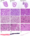
H&E stained histology was prepared from WT, Tnnt3lacZ/+ and Tnnt3lacZ/lacZ embryos harvested on E18.5. Representative micrographs of heart (A, E, I), Lung (B, F, J), liver (C, G, K) and kidney (D, H, L) tissue are shown. Note the normal appearances of the heart (I) and lung (J) tissue, and the hemorrhagic tissue (arrows) in Tnnt3lacZ/lacZ liver (K) and kidney (L). No abnormalities were detected in any of the tissue harvested from WT and Tnnt3lacZ/+ embryos. Representative histology of diaphragm tissues from WT (M), Tnnt3lacZ/+ (N) and Tnnt3lacZ/lacZ (O) embryos stained with X-gal are also presented to illustrate the markedly thinner diaphragm muscle in the Tnnt3lacZ/lacZ embryo.
Fig. 6
Fig. 6. Tnnt3 is highly expressed in the skeletal muscle attached to the PLN
X-gal staining of the PLN and surrounding fat and muscle tissue from Tnnt3lacZ/+ mice reveals that positive staining is only detected in the skeletal muscle.

References

    1. Darland DC, D'Amore PA. Cell-cell interactions in vascular development. Curr Top Dev Biol. 2001;52:107–149. - PubMed
    1. Dong Y, Jesse AM, et al. RBPjkappa-dependent Notch signaling regulates mesenchymal progenitor cell proliferation and differentiation during skeletal development. Development. 2010;137(9):1461–1471. - PMC - PubMed
    1. el-Saleh SC, Warber KD, et al. The role of tropomyosin-troponin in the regulation of skeletal muscle contraction. J Muscle Res Cell Motil. 1986;7(5):387–404. - PubMed
    1. Grcevic D, Pejda S, et al. In vivo fate mapping identifies mesenchymal progenitor cells. Stem Cells. 2012;30(2):187–196. - PMC - PubMed
    1. Hilton MJ, Tu X, et al. Tamoxifen-inducible gene deletion reveals a distinct cell type associated with trabecular bone, and direct regulation of PTHrP expression and chondrocyte morphology by Ihh in growth region cartilage. Dev Biol. 2007;308(1):93–105. - PMC - PubMed

Publication types

LinkOut - more resources