. 2013 Jul 20;31(21):2654-61.
doi: 10.1200/JCO.2012.44.2137. Epub 2013 Jun 17.

Follicular lymphoma cells induce changes in T-cell gene expression and function: potential impact on survival and risk of transformation

Affiliations

Follicular lymphoma cells induce changes in T-cell gene expression and function: potential impact on survival and risk of transformation

Shahryar Kiaii et al. J Clin Oncol. .

Abstract

Purpose: Previous studies have demonstrated the prognostic importance of the immune microenvironment in follicular lymphoma (FL). To investigate the molecular mechanisms during which tumor-infiltrating T cells (TILs) are altered in the FL microenvironment, we studied highly purified CD4 and CD8 TILs from lymph node biopsies at diagnosis in treatment-naive patients with FL compared with reactive tonsils and the peripheral blood of healthy donors.

Patients and methods: Gene expression profiling of highly purified CD4 and CD8 TILs was performed on the Affymetrix platform. Diagnostic tissue microarrays from an independent patient set (n = 172) were used to verify protein expression and analyze any impact of TIL-expressed genes on outcome. Time-lapse imaging was used to assess T-cell motility.

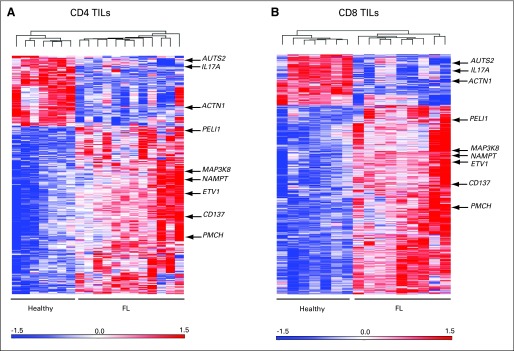
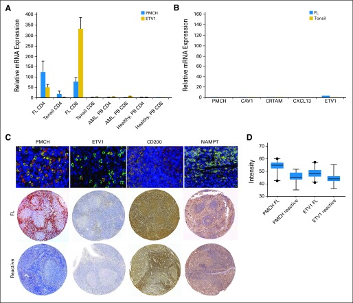
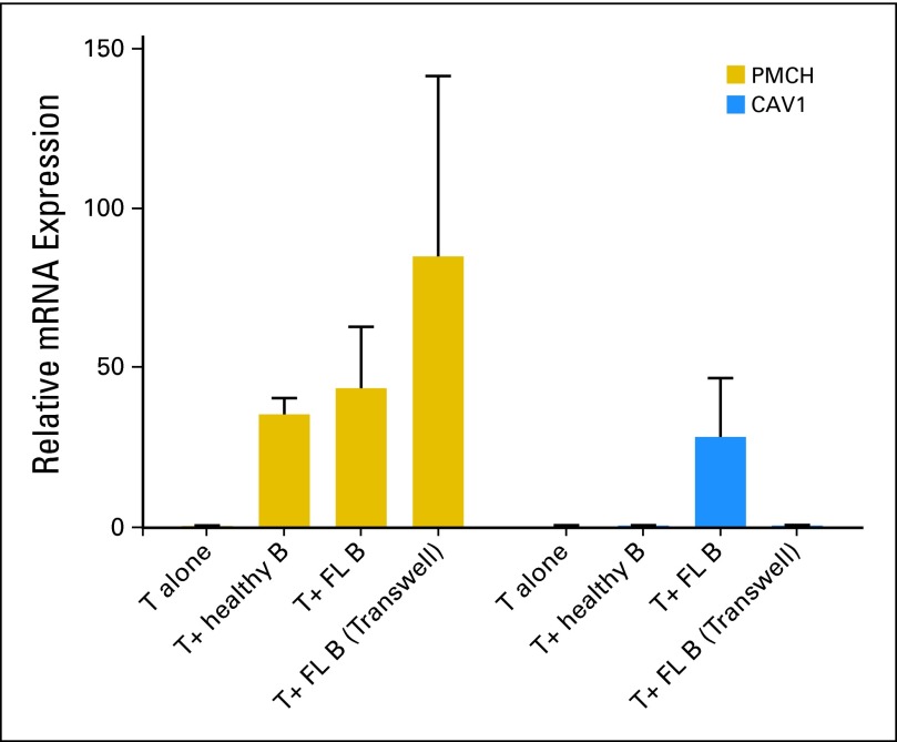
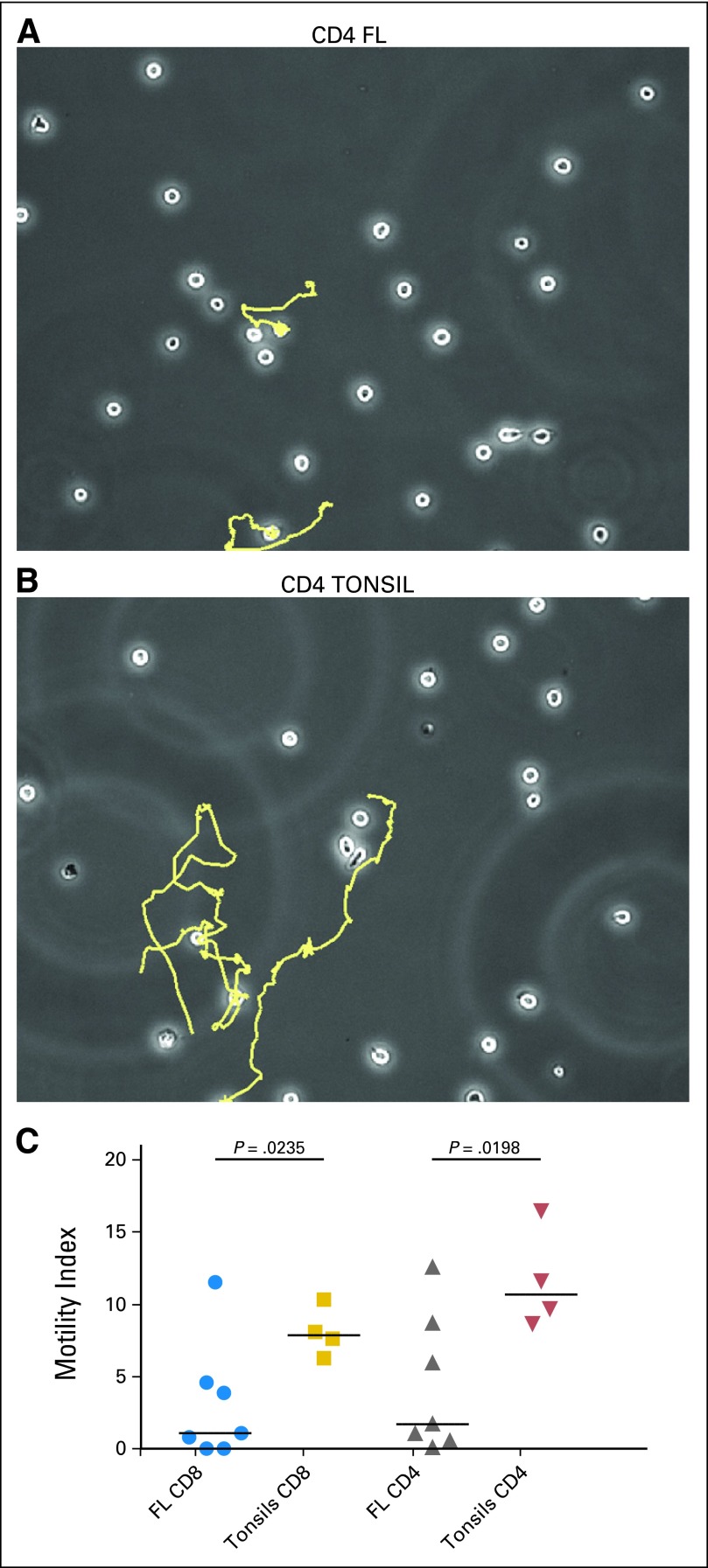
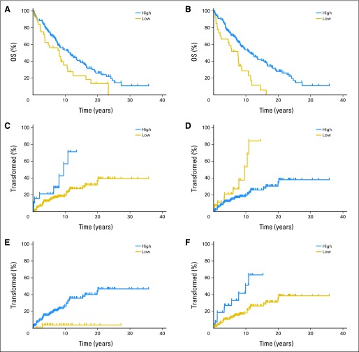
Results: The most upregulated genes in both CD4 and CD8 TILs were PMCH, ETV1, and TNFRSF9. PMCH is not expressed in peripheral blood T cells, but expression is highly induced on culture with FL. Both CD4 and CD8 TILs from patients with FL have significantly impaired motility compared with those of healthy TILs from reactive tonsils and this can be induced on healthy T cells by FL cells. During multivariate analysis, a model incorporating the number and location of T cells expressing PMCH, NAMPT, and ETV1 showed prognostic significance for overall survival and for time to transformation.

Conclusion: We showed altered gene expression in TILs in FL and demonstrated that altering the immune microenvironment in FL affects overall survival and time to transformation in this disease.


Conflict of interest statement

Authors' disclosures of potential conflicts of interest and author contributions are found at the end of this article.

Figures

Fig 1.
Fig 1.
Heat maps showing differentially expressed genes in highly pure tumor-infiltrating lymphocytes (TILs) CD8 and CD4 from patients with follicular lymphoma (FL) at diagnosis compared with healthy donors. (A) Dendrogram of differentially expressed genes by unsupervised analysis (P < .05; false discovery rates < 0.05) showed 182 genes were significantly upregulated (red) and 85 genes were significantly downregulated (blue) in CD4 cells from patients with FL. (B) CD8 cells showed 481 genes were significantly upregulated and 163 genes downregulated in patients with FL.
Fig 2.
Fig 2.
Validation of gene expression observed by microarray. The results of quantitative real time polymerase chain reaction are concordant with data seen on microarray. The figure shows results of (A) PMCH and ETV1 as representative data for five investigated genes in CD4 and CD8 tumor-infiltrating lymphocytes from previously untreated follicular lymphoma patients (FL; n = 12) compared with those from tonsil (n = 10), peripheral blood (PB) of acute myeloid leukemia patients (AML; n = 10), and healthy age-matched donors (n = 10). (B) The examined genes showed low expression in the CD19+ cells of tonsils (n = 7) and FL cells (n = 12). (C) The merged fluorescence double staining of CD3 (green) and PMCH, ETV1, CD200, or NAMPT (red) in lymph nodes (LN) of a patient with FL that represents the double staining in 10 different FL patients. (D) The gene array results were also concordant with the results of tissue microarrays (TMAs) stained for PMCH, ETV1, CD200, and NAMPT (Visfatin-1) comparing FL patients (top raw) and reactive LN (down raw). Figure is representative of 172 FL patients and 12 reactive LN analyzed. Comparison of mean intensity expression of PMCH and ETV1in FL patients (n = 172) with reactive LN (n = 12).
Fig 3.
Fig 3.
Relative fold change in mRNA expression results (by qualitative reverse-transcriptase–polymerase chain reaction) for PMCH and CAV1 genes in allogeneic healthy T cells cultured alone (n = 4) or cocultured with follicular lymphoma (FL) cells (n = 7) and healthy B cells (n = 4) for 48 hours in cell-cell contact or in transwell experiment (n = 4).
Fig 4.
Fig 4.
Expression of MCHR2 by interfollicular tissue macrophages. Staining of lymph node (LN) paraffin-embedded section using (A) anti-CD68 TexRed conjugated secondary Ab and (B) anti-MCHR2 and FITC-conjugated secondary Ab. (C) A combination of (A) and (B), which shows the specific expression of MCHR2 by interfollicular tissue macrophages. This is representative of 10 follicular lymphoma patients and five reactive LNs examined.
Fig 5.
Fig 5.
Tracking T cell motility on the intercellular adhesion molecule (ICAM-1) coated slides. Shown are representative paths of the movements of CD4 tumor-infiltrating lymphocytes (TILs) from (A) a patient with FL and (B) a healthy donor. (C) Severely impaired Motility Index, calculated via time-lapse imaging of CD4 and CD8 TILs from patients with follicular lymphoma (FL; n = 7) compared with those from reactive tonsils (n = 4).
Fig 6.
Fig 6.
Overall survival (OS) and time to transformation (TT) of patients with follicular lymphoma (FL) according to high versus low expression of examined proteins at time of diagnosis. Number of positive cells for PMCH expression (P = .03) in (A) intrafollicular and (B) interfollicular area (P = .0002). TT of patients with FL according to number of PMCH-expressing cells in (C) intrafollicular (P = .029) and (D) interfollicular area (P = .033). TT for the same patients for (E) number of ETV1-expressing cells in intrafollicular area (P = .02) and (F) mean intensity of ETV1 expression in interfollicular area (P = .0005).

Comment in

References

    1. Gribben JG. How I treat indolent lymphoma. Blood. 2007;109:4617–4626. - PubMed
    1. Montoto S, Fitzgibbon J. Transformation of indolent B-cell lymphomas. J Clin Oncol. 2011;29:1827–1834. - PubMed
    1. Federico M, Bellei M, Marcheselli L, et al. Follicular lymphoma International Prognostic Index 2: A new prognostic index for follicular lymphoma developed by the International Follicular Lymphoma Prognostic Factor project. J Clin Oncol. 2009;27:4555–4562. - PubMed
    1. Morin RD, Mendez-Lago M, Mungall AJ, et al. Frequent mutation of histone-modifying genes in non-Hodgkin lymphoma. Nature. 2011;476:298–303. - PMC - PubMed
    1. Dave SS, Wright G, Tan B, et al. Prediction of survival in follicular lymphoma based on molecular features of tumor-infiltrating immune cells. N Engl J Med. 2004;351:2159–2169. - PubMed

MeSH terms

Substances