. 2013 Jul 2;110(27):10940-5.
doi: 10.1073/pnas.1300686110. Epub 2013 Jun 17.

Mechanistic and structural studies on legumain explain its zymogenicity, distinct activation pathways, and regulation

Affiliations

Mechanistic and structural studies on legumain explain its zymogenicity, distinct activation pathways, and regulation

Elfriede Dall et al. Proc Natl Acad Sci U S A. .

Abstract

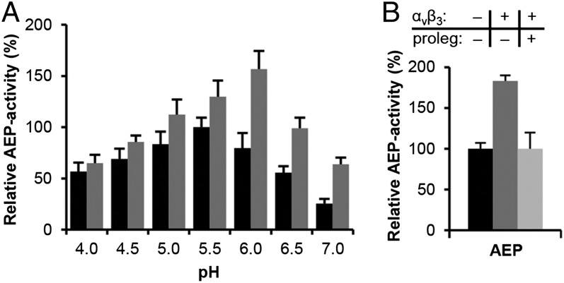
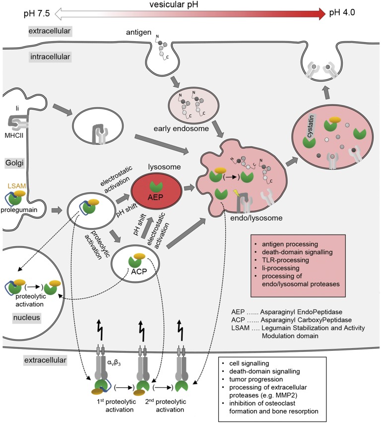
The cysteine protease legumain plays important functions in immunity and cancer at different cellular locations, some of which appeared conflicting with its proteolytic activity and stability. Here, we report crystal structures of legumain in the zymogenic and fully activated form in complex with different substrate analogs. We show that the eponymous asparagine-specific endopeptidase activity is electrostatically generated by pH shift. Completely unexpectedly, the structure points toward a hidden carboxypeptidase activity that develops upon proteolytic activation with the release of an activation peptide. These activation routes reconcile the enigmatic pH stability of legumain, e.g., lysosomal, nuclear, and extracellular activities with relevance in immunology and cancer. Substrate access and turnover is controlled by selective protonation of the S1 pocket (KM) and the catalytic nucleophile (kcat), respectively. The multibranched and context-dependent activation process of legumain illustrates how proteases can act not only as signal transducers but as decision makers.

Keywords: allostery; context-dependent activities; death domain; electrostatic stability switch; kcat-substrate specificity.


Conflict of interest statement

The authors declare no conflict of interest.

Figures

Fig. 1.
Fig. 1.
Substrate specificity and catalytic mechanism of legumain. (A) Legumain has a caspase-like overall structure. Active site residues (Cys189, His148, and Asn42) and the RGD120 motif are highlighted as green sticks, and the covalent Z-Ala-Ala-AzaAsn-chloromethylketone inhibitor in purple sticks. (B) Substrate specificity of legumain is determined by its zwitterionic S1 pocket. The Z-Ala-Ala-AzaAsn-cmk inhibitor (purple sticks) is covalently linked to the catalytic Cys189. The catalytic triad is represented by green sticks and residues forming the S1 pocket by red and blue sticks. Note the trivalent oxyanion pocket indicated by dashed lines. (C) Legumain activity critically depends on the local pKa of Cys189. The latter can be tuned by an E190K mutation, resulting in a fourfold increase in kcat at pH 5.5 as measured by the turnover of Bz-Asn-pNA. (D) KM values of active wild-type legumain (AEP) and E190K legumain toward the Bz-Asn-pNA substrate at pH 5.5 are virtually identical, indicating that the charge reversal affects exclusively the turnover number via pKa tuning of Cys189. Data in C and D are represented as mean ± SD of three experiments.
Fig. 2.
Fig. 2.
The C-terminal “propeptide” harbors a death domain like fold. (A) Prolegumain consists of a catalytic domain (green, Val18-Met286), an AP (blue, Lys287–Asn323), and a LSAM domain (wheat, Asp324–Tyr433). Cleavages at the α- (KRK289) and β-sites (N323D324) release the AP and, thus, renders the active site (C189) accessible. (B) Topology diagram of LSAM (colored in wheat) compared with classical death domains (colored in red).
Fig. 3.
Fig. 3.
The LSAM domain stabilizes legumain via electrostatic interactions. (A) Color-coded electrostatic surface potential of the catalytic domain (red; negative charge) and the LSAM domain including helix APαV (blue; positive charge) calculated at pH 7.0. The catalytic domain is rotated relative to the view in Fig. 2A by 90° around the horizontal x axis, and the LSAM domain has been separated and rotated by 180° relative to the catalytic domain. (B) Isomorphous σA-weighted difference density FpH7.5FpH5.0 of legumain (AEP) crystallized at pH 7.5 and pH 5.0. In either case, legumain was covalently inhibited with Ac-YVAD-cmk (orange sticks). Strong changes in X-ray diffraction, as evident by the difference density, reflect changes in protein dynamics and stability. These changes cluster at the area surrounding the active site that corresponds to the LSAM domain binding interface; this confined mobility suggests that LSAM stabilizes the protease domain at neutral pH. Contour level: 2.9 σ over the mean; catalytic residues are indicated as green sticks. (C) Thermal denaturation curves show a stabilization of AEP by the LSAM domain. Melting curves of AEP (lacking the LSAM domain), β-legumain (cleaved at Asn323, LSAM domain remains noncovalently bound to the catalytic domain), and prolegumain were measured at pH 6.5 by the Thermofluor method. Melting points are indicated by dashed lines.
Fig. 4.
Fig. 4.
Prolegumain can develop carboxypeptidase activity upon cleavage of the AP. (A) The AP binds substrate-like to the nonprimed substrate binding cleft. The view to the active site is identical as in Fig. 1B. Catalytic residues are indicated as green sticks, and the double Arg-motif (Arg403 and Arg342) anchors the helix APαV via a salt bridge to Asp309. (B) View to the carboxypeptidase active site upon release of the AP. Arg403 and Arg342 are ideally positioned to anchor the C terminus of the substrate and, thereby, act as a molecular ruler for monopeptidyl peptidase activity, consistent with results from the peptide competition assay (Fig. S4 B and C). (C) SDS/PAGE after trypsin activation. After 2 h incubation of prolegumain with trypsin in a 1:50 molar ratio, approximately 12% of prolegumain were converted to an active enzyme (α-legumain, ∼36 kDa, orange box) as judged by using ImageJ (http://rsbweb.nih.gov/ij). In a control experiment, legumain was autoactivated via pH shift (AEP, green box). M, molecular mass marker in kilodaltons. (D) α-legumain carboxypeptidase activity can be generated via trypsin activation. Specific activity toward the fluorescent Z-AAN-AMC substrate is displayed. Orange, trypsin activated α-legumain; green, pH activated AEP. (E) Turnover of Z-AAN-AMC by pH 4.0 activated legumain (AEP) and trypsin activated α-legumain at pH 5.5 (gray bars) and pH 6.5 (black bars). Trypsin-activated legumain is not pH sensitive. Activity was normalized to maximum activity at pH 5.5. Data in D and E are represented as mean ± SD of three experiments. (F) Strict requirement for substrates with free carboxy terminus in ACP, but not AEP. Fluorogenic ACP activity can be competed off by peptides with a free P1' C terminus, H-AlaAlaAsn-Ala-OH; a single atom replacement (H-AlaAlaAsn-Ala-NH2; gray bars) completely abolishes the binding to ACP. By contrast, this exchange does not affect binding to the endopeptidase AEP.
Fig. 5.
Fig. 5.
Legumain and prolegumain interact with, and are stabilized by, integrin αVβ3. (A) Incubation of AEP with αVβ3 shifts its pH activity optimum to the neutral. Black bars, turnover of Bz-Asn-pNA by AEP at indicated pH values; gray bars, activity after incubation of legumain with αVβ3. Activity was normalized to maximum turnover of Bz-Asn-pNA at pH 5.5 in the absence of αVβ3. (B) Binding to αVβ3 integrin results in a twofold increase in enzymatic activity at pH 6.0. Black, active legumain only (AEP); dark gray, AEP activity boost in the presence of αVβ3, suggesting direct physical interaction between AEP and αVβ3. AEP binding to αVβ3 can be outcompeted by coincubation with excess (∼20-fold) prolegumain, abolishing the stimulating effect (light gray). Data in A and B are represented as mean ± SD of three experiments.
Fig. 6.
Fig. 6.
Multiple functions at multiple locations. Enzymatically active legumain was reported to exist in locations that are incompatible with AEP stability, i.e., pH ≥ 6. This contradiction can be reconciled by legumains three-modular architecture and its allosteric stabilization by the αVβ3 integrin receptor. After its posttranslational modification within the Golgi apparatus and vesiculation, prolegumain can be loaded onto αVβ3 receptors where a first proteolytic activation to ACP can take place, as necessary for its extracellular tasks (white text box). Integrin-stabilized AEP could be obtained by a second proteolytic activation event that would have to degrade the LSAM domain. Alternatively, transfer/fusion of prolegumain to lysosomal compartments with acidic pH (<5) will result in electrostatic release, and subsequent cleavage, of the prodomain resulting in AEP. By contrast, within near neutral compartments proteolytic activation generates ACP which will participate in endolysosomal roles (red text box).

References

    1. Mason SD, Joyce JA. Proteolytic networks in cancer. Trends Cell Biol. 2011;21(4):228–237. - PMC - PubMed
    1. Chen JM, Fortunato M, Barrett AJ. Activation of human prolegumain by cleavage at a C-terminal asparagine residue. Biochem J. 2000;352(Pt 2):327–334. - PMC - PubMed
    1. Li DN, Matthews SP, Antoniou AN, Mazzeo D, Watts C. Multistep autoactivation of asparaginyl endopeptidase in vitro and in vivo. J Biol Chem. 2003;278(40):38980–38990. - PubMed
    1. Dall E, Brandstetter H. Activation of legumain involves proteolytic and conformational events, resulting in a context- and substrate-dependent activity profile. Acta Crystallogr Sect F Struct Biol Cryst Commun. 2012;68(Pt 1):24–31. - PMC - PubMed
    1. Manoury B, et al. An asparaginyl endopeptidase processes a microbial antigen for class II MHC presentation. Nature. 1998;396(6712):695–699. - PubMed

Publication types

MeSH terms