. 2013 Jul 2;110(27):11133-8.
doi: 10.1073/pnas.1309070110. Epub 2013 Jun 17.

Architecture of respiratory syncytial virus revealed by electron cryotomography

Affiliations

Architecture of respiratory syncytial virus revealed by electron cryotomography

Lassi Liljeroos et al. Proc Natl Acad Sci U S A. .

Abstract

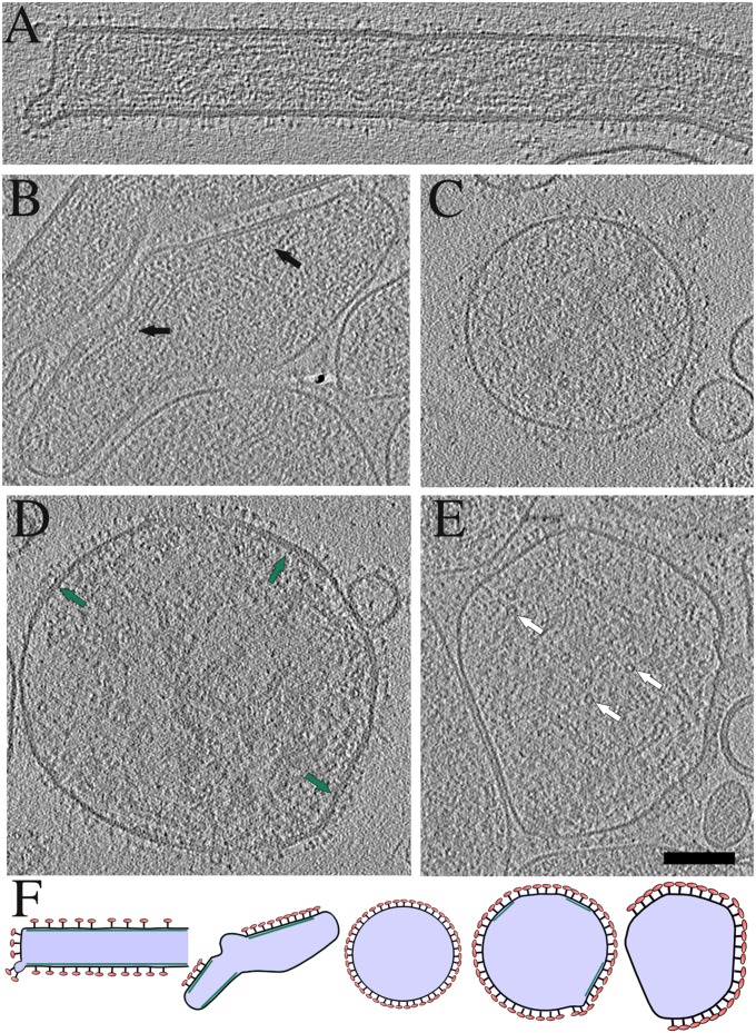
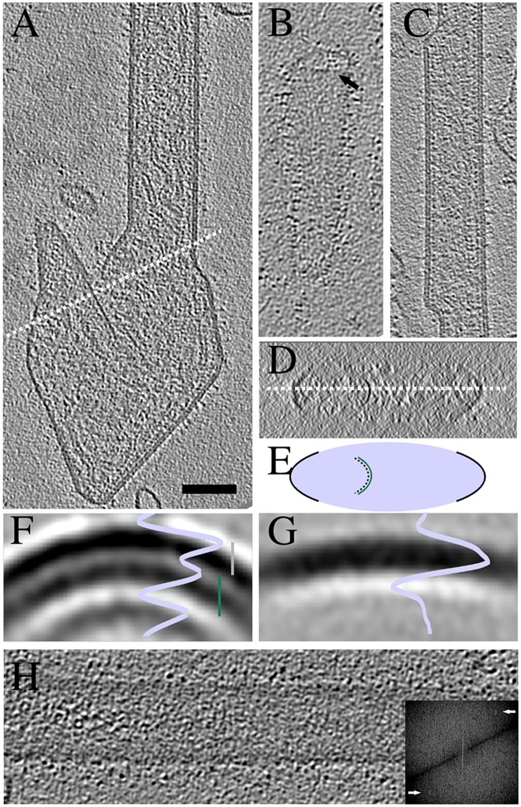
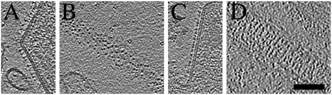
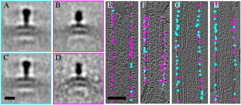
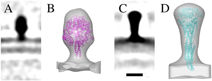
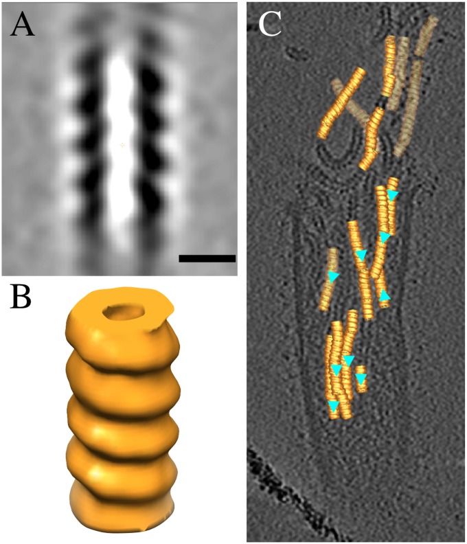
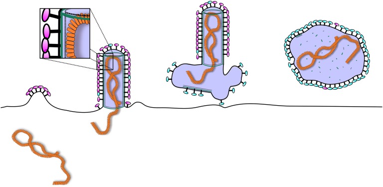
Human respiratory syncytial virus is a human pathogen that causes severe infection of the respiratory tract. Current information about the structure of the virus and its interaction with host cells is limited. We carried out an electron cryotomographic characterization of cell culture-grown human respiratory syncytial virus to determine the architecture of the virion. The particles ranged from 100 nm to 1,000 nm in diameter and were spherical, filamentous, or a combination of the two. The filamentous morphology correlated with the presence of a cylindrical matrix protein layer linked to the inner leaflet of the viral envelope and with local ordering of the glycoprotein spikes. Recombinant viruses with only the fusion protein in their envelope showed that these glycoproteins were predominantly in the postfusion conformation, but some were also in the prefusion form. The ribonucleocapsids were left-handed, randomly oriented, and curved inside the virions. In filamentous particles, they were often adjacent to an intermediate layer of protein assigned to M2-1 (an envelope-associated protein known to mediate association of ribonucleocapsids with the matrix protein). Our results indicate important differences in structure between the Paramyxovirinae and Pneumovirinae subfamilies within the Paramyxoviridae, and provide fresh insights into host cell exit of a serious pathogen.

Keywords: cryo-ET; paramyxovirus; virus structure.


Conflict of interest statement

The authors declare no conflict of interest.

Figures

Fig. 1.
Fig. 1.
Tomography of A2 HRSV virions. Virion morphology ranges from completely filamentous (A) to completely spherical (C) with intermediate forms (B and D) that have some tubularly curved parts but are otherwise spherically curved. Spherical particles are highly deformable when in the proximity of other particles and the membrane proximal to the neighboring particle is free of glycoprotein spikes (E). Virions in A–E are illustrated schematically in F in alphabetical order. Black arrows: side views of the RNP; white arrows: top views of the RNP; green arrows: secondary density layer under the membrane in a spherical particle. (Scale bar, 100 nm.) Tomographic slices are 3.8-nm-thick.
Fig. 2.
Fig. 2.
Assembly of M causes curvature of the virion membrane. (A) A tomographic slice of a filamentous A2 virion with a large irregular appendage at one end. White dotted line cuts through a curved M layer detached from the membrane. An orthogonal view around the dotted line in A is shown in D and schematically in E. (B) A tomographic slice of an A2 virion without M layer in the particle tip viewed from the top at the level of the M layer. Arrow indicates the end of the M layer. (C) A tomographic slice of an rgRSVΔG filamentous virion with varying diameter. (F) A radial profile and a slice of a subvolume average from A2 filamentous particles. Distance from membrane to M layer (gray bar) is 6.1 nm and from M to the third, innermost layer (green bar) 6.9 nm. (G) A radial profile and a slice of a subvolume average from A2 spherical particles. (H) A tomographic slice from the top of a filamentous virion. Fourier transform of the corresponding slice is shown in the Inset. Peaks corresponding to a layer line at ∼8 nm are shown with arrows. Slices are 0.77-nm-thick in A–C, F, and G; 11.5 nm in D, and 3.8 nm in H. (Scale bar, 100 nm for A–D and H.)
Fig. 3.
Fig. 3.
Virions have two types of spikes that are organized differently on the surface. Central tomographic slices from virions with long (A) or short (C) spikes. Top slices from virions with long (B) or short (D) spikes. Slices are 0.77-nm-thick. (Scale bar, 100 nm.)
Fig. 4.
Fig. 4.
F occurs in two different conformations on the virions. Averages of class 1 (253 subvolumes) representing the long (A) and class 2 (226 subvolumes) representing the short spike (B) from subvolumes extracted from both A2 and rgΔGΔSH virions. Class averages 1 (224 subvolumes) and 2 (65 subvolumes) including only subvolumes from rgΔGΔSH are shown (C and D). Positions of the classified spikes on the A2 virions (E and F) and on the rgΔGΔSH virions (G and H). Tomographic slices in E–H are transparent to show all spike positions. Cyan spheres correspond to class 1 spikes and magenta spheres to class 2 spikes. (AD) Scale bar in (C) is 10 nm. (EH) Scale bar in (E) is 100 nm.
Fig. 5.
Fig. 5.
F spike structures in pre- and postfusion conformation. Central sections (0.77-nm-thick) of the subvolume averages of F from A2(s) (A) and A2(l) (C). Crystal structures of PIV5 F in the prefusion conformation (PDB ID code 4GIP) (21) and HRSV F in the postfusion conformation (PDB ID code 3RRT) (14) fitted into the subvolume average density of the short (B) and the long (D) spike. (Scale bar, 10 nm.) The isosurfaces in B and D were rendered at 2 σ and at 5 σ from the mean, respectively.
Fig. 6.
Fig. 6.
The RNP of HRSV is left-handed. A central section (A) and an isosurface representation of the HRSV RNP (B). (Scale bar, 10 nm.) (C) A transparent tomographic slice showing the refined positions and orientations of the RNP subvolumes on the tomogram. Cyan triangles indicate the direction of the RNP helix. The isosurface was rendered at 1 σ from the mean.
Fig. 7.
Fig. 7.
Schematic model of assembly of HRSV. Initially, the F glycoproteins (magenta) gather at the plasma membrane enriching in lipid rafts and initiate a bud. M protein (green) is recruited to the budding site via interactions with the glycoprotein tails and the membrane. Interactions between M and G promote incorporation of G into the bud. Assembly of M into tubular structure provides the force for the elongation of the virion and recruits the RNP into the nascent filament. This recruitment can be mediated by the M2-1 protein (dashed line). Once the RNP is inside the budding virion, release occurs via a currently unknown endosomal sorting complexes required for transport-independent mechanism. The virion draws a variable amount of membrane with it and this membrane remains as an appendage or a larger membrane sack in the end of the filament. Some of the virions then convert into roughly spherical forms as the M disassembles from the membrane with conversion of F to the postfusion-like conformation (cyan).

References

    1. Falsey AR, Hennessey PA, Formica MA, Cox C, Walsh EE. Respiratory syncytial virus infection in elderly and high-risk adults. N Engl J Med. 2005;352(17):1749–1759. - PubMed
    1. Nair H, et al. Global burden of acute lower respiratory infections due to respiratory syncytial virus in young children: A systematic review and meta-analysis. Lancet. 2010;375(9725):1545–1555. - PMC - PubMed
    1. Group TI-RS Palivizumab, a humanized respiratory syncytial virus monoclonal antibody, reduces hospitalization from respiratory syncytial virus infection in high-risk infants. The IMpact-RSV Study Group. Pediatrics. 1998;102(3 Pt 1):531–537. - PubMed
    1. Battisti AJ, et al. Structure and assembly of a paramyxovirus matrix protein. Proc Natl Acad Sci USA. 2012;109(35):13996–14000. - PMC - PubMed
    1. Liljeroos L, Huiskonen JT, Ora A, Susi P, Butcher SJ. Electron cryotomography of measles virus reveals how matrix protein coats the ribonucleocapsid within intact virions. Proc Natl Acad Sci USA. 2011;108(44):18085–18090. - PMC - PubMed

Publication types

MeSH terms

Substances