. 2013 Jun 12;8(6):e63676.
doi: 10.1371/journal.pone.0063676. Print 2013.

Ataxia-telangiectasia group D complementing gene (ATDC) promotes lung cancer cell proliferation by activating NF-κB pathway

Affiliations

Ataxia-telangiectasia group D complementing gene (ATDC) promotes lung cancer cell proliferation by activating NF-κB pathway

Zhong-Ping Tang et al. PLoS One. .

Abstract

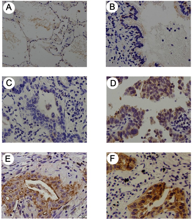
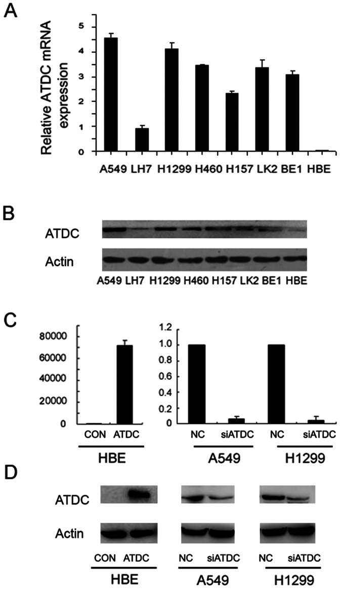
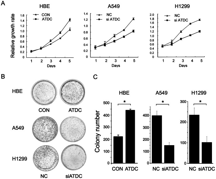
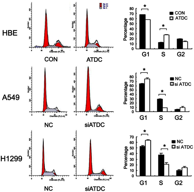
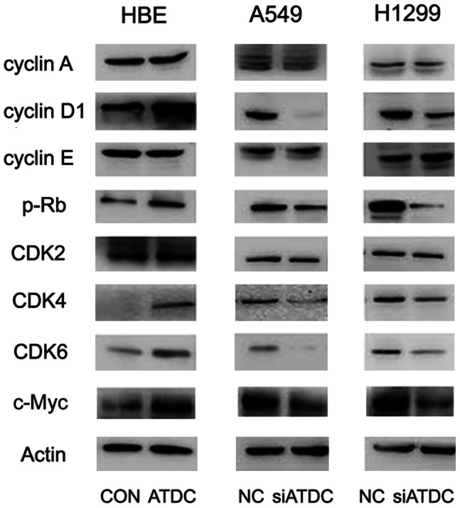
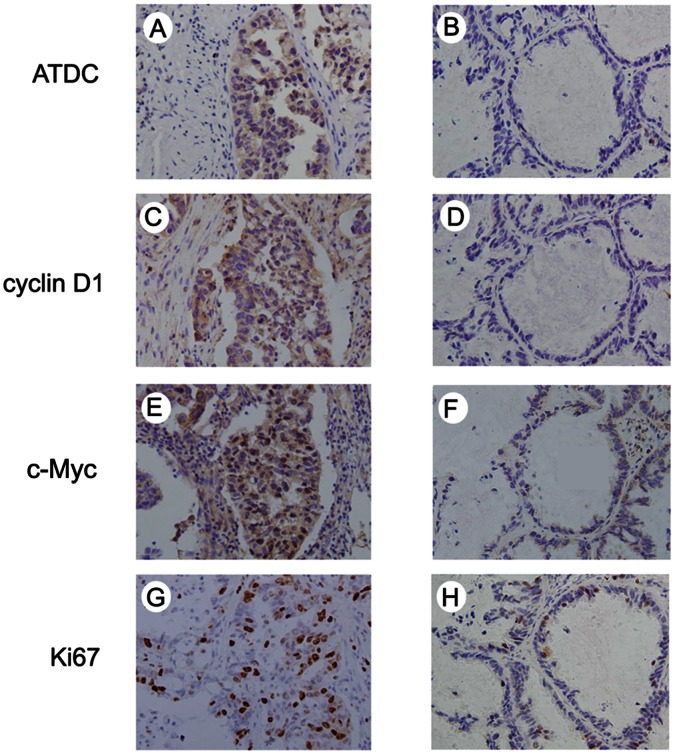
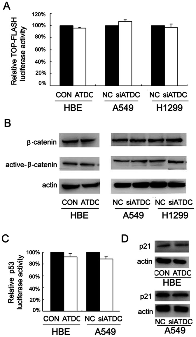
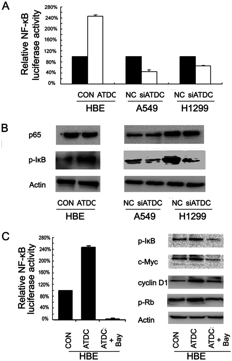
Previous studies suggested Ataxia-telangiectasia group D complementing gene (ATDC) as an oncogene in many types of cancer. However, its expression and biological functions in non-small cell lung cancer (NSCLC) remain unclear. Herein, we investigated its expression pattern in 109 cases of human NSCLC samples by immunohistochemistry and found that ATDC was overexpressed in 62 of 109 NSCLC samples (56.88%). ATDC overexpression correlated with histological type (p<0.0001), tumor status (p = 0.0227) and histological differentiation (p = 0.0002). Next, we overexpressed ATDC in normal human bronchial epithelial cell line HBE and depleted its expression in NSCLC cell lines A549 and H1299. MTT and colony formation assay showed that ATDC overexpression promoted cell proliferation while its depletion inhibited cell growth. Furthermore, cell cycle analysis showed that ATDC overexpression decreased the percentage of cells in G1 phase and increased the percentage of cells in S phase, while ATDC siRNA treatment increased the G1 phase percentage and decreased the S phase percentage. Further study revealed that ATDC overexpression could up-regulate cyclin D1 and c-Myc expression in HBE cells while its depletion down-regulated cyclin D1 and c-Myc expression in A549 and H1299 cells. In addition, ATDC overexpression was also associated with an increased proliferation index, cyclin D1 and c-Myc expression in human NSCLC samples. Further experiments demonstrated that ATDC up-regulated cyclin D1 and c-Myc expression independent of wnt/β-catenin or p53 signaling pathway. Interestingly, ATDC overexpression increased NF-κB reporter luciferase activity and p-IκB protein level. Correspondingly, NF-κB inhibitor blocked the effect of ATDC on up-regulation of cyclin D1 and c-Myc. In conclusion, we demonstrated that ATDC could promote lung cancer proliferation through NF-κB induced up-regulation of cyclin D1 and c-Myc.


Conflict of interest statement

Competing Interests: The authors have declared that no competing interests exist.

Figures

Figure 1
Figure 1. Immunohistochemical staining of ATDC in lung cancer tissue and paired normal lung tissue.
A. Negative staining in normal pneumocytes in the alveoli of non-neoplastic lung tissue. B. Negative staining in normal bronchial epithelium in non-neoplastic lung tissue. C. Negative ATDC staining in lung adenocarcinoma. D. Weak ATDC staining in lung adenocarcinoma. E. Weak ATDC staining in lung squamous cell carcinoma. F. Strong ATDC staining in lung squamous cell carcinoma.
Figure 2
Figure 2. ATDC overexpression and knockdown in lung cancer cell lines.
A. mRNA expression levels of ATDC analyzed by Real-time PCR in a panel of lung cancer cell lines. Note the highest expression is observed in A549 cell line and the lowest is seen in HBE cell. B. protein expression levels of ATDC analyzed by Western blot in a panel of lung cancer cell lines. Note a direct correlation of the protein level (B) with the transcripts (A) in each cell line. C. Real-time PCR analysis of ATDC overexpression efficiency in HBE cells and depletion efficiency in A549 and H1299 cells at a transcriptional level. D. Western blot analysis of ATDC overexpression efficiency in HBE cells and depletion efficiency in A549 and H1299 cells at a protein level. The band of lower MW in H1299 cells is a protein degradation product. Note a direct correlation of the protein level (D) with the transcripts (C) in each cell line.
Figure 3
Figure 3. ATDC promotes cell proliferation in lung cancer cell lines.
A. MTT assay shows that ATDC transfection in HBE cells promotes cell growth and ATDC knockdown in A549 and H1299 cells inhibits cell proliferation. B–C. Assessment of clonogenic potentials of the ATDC overexpressing cells and ATDC depleted cells by counting colony numbers. A remarkable increase in colony numbers was observed in the HBE cells with ATDC overexpression in comparison with control, and the number of colonies formed by A549 and H1299 cells treated with ATDC siRNA was far less than that of control cells. Columns in C represent the mean value of 3 duplicates; bars represent standard deviation . * indicates a statistical significance in difference between the indicated 2 bar values (p<0.05).
Figure 4
Figure 4. ATDC regulates cell cycle progression in lung cancer cells.
Note ATDC overexpression in HBE cells increases S phase cells and decreases G1 phase cells (p<0.05 compared with control). ATDC knockdown in A549 and H1299 cells increases G1 phase cells and decreases S phase cells (p<0.05 compared with control).
Figure 5
Figure 5. ATDC upregulates Cyclin D1 and c-Myc expression in lung cancer cells.
Western blotting analysis reveals that ATDC transfection increases Cyclin D1 and c-Myc expression in HBE cells and knockdown of ATDC decreases the protein levels of cyclin D1 and c-Myc in both A549 and H1299 cells, without significant changes in Cyclin A and Cyclin E expression.
Figure 6
Figure 6. ATDC expression correlates directly with index of Ki-67 labeling and expression of cyclinD1 and c-Myc in NSCLC tissues.
Immunohistochemical staining of ATDC (A and B), cyclin D1 (C and D), c-Myc (E and F) and Ki67 (G and H) in NSCLC specimens. A, C, E and G indicate a representative case with positive ATDC demonstrating a high level of cyclin D1 and c-Myc, a high labeling index of ki67, while B, D, F and H represent a case with negative ADTC and corresponding negative cyclin D1 and c-Myc staining or low level of ki67 labeling.
Figure 7
Figure 7. ATDC does not seem to be involved in regulating Wnt or p53 activity in lung cancer cell lines.
A. There was no significant change of Topflash luciferase activity after ATDC transfection in HBE cells and after siRNA treatment in A549 and H1299 cells. B. There was no significant change of β-catenin and active β-catenin protein levels after ATDC transfection in HBE cells and after siRNA treatment in A549 and H1299 cells. C. ATDC transfection in HBE cells or its depletion in A549 cells did not change the level of p53 luciferase activity. D. ATDC transfection in HBE or its depletion in A549 cells did not change the protein level of p53 target gene p21.
Figure 8
Figure 8. ATDC up-regulates Cyclin D1 and c-Myc via activation of NF-κB signaling pathway.
A. ATDC overexpression up-regulated NF-κB reporter luciferase activity in HBE cells and ATDC depletion inhibited NF-κB reporter luciferase activity in both A549 and H1299 cells. B. ATDC transfection increased p-IκB expression in HBE cells and ATDC depletion decreased the level of p-IκB in A549 and H1299 cells. C. NF-κB inhibitor Bay 11-7082 completely blocked NF-κB reporter luciferase activity and reversed the effect of ATDC on cyclin D1, c-Myc and p-Rb up-regulation.

References

    1. Minna JD, Roth JA, Gazdar AF (2002) Focus on lung cancer. Cancer Cell 1: 49–52. - PubMed
    1. Jemal A, Siegel R, Xu J, Ward E Cancer statistics, 2010. CA Cancer J Clin 60: 277–300. - PubMed
    1. Poste G, Fidler IJ (1980) The pathogenesis of cancer metastasis. Nature 283: 139–146. - PubMed
    1. Schiller JH, Harrington D, Belani CP, Langer C, Sandler A, et al. (2002) Comparison of four chemotherapy regimens for advanced non-small-cell lung cancer. N Engl J Med 346: 92–98. - PubMed
    1. Reymond A, Meroni G, Fantozzi A, Merla G, Cairo S, et al. (2001) The tripartite motif family identifies cell compartments. EMBO J 20: 2140–2151. - PMC - PubMed

Publication types

MeSH terms