. 2013 Jun 12;8(6):e65235.
doi: 10.1371/journal.pone.0065235. Print 2013.

Inhibition of fast axonal transport by pathogenic SOD1 involves activation of p38 MAP kinase

Affiliations

Inhibition of fast axonal transport by pathogenic SOD1 involves activation of p38 MAP kinase

Gerardo A Morfini et al. PLoS One. .

Abstract

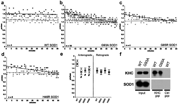
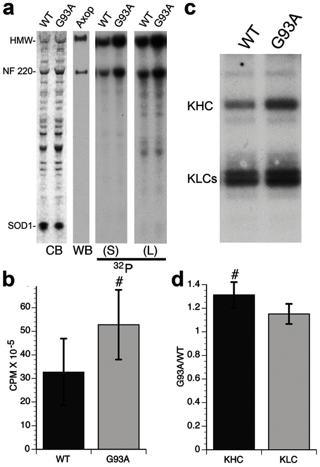
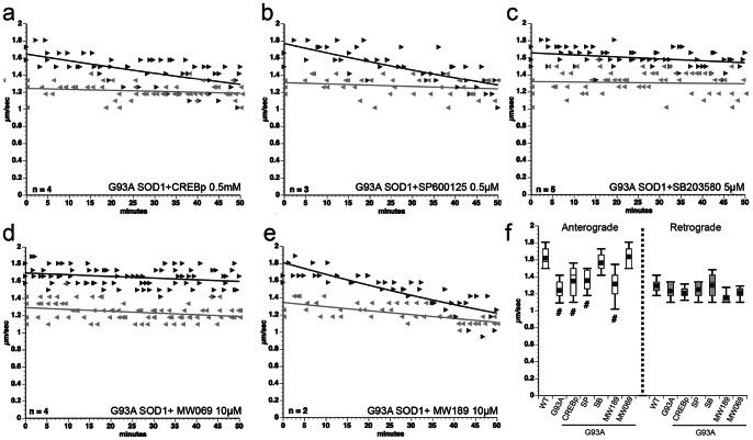
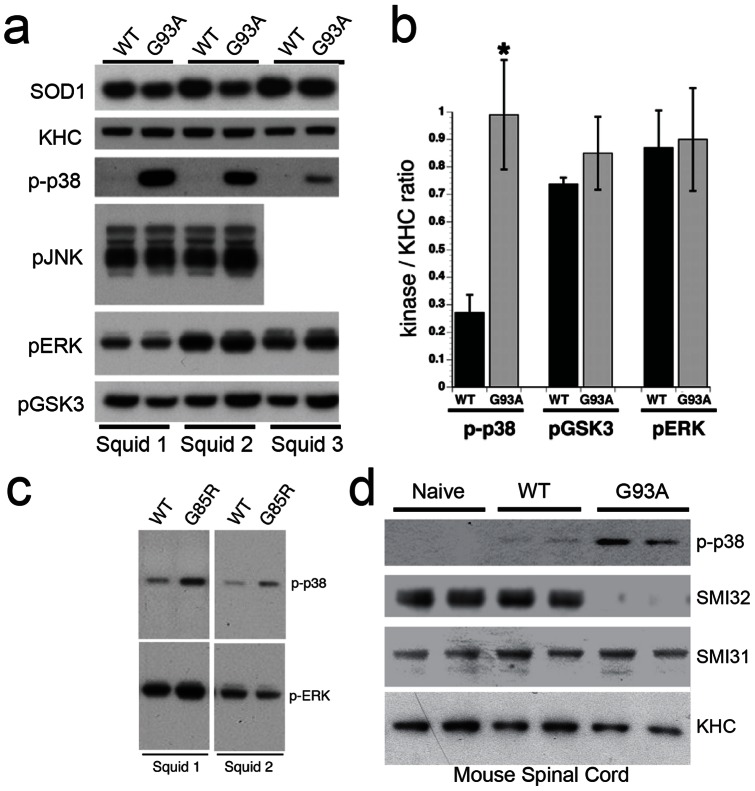
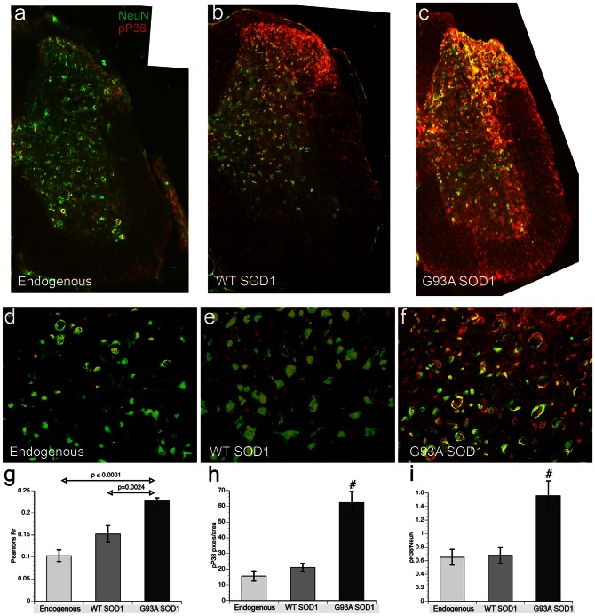
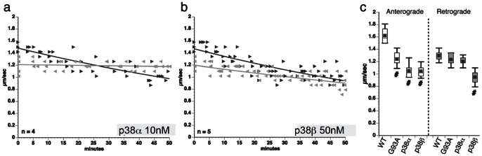
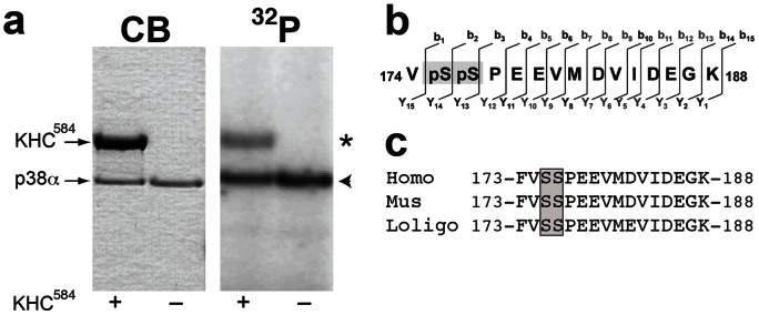
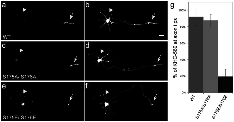
Dying-back degeneration of motor neuron axons represents an established feature of familial amyotrophic lateral sclerosis (FALS) associated with superoxide dismutase 1 (SOD1) mutations, but axon-autonomous effects of pathogenic SOD1 remained undefined. Characteristics of motor neurons affected in FALS include abnormal kinase activation, aberrant neurofilament phosphorylation, and fast axonal transport (FAT) deficits, but functional relationships among these pathogenic events were unclear. Experiments in isolated squid axoplasm reveal that FALS-related SOD1 mutant polypeptides inhibit FAT through a mechanism involving a p38 mitogen activated protein kinase pathway. Mutant SOD1 activated neuronal p38 in mouse spinal cord, neuroblastoma cells and squid axoplasm. Active p38 MAP kinase phosphorylated kinesin-1, and this phosphorylation event inhibited kinesin-1. Finally, vesicle motility assays revealed previously unrecognized, isoform-specific effects of p38 on FAT. Axon-autonomous activation of the p38 pathway represents a novel gain of toxic function for FALS-linked SOD1 proteins consistent with the dying-back pattern of neurodegeneration characteristic of ALS.


Conflict of interest statement

Competing Interests: The authors have declared that no competing interests exist.

Figures

Figure 1
Figure 1. FALS-linked mutant SOD1 proteins selectively inhibit anterograde, conventional kinesin-dependent FAT.
(a–d) Vesicle motility assays in isolated squid axoplasm. Individual velocity (µm/sec) rate measurements (arrowheads) are plotted as a function of time (minutes). Dark arrowheads and lines represent anterograde, conventional kinesin-dependent rates. Grey arrows and lines represent retrograde, cytoplasmic dynein-dependent rates. Perfusion of WT-SOD1 protein (a) in axoplasm shows no effect on either direction. In contrast, perfusion of G93A-SOD1 specifically inhibits anterograde, but not retrograde FAT (b). Similar results were obtained after perfusion of SOD1-G85R (c) and SOD1-H46R (d). n: number of experiments. (e) 30 to 50 minutes after perfusion, anterograde FAT rates were significantly lower in axoplasms perfused with mSOD1, than in axoplasms perfused with either WT-SOD1 (#: p≤0.01) or control buffer (not shown). All pathogenic SOD1 proteins tested had no effect on retrograde FAT. (f) Kinesin-1 (KHC IPP) was immunoprecipitated from spinal cord lysates of transgenic mice expressing either WT-SOD1 (WT) or G93A-SOD1 (G93A). An aliquot of lysate (Input) was included as positive control. Immunoblots using anti-kinesin-1 antibody (KHC) demonstrated effective immunoprecipitation from lysates, regardless of genotype. Specificity was confirmed by the absence of kinesin-1 on control immunoprecipitates with non-immune mouse IgG (Ctrl IPP). Immunoblot with an anti-SOD1 monoclonal antibody (D3H5) failed to detect SOD1 in kinesin-1 immunoprecipitates, suggesting that neither WT SOD1 nor G93A-SOD1 interact directly with kinesin-1.
Figure 2
Figure 2. Pathogenic SOD1 increases neurofilament phosphorylation.
Phosphorylation of squid neurofilaments (NF) in isolated “sister” axoplasms (see Methods) was analyzed using metabolic labeling experiments with 32P-γ-ATP. (a) Coomassie Blue staining (CB) shows similar levels of perfused WT-SOD1, G93A-SOD1 and total axoplasmic proteins. Immunoblot analysis (WB) with the NFH antibody SMI-31 confirmed the identity of major phosphorylated bands as NF220 and HMW, major NF subunits in squid axoplasm . Short (S) and long (L) exposure of autoradiograms (32P) show increased phosphorylation of NF220 and HMW NF subunits in axoplasms perfused with G93A-SOD1, compared to WT-SOD1. (b) Quantitation of squid NF phosphorylation showed ⋍70% increase in G93A-SOD1 treated axoplasms, compared to those treated with WT-SOD1 (p≤0.01 (#) in a paired t-test). (c) In parallel experiments, kinesin-1 was immunoprecipitated from axoplasms labeled with γ-32P-ATP in the presence of WT-SOD1 or G93A-SOD1. Both heavy (KHC) and light (KLC) chains of conventional kinesin were phosphorylated. (d) The ratio of counts for G93A-SOD1/WT-SOD1 indicates that KHC labeling increased 31% in G93A-SOD1 axoplasms, compared to WT-SOD1 (significant at p≤0.05 by paired t-test, #). KLC phosphorylation increased by 15%, but was not statistically significant (p = 0.123). n = 7.
Figure 3
Figure 3. p38 MAPK mediates the inhibition of anterograde FAT by pathogenic SOD1.
(a–e) Co-perfusion of G93A-SOD1 with either CREBp (a competitive inhibitor of GSK3) (a) or SP600125 (a pharmacological inhibitor of JNK) (b) did not block effects of G93A-SOD1 on anterograde FAT, but co-perfusion with SB203580 (an inhibitor of p38 and JNK2/3) completely blocked effects of mutant G93A-SOD1 on anterograde FAT (c). MW01-2-069SRM (MW069) , a selective inhibitor of p38α structurally unrelated to SB203580 also blocked inhibition of FAT (d), but an inactive MW069 analog MW01-6-189WH (MW189) did not (e). n = number of experiments. (f) Quantitation of a–e shows that SB203580 (SB) and MW069 compounds, but not CREBp, SP600125 (SP) or MW189 compounds blocked the effects of G93A-SOD1 on anterograde FAT (# indicates different from WT SOD1 at p≤0.001 by pooled t-test). Plots show the mean, standard deviation, maximum and minimum of FAT rates recorded 30–50 min for each experimental condition.
Figure 4
Figure 4. Activation of p38 MAPK by pathogenic SOD1 in axoplasm and mouse spinal cord.
(a) Immunoblots with phosphoantibodies show activation of p38 (p-p38) in axoplasms perfused with G93A-SOD1 (G93A), compared to WT-SOD1 (WT). No changes were found in activities of GSK3 (p-GSK3), JNK (p-JNK) or ERK (p-ERK) with perfusion of SOD1. An SOD1 antibody confirmed that similar levels of WT-SOD1 and G93A-SOD1 were perfused, and kinesin-1 (KHC) antibodies serve as a loading control for axoplasmic protein. Representative results from three independent experiments (Squids 1–3) are shown. (b) Quantitation of blots reveals a 3–4 fold increase in p-p38 with G93A SOD1, compared to WT-SOD1 (n = 8; p≤0.05 (#) by a t-test). No significant differences were found in levels of activated GSK3 (n = 3) or ERK (n = 3) (c) Increased activation of p38, but not ERK was also seen in axoplasms perfused with G85R-SOD1 (G85R) polypeptides. (d) Phosphorylation of both neurofilaments (NF) and p38 MAPK was analyzed in spinal cords of age-matched (50 days old) non-transgenic (Naïve), human WT-SOD1 (WT) transgenic and human G93A-SOD1 (G93A) transgenic mice using phosphorylation sensitive antibodies. Kinesin heavy chain (KHC) blots show similar levels of protein loading. SMI32 antibodies recognize a dephosphorylated epitope in NFH that can be phosphorylated by MAPKs, whereas SMI31 recognizes an epitope not affected by phosphorylation with MAPKs . SMI31 immunoreactivity showed similar levels in all mice and serves as a second loading control. In contrast, SMI32 reactivity is reduced in G93A-SOD1 mice, but not naïve and WT-SOD1 mice, suggesting increased phosphorylation of NFs by MAPKs in FALS. Accordingly, p38 activity (p-p38) was increased in spinal cord of G93A-SOD1 mice, with a slight activation in WT-SOD1 mice.
Figure 5
Figure 5. Increased Phospho-p38 in ventral horn of lumbar spinal cord in presymptomatic G93A-SOD1 mice.
Spinal cords from 60 days old naïve nontransgenic mice (endogenous) and transgenic mice overexpressing either human WT- or G93A-SOD1 were analyzed by immunohistochemistry for cellular distribution of activated p38 MAPK. (a–c) Low magnification (10X objective) images of lumbar spinal cord double-labeled with a neuronal marker (NeuN, in green) and activated p38 MAPK (pP38, in red). (d–f) Higher magnification images (25× objective) of ventral horns. In naïve mice spinal cords with only endogenous mouse SOD1 (a, d), neurons are readily visible in the both dorsal and ventral horns (green), but p38 MAPK activity (red) is low with a few neurons positive for both markers (yellow), primarily in the dorsal horn. In transgenic WT-SOD1 spinal cord (b, e), neuronal staining is comparable to naïve mice, but p-p38 immunoreactivity clearly increases in regions with minimal NeuN staining, suggesting increased p38 MAPK activation in glia. In G93A-SOD1 spinal cord (c, f),p-p38 immunoreactivity is much higher in both white and grey matter regions,. An increase in pP38 co-localization with NeuN, is consistent with activation of p38 MAPK in neurons. (g–i) Semi-quantitative fluorescence analysis of high magnification sections shows an increase in co-localization of pP38 and NeuN immunoreactivity with G93A-SOD mice (n = 9 for all conditions). (g) Pearson's correlation coefficient (PCC) (showing co-localization, but not intensity levels) of NeuN and pP38 significantly increased in both transgenic mice relative to naïve mice, not quite rising to significance between naïve and WT-SOD1 (p = 0.058). In G93A-SOD1 mice, PPC values were significantly increased over both naïve and WT-SOD1 mice (p<0.0001). (h) To address relative levels of pP38 immunoreactivity, we compared red pixels/area. The difference between naïve and WT-SOD1 mice was not significant, but G93A-SOD1 mice was significantly different from both naïve and WT-SOD1 mice at p≤0.0001 (#) (i) An increased ratio of pP38/NeuN (R/G ratio) for G93A-SOD1 mice suggests that mutant SOD1 activates p38 in neurons. Differences between G93A-SOD1 and both naïve and WT-SOD1 mice were significant at p≤0.005 (#).
Figure 6
Figure 6. Active p38 α mimics the effects of pathogenic SOD1 on anterograde FAT.
Effects of active, recombinant p38 isoforms on FAT were evaluated using vesicle motility assays in isolated squid axoplasm. P38α and P38β were perfused at a constant specific activity based on in vitro kinase assays with the ATF-2 substrate. (a) Perfusion of active p38α in axoplasm selectively inhibited anterograde FAT, as did pathogenic SOD1 (compare to Fig. 1b-d). (b) Unlike p38α, p38β inhibited both anterograde and retrograde FAT. (c) Quantitation of values obtained between 30-50 minutes shows that p38α most closely mimicked effects of pathogenic SOD1, suggesting this isoform mediates the effects of mSOD1 on FAT in axoplasm (#: difference significant from WT-SOD1 at p<0.01 by t-test).
Figure 7
Figure 7. Active p38 α directly phosphorylates kinesin-1 at serines 175–176.
(a) Recombinant p38α was incubated with γ-32P-ATP in the presence (+) or absence (–) of a recombinant protein construct comprising the first 584 amino acids of kinesin-1 (KHC584). Coomassie blue staining of gels shows the position of KHC584 and p38α. Autoradiogram (32P) shows 32P incorporation into KHC584 (asterisk), as well as autophosphorylation of p38α (arrowhead). (b) Mass spectrometry studies identified a peptide within the motor domain of kinesin-1 (amino acids 174–188) showing unequivocal evidence of phosphorylation by p38α. Tandem mass spectrometry analysis (MS/MS) by collision-induced dissociation further mapped phosphorylation on both Ser175 and Ser176 (grey box). (c) Sequence alignment shows that serines 175 and 176 (grey box) are conserved among human, mouse and squid sequences for kinesin-1.
Figure 8
Figure 8. Pseudophosphorylation of kinesin-1 at S175/S176 inhibits movement of kinesin-1.
To determine the effects of modifying S175 and S176 on kinesin-1function, recombinant GFP-tagged kinesin (KHC559) was modified to preclude phosphorylation at these sites (S175AS176A) or to mimic phosphorylation (S175ES176E). (a–f) Stage 3 hippocampal neurons were examined 5 h after co-transfection with GFP-tagged KHC559 constructs and a tdTomato construct, which diffuses throughout the cell and allows visualization of neurites (b, d, f). Both wild-type kinesin-1 (KHC559 WT, a) and a non-phosphorylatable mutant (KHC559 S175A/S176A, c) accumulated efficiently at axonal tips (labeled by arrows) with minimal steady-state labeling of cell bodies (arrowheads). In contrast, pseudophosphorylated mutant KHC559 S175E/S176E, e) was mainly present in neuronal cell bodies. Quantitative immunofluorescence analysis shows fraction of total KHC559 fluorescence at axon tips for all constructs (g). Far less phosphomimicking KHC559 S175E/S176E constructs accumulated at axon tips than KHC559 WT or KHC559 S175A/S176A (#: p<0.001; n: 27–43 cells per condition). Bars show mean and standard deviation. Scale bar  = 20 µm.
Figure 9
Figure 9. Inhibition of anterograde FAT induced by mSOD1 depends on specific MKKK-MKK interactions.
Co-perfusion of G93A-SOD1 with DVD peptide (a), but not with the Mixed-Lineage Kinase inhibitor CEP11004 (b), prevents inhibition of FAT induced by G93A-SOD1. DVD peptide prevents activation of MKKs by some MKKKs (n =  number of axoplasms) . These data suggest that the activation of p38 and the inhibition of FAT induced by G93A-SOD1 involves activation of one or more MAPKKKs other than MLKs. (c) The DVD peptide also blocks inhibition of FAT by oxidized WT-SOD1 suggesting that FALS mutant SOD1 and misfolded WT-SOD1 activate a common p38 MAPK pathway .

References

    1. Bruijn LI, Miller TM, Cleveland DW (2004) Unraveling the mechanisms involved in motor neuron degeneration in ALS. Annu Rev Neurosci 27: 723–749. - PubMed
    1. DeJesus-Hernandez M, Mackenzie IR, Boeve BF, Boxer AL, Baker M, et al. (2011) Expanded GGGGCC hexanucleotide repeat in noncoding region of C9ORF72 causes chromosome 9p-linked FTD and ALS. Neuron 72: 245–256. - PMC - PubMed
    1. Renton AE, Majounie E, Waite A, Simon-Sanchez J, Rollinson S, et al. (2011) A hexanucleotide repeat expansion in C9ORF72 is the cause of chromosome 9p21-linked ALS-FTD. Neuron 72: 257–268. - PMC - PubMed
    1. Dion PA, Daoud H, Rouleau GA (2009) Genetics of motor neuron disorders: new insights into pathogenic mechanisms. Nat Rev Genet 10: 769–782. - PubMed
    1. Pasinelli P, Brown RH (2006) Molecular biology of amyotrophic lateral sclerosis: insights from genetics. Nat Rev Neurosci 7: 710–723. - PubMed

Publication types

MeSH terms

Supplementary concepts