. 2013 Jun 11;8(6):e66029.
doi: 10.1371/journal.pone.0066029. Print 2013.

Cholecystokinin-mediated RhoGDI phosphorylation via PKCα promotes both RhoA and Rac1 signaling

Affiliations

Cholecystokinin-mediated RhoGDI phosphorylation via PKCα promotes both RhoA and Rac1 signaling

Maria Eugenia Sabbatini et al. PLoS One. .

Abstract

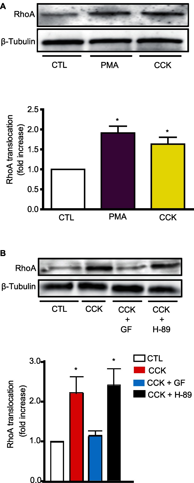
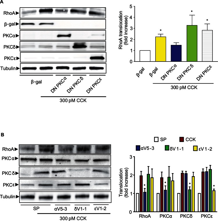
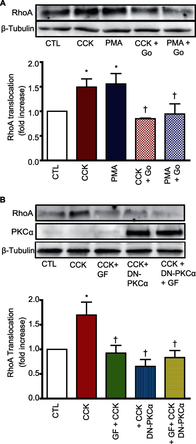
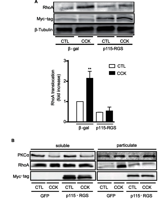
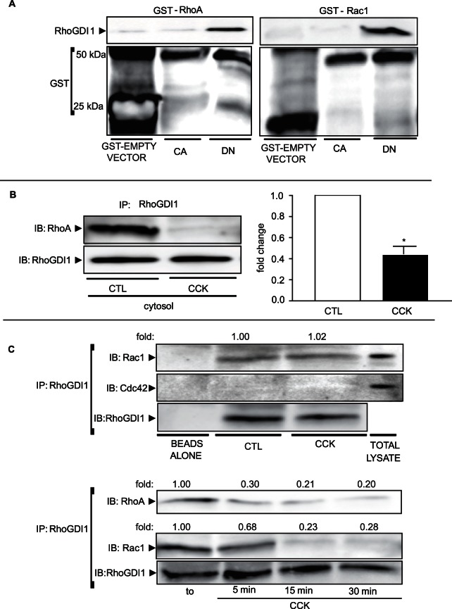
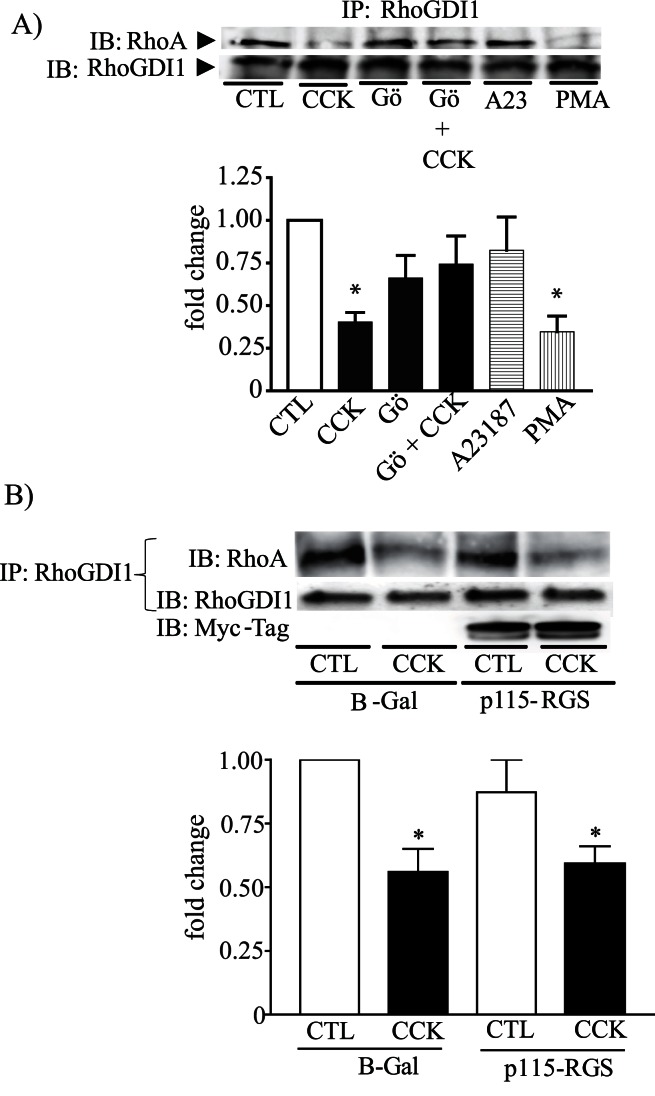
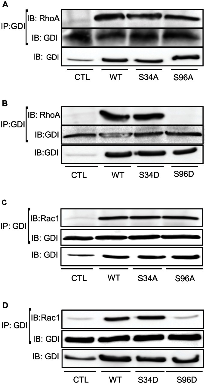
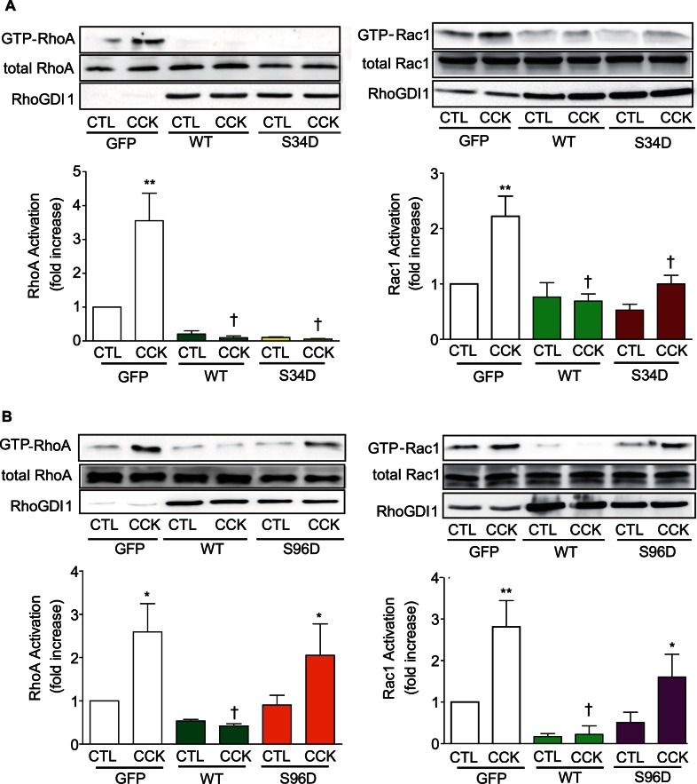
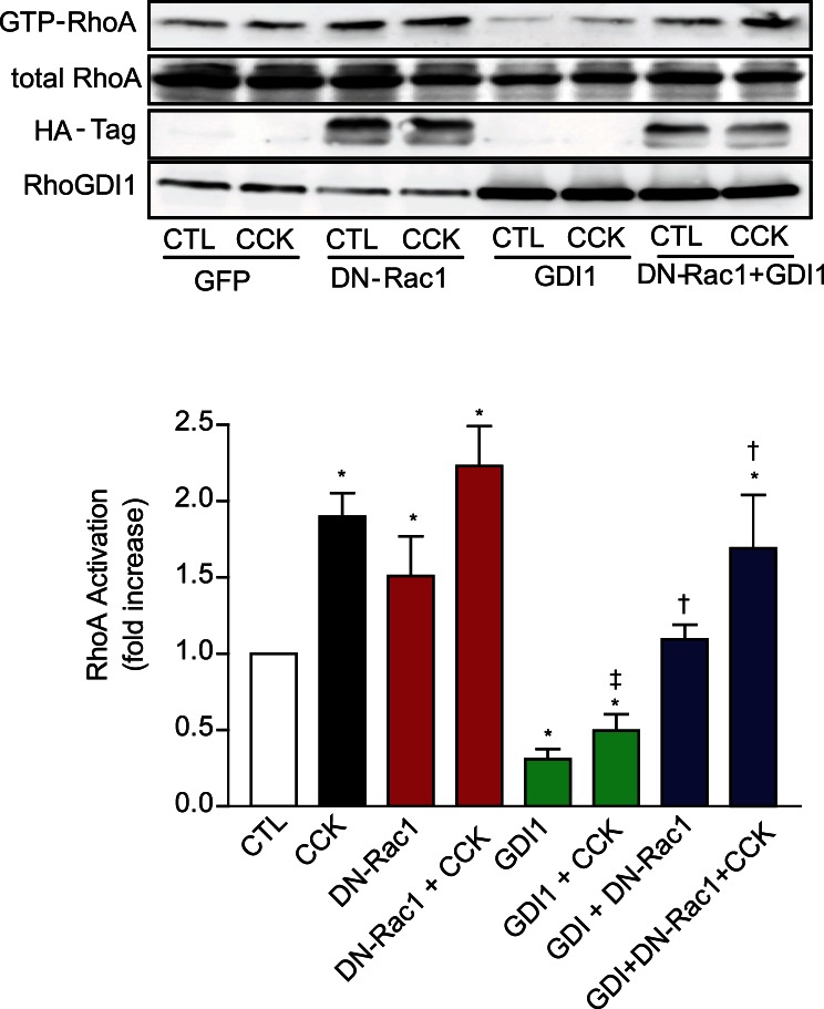
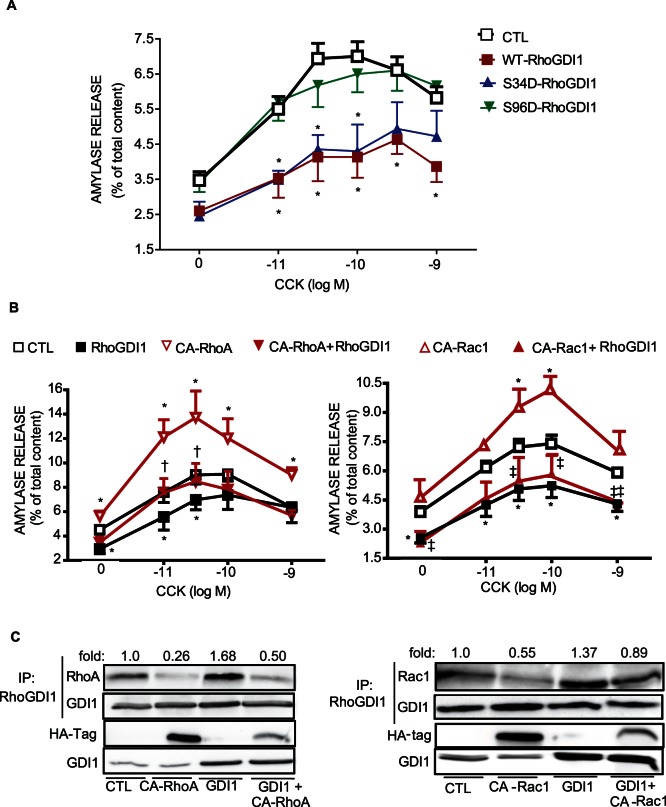
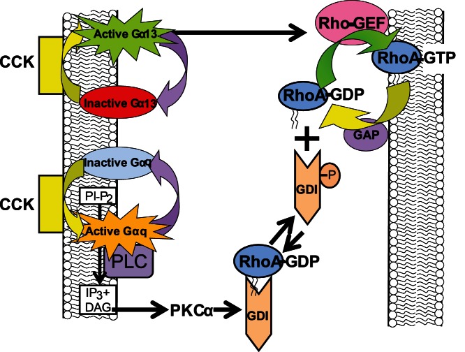
RhoA and Rac1 have been implicated in the mechanism of CCK-induced amylase secretion from pancreatic acini. In all cell types studied to date, inactive Rho GTPases are present in the cytosol bound to the guanine nucleotide dissociation inhibitor RhoGDI. Here, we identified the switch mechanism regulating RhoGDI1-Rho GTPase dissociation and RhoA translocation upon CCK stimulation in pancreatic acini. We found that both Gα13 and PKC, independently, regulate CCK-induced RhoA translocation and that the PKC isoform involved is PKCα. Both RhoGDI1 and RhoGDI3, but not RhoGDI2, are expressed in pancreatic acini. Cytosolic RhoA and Rac1 are associated with RhoGDI1, and CCK-stimulated PKCα activation releases the complex. Overexpression of RhoGDI1, by binding RhoA, inhibits its activation, and thereby, CCK-induced apical amylase secretion. RhoA translocation is also inhibited by RhoGDI1. Inactive Rac1 influences CCK-induced RhoA activation by preventing RhoGDI1 from binding RhoA. By mutational analysis we found that CCK-induced PKCα phosphorylation on RhoGDI1 at Ser96 releases RhoA and Rac1 from RhoGDI1 to facilitate Rho GTPases signaling.


Conflict of interest statement

Competing Interests: The authors have declared that no competing interests exist.

Figures

Figure 1
Figure 1. PKC is required for CCK-induced RhoA translocation.
A) Freshly isolated pancreatic acini were stimulated with PMA (500 nM) or CCK (300 pM) for 5 min and RhoA translocation determined. B) Acini were pretreated with the PKC inhibitor GF-109203X (GF, 5 µM) or the PKA inhibitor H-89 (10 µM) for 30 min and then stimulated with 300 pM CCK for 5 min. RhoA translocation was determined. For both A) and B) a representative immunoblot for RhoA of acinar particulate fraction and a quantitative analysis of RhoA translocation are shown. Equivalent loading was confirmed using β-tubulin. All values shown are means ± SE (n  = 4 experiments). * p<0.05 vs. control (CTL).
Figure 2
Figure 2. PKCα is the isoform required for CCK-induced RhoA translocation.
A) β-Gal, DN-PKCα, DN-PKCδ, and DN-PKCε were expressed in isolated pancreatic acini by means of recombinant adenoviruses with overnight incubation. Acini were stimulated with CCK (300 pM) for 5 min and RhoA translocation to the particulate fraction was evaluated. The expression of β-Gal and each DN-PKC isoform was confirmed by Western-blotting. B) Freshly pancreatic acini were incubated for 2 h with PKC peptide inhibitors of PKCα (αV5-3, 1 µM), PKCδ (δV1-1, 1 µM), PKCε (εV1-2, 1 µM) or scrambled peptide (SP) (1 µM) and then stimulated with CCK (300 pM) for 5 min. Western-blotting analysis of soluble and particulate fractions from isolated pancreatic acini was carried out to demonstrate isozyme-selective inhibition. For both A) and B): The left panel shows a representative immunoblot for RhoA. Only PKCα is required for CCK-stimulated RhoA translocation. The right panel shows a quantitative analysis of translocation. Equivalent loading was confirmed using β-tubulin. Values in both A) and B) are means ± SE (n  = 4 experiments). * p<0.05 vs. control or scrambled peptide.
Figure 3
Figure 3. No additional conventional PKC isoform is involved in the response to CCK.
A) Freshly isolated pancreatic acini were pre-treated with the conventional PKC isoform inhibitor Gö6976 (Go, 2 µM) and stimulated with PMA (500 nM) or CCK (300 pM) for 5 min. RhoA translocation was determined. B) Pancreatic acini expressing DN-PKCα or β-gal (control) were pre-treated with the broad spectrum PKC inhibitor GF-109203X (GF, 5 µM). Acini were stimulated with CCK (300 pM) for 5 min and then RhoA translocation was evaluated. For both A) and B) a representative immunoblot for RhoA of acinar particulate fraction and a quantitative analysis of RhoA translocation are shown. Equivalent loading was confirmed using β-tubulin. The expression of DN-PKCα was confirmed by Western-blotting of the PKCα. All values shown are means ± SE (n  = 5 experiments). * p<0.05 vs. control (CTL), †: p<0.05 vs CCK or PMA.
Figure 4
Figure 4. Gα13 is involved in CCK-induced RhoA translocation in a PKCα-independent manner
. A) Overnight incubated pancreatic acini expressing p115-RGS or β-Gal (control) were stimulated with or without 300 pM CCK for 5 min and RhoA translocation was determined. A representative immunoblot shows that the expression of p115-RGS inhibits CCK-induced RhoA translocation. The expression of p115-RGS was confirmed by Western-blotting of the Myc-tag. Equivalent loading was confirmed using β-tubulin. Bottom: quantitative analysis of RhoA translocation. Values are means ± SE (n  = 4 experiments). ** p<0.01 vs. control. B) Overnight incubated pancreatic acini expressing Myc-p115-RGS were stimulated with 300 pM CCK for 5 min and then the translocation of both PKCα and RhoA was evaluated. Representative immunoblots for PKCα and RhoA show that Gα13 is not required for the translocation of PKCα induced by CCK, though it is necessary for the translocation of RhoA induced by CCK. The expression of p115-RGS was confirmed by Western-blotting of the Myc-tag.
Figure 5
Figure 5. RhoGDI1 interacts with inactive RhoA and Rac1 and upon CCK stimulation both complexes are dissociated.
A) Total lysates were incubated with GST-CA-RhoA, GST-DN-RhoA, GST-CA-Rac1 and GST-DN-Rac1. GST-empty vector was used as a control. The beads were eluted with elution buffer containing reduced glutathione, and then, total lysates, as well as elutes were subjected to Western-blotting for RhoGDI1. A representative immunoblot for RhoGDI1 shows that only GST-DN-RhoA and GST-DN-Rac1 are able to interact with RhoGDI1. Comparable amount of GST-fusion proteins was shown by Western-blot using anti-GST antibody. B) Isolated pancreatic acini were stimulated with 300 pM CCK for 5 min. A cytosolic fraction was obtained using differential centrifugation and co-immunoprecipitated with anti-RhoGDI1. Left: representative immunoblots for RhoA and RhoGDI1 shows that upon CCK stimulation, RhoA is released from RhoGDI1. Comparable amount of immunoprecitated RhoGDI1 was confirmed by Western-blotting using anti-RhoGDI1 antibody. Right: quantitative analysis of RhoGDI1 associated with RhoA. Data are representative of 4 independent experiments; the dissociation levels were normalized to control  = 1.0 in each assay and expressed as fold change.Values are means ± SE. *: p<0.05 vs control. C) Top: Acini were stimulated with CCK (300 pM) for 5 min. A representative immunoblot for Rac1 and Cdc42 shows Rac1, but not Cdc42, is associated with RhoGDI1. Bottom: Acini were stimulated with CCK (300 pM) for 5, 15 or 30 min. A representative immunoblot for RhoA and Rac1 shows that RhoA is dissociated from RhoGDI1 faster than Rac1 (5 min vs 15 min). Comparable amount of immunoprecitated RhoGDI1 was confirmed by Western-blotting using anti-RhoGDI1 antibody. Data are representative of 3 independent experiments; the dissociation levels were normalized to control  = 1.0 in each assay and expressed as fold change.
Figure 6
Figure 6. PKCα, but not Gα13, promotes the release of RhoA from RhoGDI1.
(A) Isolated pancreatic acini were pretreated with the PKCα inhibitor Gö6976 for 30 min and then stimulated with 300 pM CCK for 5 min. Another group of acini were stimulated with either 2 µM A-23187 (A23) or 500 nM PMA. (B) Overnight incubated pancreatic acini expressing p115-RGS or β-Gal (control) were stimulated with or without 300 pM CCK for 5 min. In both cases, acini were lysed and immunoprecipitated with rabbit anti-RhoGDI1. A, B) Top: A representative immunoblot for RhoA shows that the PKCα, but not Gα13, is requires for RhoA-RhoGDI1 complex dissociation. Comparable amount of immunoprecitated RhoGDI1 was confirmed by Western-blotting using anti-RhoGDI1 antibody. Bottom: A quantitative analysis of RhoGDI1 associated with RhoA. Values are means ± SE (n  = 4 experiments). *: p<0.05 vs control.
Figure 7
Figure 7. Pancreatic acini overexpressing mutant S96D-RhoGDI1 exhibit a RhoA and Rac1-binding deficiency.
Wild-type-RhoGDI1 (WT), the phosphodefective mutants S34A-RhoGDI1 and S96A-RhoGDI1, as well as the phosphomimetic mutants S34D-RhoGDI1 and S96D-RhoGDI1 were expressed in pancreatic acini using adenoviral delivery. The association between either RhoA (A, B) or Rac1 (C, D) with RhoGDI1 was studied in total lysates using co-immunoprecipitation. Comparable expression of the WT-RhoGDI1, as well as mutants was analyzed by Western-blotting using anti-RhoGDI1 antibody. A representative immunoblot shows that only in acini expressing the phosphomimetic mutant S96D-RhoGDI1, the complex formation was inhibited. Comparable amount of immunoprecitated RhoGDI1 was confirmed by Western-blotting using anti-RhoGDI1 antibody (n  = 4 experiments).
Figure 8
Figure 8. Phosphorylation at Ser96 in RhoGDI1 is required for CCK-induced RhoA and Rac1 activation.
WT-RhoGDI1 and the phosmomimetic mutants S34D-RhoGDI and S96D-RhoGDI were expressed in pancreatic acini using adenoviral delivery. The expression of GFP was used as a control. Overnight incubated acini were stimulated with CCK (300 pM) for 5 min and then RhoA activation or 10 min and then Rac1 activation were determined using a pull-down assay. Top: A representative immunoblot shows that the expression of WT-RhoGDI1 inhibits CCK-induced RhoA activation or Rac1 activation. Only the expression of phosphomimetic mutant S96D-RhoGDI1 induced CCK-stimulated GTP-RhoA and GTP-Rac1 levels (Representative of 4 experiments). Comparable expression of WT-RhoGDI1, as well as mutants was analysed by Western-blotting using an anti-RhoGDI1 antibody. Bottom: Quatitative analysis of either RhoA or Rac1 activation. Values are means ± SE (n  = 4 experiments). *: p<0.05 vs control and †: p<0.05 vs CCK.
Figure 9
Figure 9. Inactive Rac1 reverses the inhibitory effect of RhoGDI1 on RhoA activation.
DN-Rac1, WT-RhoGDI1 or combination of both were expressed in pancreatic acini using adenoviral delivery. Overnight pancreatic acini were stimulated with CCK (300 pM) for 5 min and then RhoA activation was determined by pull-down assay. Top: A representative immunoblot for RhoA shows that the expression of DN-Rac1 increases the amount of GTP-RhoA in both non-stimulated and stimulated conditions. The expression of WT-RhoGDI1 inhibits RhoA activation by sequestering inactive RhoA. The co-expression of DN-Rac1 and WT-RhoGDI1 prevents the inhibitory effect of RhoGDI1 on RhoA activation. Comparable expression of DN-Rac1 and WT-RhoGDI1 was analyzed using anti-HA-tag and anti-RhoGDI1 antibodies, respectively. Bottom: A quantitative analysis of RhoA activation. Values are means ± SE (n  = 4 experiments). *: p<0.05 vs control, †: p<0.05 vs RhoGDI1 and ‡: p<0.05 vs CCK.
Figure 10
Figure 10. The overexpression of WT-RhoGDI1 or S34D-RhoGDI1 inhibits CCK-induced amylase secretion whereas S96D-RhoGDI1 does not.
A) Isolated pancreatic acini expressing β-Gal, WT-RhoGDI1 and the phosphomimetic mutants S34D and S96D were stimulated with different concentrations of CCK for 30 min, and amylase release was measured. RhoGDI1 plays a negative regulatory role on amylase secretion by sequestering inactive RhoA. B) CA-RhoA, CA-Rac1 and WT-RhoGDI1 were co-expressed in pancreatic acini. Isolated pancreatic acini expressing WT-RhoGDI1, CA-RhoA, CA-Rac1 or the combination of both were stimulated with different concentration of CCK for 30 min, and amylase release was measured. The expression of CA-RhoA (left panel) reversed the inhibitory effect of overexpressed WT-RhoGDI1 on amylase secretion, whereas the expression of CA-Rac1 (right panel) does not. Values are means ± SE (n  = 3–4 experiments) of amylase release expressed as a percentage of total. * p<0.05 vs. control acini; † p<0.05 vs. RhoGDI1. Active RhoA and active Rac1 do not interact with RhoGDI1. C) The association between CA-RhoA or CA-Rac1 with RhoGDI1 was analyzed in total lysates using co-immunoprecipitation. The expression of CA-RhoA, CA-Rac1 and WT-RhoGDI1 was analyzed by Western-blotting using anti-HA-tag and anti-RhoGDI1 antibodies, respectively. A representative immunoblot shows that RhoGDI1 does not form a complex with CA-RhoA or CA-Rac1. Comparable amount of immunoprecitated RhoGDI1 was confirmed by Western-blotting using anti-RhoGDI1 antibody. Data are representative of 4 independent experiments; the dissociation levels were normalized to control  = 1.0 in each assay and expressed as fold change.
Figure 11
Figure 11. A schematic presentation describing the different signaling components involved in the effect of CCK on RhoA signaling.
Inactive RhoA is localized in the cytosol complexed to RhoGDI1, which masks the geranylgeranylated group. Upon CCK stimulation, two pathways are activated: Gα13 pathway and PKCα pathway. RhoGDI1 is phosphorylated at Ser96 by PKCα, and thereby, releases inactive RhoA. Once inactive RhoA is dissociated, inactive RhoA is able to translocate to membranes and be activated by RhoGEF, in a mechanism which dependent on active Gα13. Finally, GAP inactivates RhoA, which is able to associate with cytosolic RhoGDI1.

References

    1. Garcia-Mata R, Boulter E, Burridge K (2011) The 'invisible hand': regulation of RHO GTPases by RHOGDIs. Nat Rev Mol Cell Biol 12: 493–504. - PMC - PubMed
    1. Hori Y, Kikuchi A, Isomura M, Katayama M, Miura Y, et al. (1991) Post-translational modifications of the C-terminal region of the rho protein are important for its interaction with membranes and the stimulatory and inhibitory GDP/GTP exchange proteins. Oncogene 6: 515–522. - PubMed
    1. Dovas A, Couchman JR (2005) RhoGDI: multiple functions in the regulation of Rho family GTPase activities. Biochem J 390: 1–9. - PMC - PubMed
    1. Knezevic N, Roy A, Timblin B, Konstantoulaki M, Sharma T, et al. (2007) GDI-1 phosphorylation switch at serine 96 induces RhoA activation and increased endothelial permeability. Mol Cell Biol 27: 6323–6333. - PMC - PubMed
    1. Dovas A, Choi Y, Yoneda A, Multhaupt HA, Kwon SH, et al. (2010) Serine 34 phosphorylation of rho guanine dissociation inhibitor (RhoGDIalpha) links signaling from conventional protein kinase C to RhoGTPase in cell adhesion. J Biol Chem 285: 23296–23308. - PMC - PubMed

Publication types

MeSH terms