. 2013 Jun 12;8(6):e66131.
doi: 10.1371/journal.pone.0066131. Print 2013.

A novel strategy to increase the proliferative potential of adult human β-cells while maintaining their differentiated phenotype

Affiliations

A novel strategy to increase the proliferative potential of adult human β-cells while maintaining their differentiated phenotype

Haytham Aly et al. PLoS One. .

Abstract

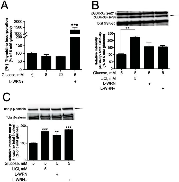
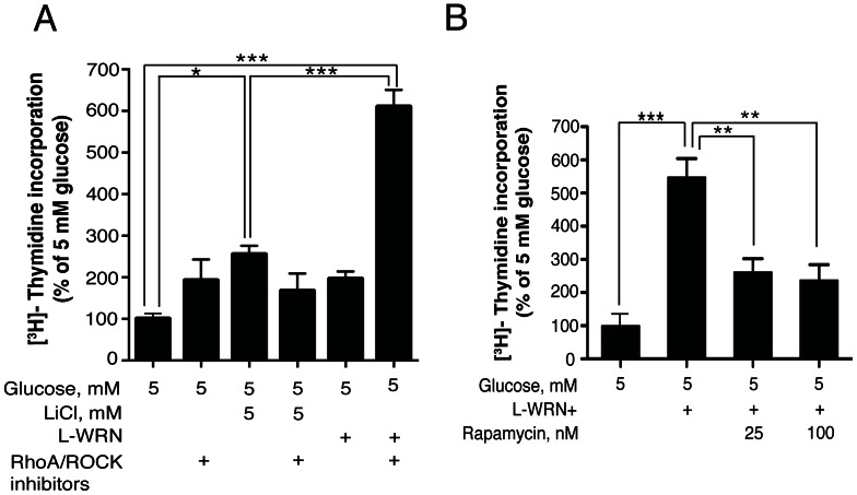
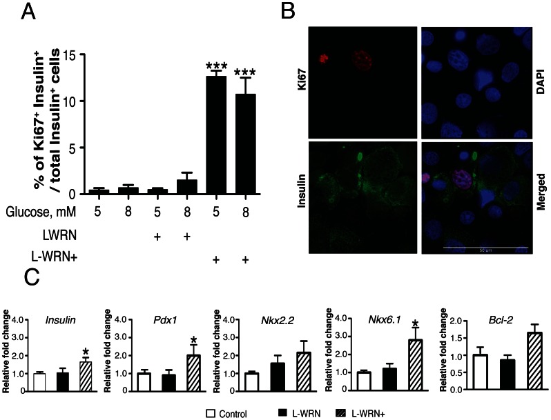
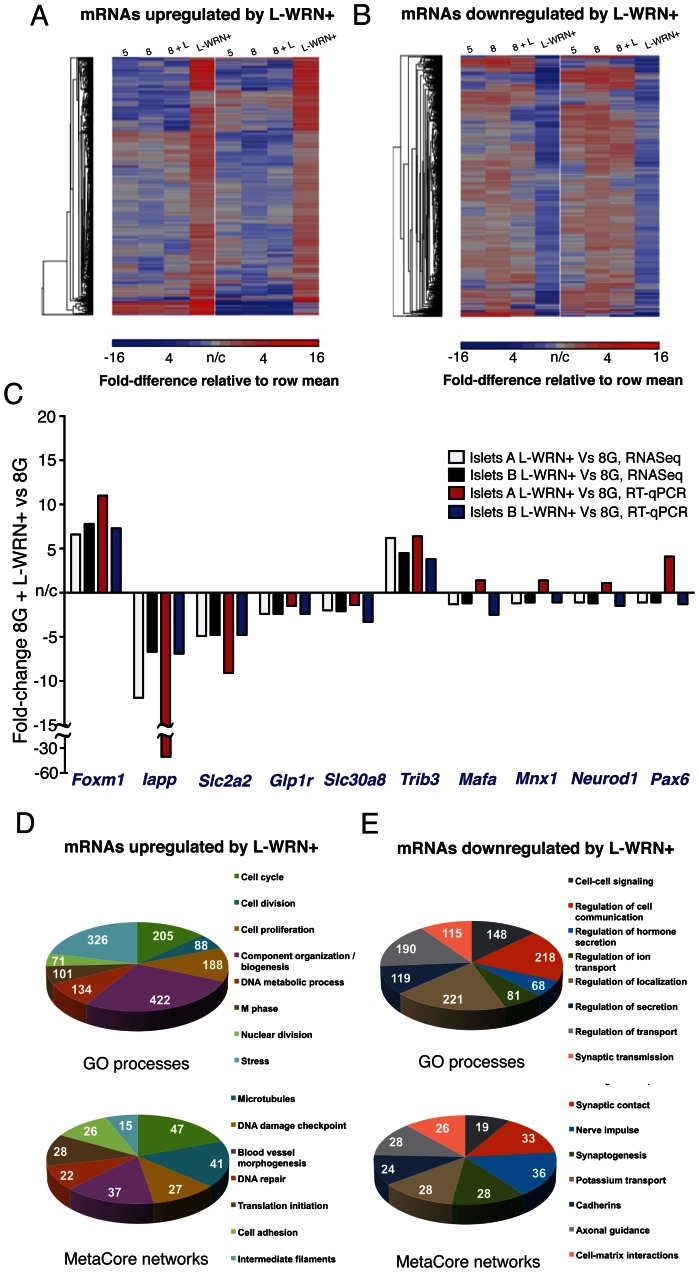
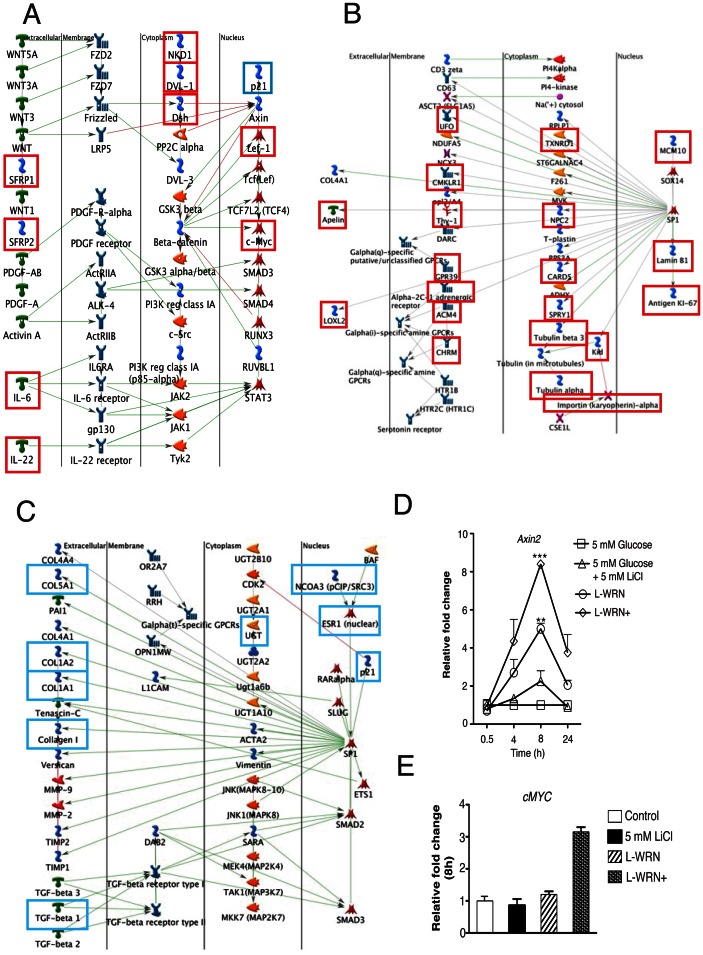
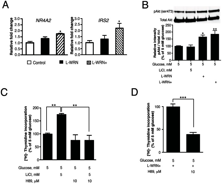
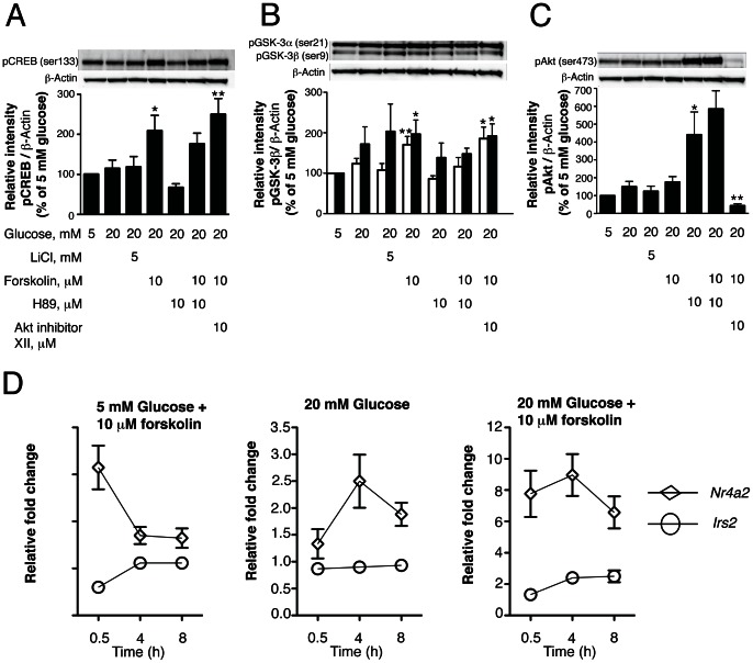
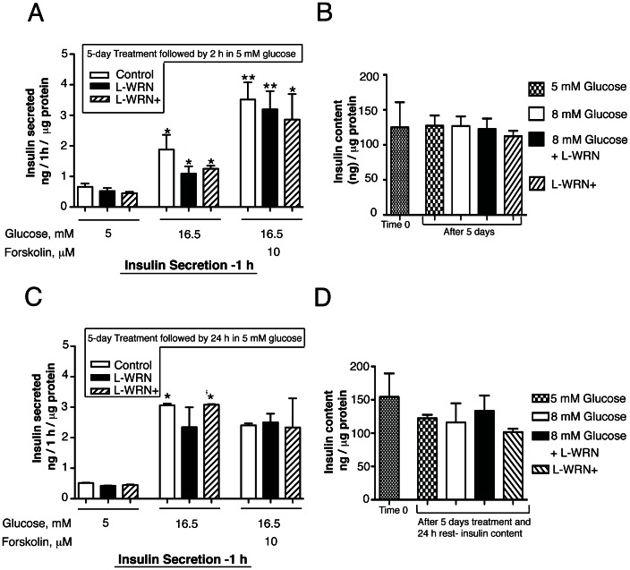
Our previous studies demonstrated that Wnt/GSK-3/β-catenin and mTOR signaling are necessary to stimulate proliferative processes in adult human β-cells. Direct inhibition of GSK-3, that engages Wnt signaling downstream of the Wnt receptor, increases β-catenin nuclear translocation and β-cell proliferation but results in lower insulin content. Our current goal was to engage canonical and non-canonical Wnt signaling at the receptor level to significantly increase human β-cell proliferation while maintaining a β-cell phenotype in intact islets. We adopted a system that utilized conditioned medium from L cells that expressed Wnt3a, R-spondin-3 and Noggin (L-WRN conditioned medium). In addition we used a ROCK inhibitor (Y-27632) and SB-431542 (that results in RhoA inhibition) in these cultures. Treatment of intact human islets with L-WRN conditioned medium plus inhibitors significantly increased DNA synthesis ∼6 fold in a rapamycin-sensitive manner. Moreover, this treatment strikingly increased human β-cell proliferation ∼20 fold above glucose alone. Only the combination of L-WRN conditioned medium with RhoA/ROCK inhibitors resulted in substantial proliferation. Transcriptome-wide gene expression profiling demonstrated that L-WRN medium provoked robust changes in several signaling families, including enhanced β-catenin-mediated and β-cell-specific gene expression. This treatment also increased expression of Nr4a2 and Irs2 and resulted in phosphorylation of Akt. Importantly, glucose-stimulated insulin secretion and content were not downregulated by L-WRN medium treatment. Our data demonstrate that engaging Wnt signaling at the receptor level by this method leads to necessary crosstalk between multiple signaling pathways including activation of Akt, mTOR, Wnt/β-catenin, PKA/CREB, and inhibition of RhoA/ROCK that substantially increase human β-cell proliferation while maintaining the β-cell phenotype.


Conflict of interest statement

Competing Interests: The authors have declared that no competing interests exist.

Figures

Figure 1
Figure 1. L-WRN conditioned medium activates the Wnt/β-catenin signaling pathway.
(A) 100 islets were cultured for 4 days in CMRL-1066 medium containing 10% FBS and 5, 8 or 20 mM glucose or in 50% Wnt3a, R-spondin-3 and Noggin conditioned medium (L-WRN)+Y-27632 and SB-431542 inhibitors. 3H-thymidine was added 24 h before the end of the 4-day period. (B) & (C): islets (150) were treated for 24 h and 72 h, respectively, in CMRL-1066 medium containing 10% FBS and 5 mM glucose ± 5 mM LiCl or in 50% L-WRN ± Y-27632 and SB-431542 inhibitors. Samples were processed for Western blotting and quantitated by Image Lab™ software. pGSK-3β and non-p-β-catenin proteins were normalized to total GSK-3β and total β-catenin proteins, respectively. Data are the means ± SE of n = 3. *P<0.05, **P<0.01 and ***P<0.001 denote significant differences between the treatment condition and the control.
Figure 2
Figure 2. Activation of Wnt and inhibition of RhoA/ROCK signaling significantly increase DNA synthesis in human islets and is mTOR-dependent.
(A & B) Islets (100) were cultured for 4 days in CMRL-1066 medium containing 10% FBS and 5 mM glucose or in 50% Wnt3a, R-spondin-3 and Noggin conditioned medium (L-WRN) ± 5 mM LiCl ± Y-27632 and SB-431542 inhibitors ±25 or 100 nM rapamycin as indicated. 3H-thymidine was added 24 h before the end of the 4-day period. Data are the means ± SE of n = 3. *P<0.05, **P<0.01 and ***P<0.001 denote significant differences between the treatment condition and the control.
Figure 3
Figure 3. Activation of Wnt and inhibition of RhoA/ROCK signaling significantly increase proliferation of adult human β-cells.
Islets (65) were cultured for 5 days in CMRL-1066 medium containing 10% FBS and 5 or 8 mM glucose or in 50% Wnt3a, R-spondin-3 and Noggin conditioned medium (L-WRN) ± 5 or 8 mM glucose ± Y-27632 and SB-431542 inhibitors as indicated. Samples were processed for immunohistochemistry as described in Materials and Methods. (A) Quantitative measurements of the % of Ki-67+and insulin+ cells/total insulin+ cells. For each experimental treatment 4500–7500 β-cells were counted in three independent experiments. (B) Representative immunofluorescence image of human islets treated for 5 days with L-WRN plus Y-27632 and SB-431542 inhibitors then dispersed into single cells and stained. Pink nucleus, Ki-67+; green, insulin+; and blue, nuclei stained with DAPI. (C) 75 islets were cultured for 8 h in CMRL-1066 medium containing 10% FBS and 5 mM glucose or in 50% Wnt3a, R-spondin-3 and Noggin conditioned medium (L-WRN) ± Y-27632 and SB-431542 inhibitors as indicated. Samples were processed for qRT-PCR and levels of mRNA for Insulin, Pdx1, Nkx2.2, Nkx6.1 and Bcl2 were determined as described in Materials and Methods. Transcripts were normalized to β-actin at the corresponding time point. Data are the means ± SE of n = 3. *P<0.05, **P<0.01 and ***P<0.001 denote significant differences between the treatment condition and the control.
Figure 4
Figure 4. Treatment of human islets with L-WRN+ regulates large numbers of proliferative genes.
(A) mRNAs upregulated in response to L-WRN+ by 1.5-fold vs treatment with 8 mM glucose alone, in islets sourced from two different donors. Relative mRNA levels in 5 mM glucose (5), 8 mM glucose (8), 8 mM glucose+LiCl (8+L) and L-WRN+-treated islet aliquots are shown; blue indicates lower abundance, red indicates higher abundance. (B) as for (A), but showing those mRNAs downregulated by 1.5-fold in response to L-WRN+ treatment. (C) qRT-PCR validation of six regulated and four nonregulated mRNAs (Mafa, Mnx1, Neurod1, Pax6); fold-changes of L-WRN+ treatment vs 8 mM glucose are shown. Individual gene expression values were normalized to the geometric mean of B2m, Gapdh, Hmbs and Ywhaz . White and black bars, fold-changes observed with RNA-sequencing; red and blue bars, fold-changes observed with qRT-PCR. (D, upper) Classification of upregulated genes into Gene Ontology categories; the most significantly over-represented categories are shown. (D, lower) Classification of upregulated genes into MetaCore custom-defined categories. (E, upper and lower) as for (D), but showing downregulated genes. Further details of Gene Ontology and MetaCore assignments are provided in Tables S2 and S3.
Figure 5
Figure 5. Inclusion of L-WRN+-regulated mRNAs in Wnt-related, proliferative and TGF-β signaling networks.
(A) Partial network of Wnt signaling; red squares designate mRNAs upregulated by at least 1.5-fold in response to L-WRN+. (B) Proliferative signaling network focused on Ki-67 and Sp1. (C) Partial network of TGF-β signaling. Up and downregulated mRNAs in panels A–C were defined using RNA-sequencing; red squares designate mRNAs upregulated by at least 1.5-fold in respone to L-WRN+; blue squares designate downregulated mRNAs. Green connecting lines indicate activation, red connecting lines indicate inhibition, and gray lines indicate mixed or uncertain effects, according to previously published studies contained in the curated MetaCore database. (D & E) Wnt pathway targets Axin2 and c-Myc compared amongst treated islets using qRT-PCR. Islets (75) were cultured for indicated times in CMRL-1066 medium containing 10% FBS and 5 mM glucose ± 5 mM LiCl or in 50% Wnt3a, R-spondin-3 and Noggin conditioned medium (L-WRN) ± Y-27632 and SB-431542 inhibitors as indicated. Samples were processed for qRT-PCR and levels of mRNA for Axin2 and c-Myc were determined as described in Materials and Methods. Transcripts were normalized to β-actin at the corresponding time point. Data are the means ± SE of n = 3 biological replicates. *P<0.05, **P<0.01 and ***P<0.001 denote significant differences between the treatment condition and the control.
Figure 6
Figure 6. L-WRN+ promotes crosstalk between Wnt and PKA/CREB and Akt signaling in human islets.
(A) Islets (75) were incubated for 8 h in CMRL-1066 medium containing 10% FBS and 5 mM glucose or in 50% Wnt3a, R-spondin-3 and Noggin conditioned medium (L-WRN)+Y-27632 and SB-431542 inhibitors as indicated. The levels of mRNA for Nr4a2 and Irs2 were determined as described in Material and Method. The values represent the –fold change due the indicated treatment over 5 mM glucose control treatment. (B) Islets (150) were treated for 24 h in CMRL-1066 medium containing 10% FBS and 5 mM glucose ± 5 mM LiCl or in 50% Wnt3a, R-spondin-3 and Noggin conditioned medium (L-WRN) ± Y-27632 and SB-431542 inhibitors. Samples were processed for Western blotting and quantitated by Image Lab™ software. pAkt protein was normalized to total Akt protein. Data are the means ± SE of n = 3. *P<0.05 and **P<0.01 denote significant differences between the treatment condition and respective control. (C) & (D) islets (100) were cultured for 4 days as indicated. 3H-thymidine was added 24 h before the end of the 4-day period. Data are the means ± SE of n = 3 with duplicate samples in each experiment. **P<0.01 and ***P<0.001 denote significant differences between the bracketed condition.
Figure 7
Figure 7. Activation of cAMP/PKA/CREB engages canonical Wnt and IRS2/Akt signaling in human islets.
(A) & (B) & (C) Human islets (150) were treated for 3 h with CMRL-1066 medium containing 10% FBS, 5 or 20 mM glucose, ± 10 µM forskolin ±5 mM LiCl as indicated. The PKA inhibitor (H-89, 10 µM) and Akt inhibitor (Akt inhibitor XII, 10 µM) were added to the culture where indicated 1 h before stimulation. Cell lysates were prepared and analyzed for CREB-, Akt- and GSK-3 phosphorylation using specific antibodies as described in Materials and Methods. In (B) open columns represent pGSK-3α and black columns represent pGSK-3β. (D) Human islets (75) were incubated for 30 min, 4 and 8 h with 5 or 20 mM glucose, ±10 µM forskolin. The levels of mRNA for Nr4a2 and Irs2 were determined as described in Materials and Methods. The values represent the fold change due to the indicated treatment over 5 mM glucose control treatment. Data are the means ± SE of n = 3 experiments. *P<0.05 **P<0.01 and ***P<0.001 denote significant differences between treatment conditions and the respective control (5 mM glucose).
Figure 8
Figure 8. L-WRN+ treatment does not impair insulin secretion.
Islets (100) were cultured for 5 days in CMRL-1066 medium containing 10% FBS and 8 mM glucose or in 50% Wnt3a, R-spondin-3 and Noggin conditioned medium (L-WRN) ±8 mM glucose ± Y-27632 and SB-431542 inhibitors as indicated. (A) & (C) Following 5 days incubation, treatments were removed and islets were incubated for 2 h or 24 h, respectively, in cCMRL with 5 mM glucose. Insulin secretion was measured by a static incubation for 1 h in cCMRL containing 5, 16.5 mM glucose ± forskolin. Quadruplicates of 10 islets were assayed. (B) & (D) Insulin content was measured from 20 islets in triplicate at the start of the incubation period (Time 0) and after 5 or 6 days of incubation, respectively, under the conditions shown. Insulin content in islets was extracted by the acid/ethanol method. Insulin secretion and content were detected using Immulite 1000 system and their values were normalized to islet protein. Data are the means ± SE of n = 3 with duplicate samples in each experiment. *P<0.05 and **P<0.01 denote significant differences between the treatment condition and respective control.
Figure 9
Figure 9. Proposed regulation of Wnt and Rho/ROCK signaling pathways by L-WRN+ in human islets.

Comment in

References

    1. Dor Y, Brown J, Martinez OI, Melton DA (2004) Adult pancreatic beta-cells are formed by self-duplication rather than stem-cell differentiation. Nature 429: 41–46. - PubMed
    1. Meier JJ, Butler AE, Saisho Y, Monchamp T, Galasso R, et al. (2008) Beta-cell replication is the primary mechanism subserving the postnatal expansion of beta-cell mass in humans. Diabetes 57: 1584–1594. - PMC - PubMed
    1. Teta M, Rankin MM, Long SY, Stein GM, Kushner JA (2007) Growth and regeneration of adult beta cells does not involve specialized progenitors. Dev Cell 12: 817–826. - PubMed
    1. Rutti S, Sauter NS, Bouzakri K, Prazak R, Halban PA, et al. (2012) In vitro proliferation of adult human beta-cells. PLoS One 7: e35801. - PMC - PubMed
    1. Nielsen JH, Brunstedt J, Andersson A, Frimodt-Moller C (1979) Preservation of beta cell function in adult human pancreatic islets for several months in vitro . Diabetologia 16: 97–100. - PubMed

Publication types

MeSH terms