. 2013 Jun 12;8(6):e67356.
doi: 10.1371/journal.pone.0067356. Print 2013.

Host cell poly(ADP-ribose) glycohydrolase is crucial for Trypanosoma cruzi infection cycle

Affiliations

Host cell poly(ADP-ribose) glycohydrolase is crucial for Trypanosoma cruzi infection cycle

Salomé C Vilchez Larrea et al. PLoS One. .

Abstract

Trypanosoma cruzi, etiological agent of Chagas' disease, has a complex life cycle which involves the invasion of mammalian host cells, differentiation and intracellular replication. Here we report the first insights into the biological role of a poly(ADP-ribose) glycohydrolase in a trypanosomatid (TcPARG). In silico analysis of the TcPARG gene pointed out the conservation of key residues involved in the catalytic process and, by Western blot, we demonstrated that it is expressed in a life stage-dependant manner. Indirect immunofluorescence assays and electron microscopy using an anti-TcPARG antibody showed that this enzyme is localized in the nucleus independently of the presence of DNA damage or cell cycle stage. The addition of poly(ADP-ribose) glycohydrolase inhibitors ADP-HPD (adenosine diphosphate (hydroxymethyl) pyrrolidinediol) or DEA (6,9-diamino-2-ethoxyacridine lactate monohydrate) to the culture media, both at a 1 µM concentration, reduced in vitro epimastigote growth by 35% and 37% respectively, when compared to control cultures. We also showed that ADP-HPD 1 µM can lead to an alteration in the progression of the cell cycle in hydroxyurea synchronized cultures of T. cruzi epimastigotes. Outstandingly, here we demonstrate that the lack of poly(ADP-ribose) glycohydrolase activity in Vero and A549 host cells, achieved by chemical inhibition or iRNA, produces the reduction of the percentage of infected cells as well as the number of amastigotes per cell and trypomastigotes released, leading to a nearly complete abrogation of the infection process. We conclude that both, T. cruzi and the host, poly(ADP-ribose) glycohydrolase activities are important players in the life cycle of Trypanosoma cruzi, emerging as a promising therapeutic target for the treatment of Chagas' disease.


Conflict of interest statement

Competing Interests: The authors have declared that no competing interests exist.

Figures

Figure 1
Figure 1. Amino acid sequence alignment of the PARG signature from different organisms.
The multiple alignment of the PARG signature amino acid sequences corresponding to T. cruzi PARG (accession number ABG73229); T. brucei PARG (GeneDB Systematic Name: Tb09.211.3760); C. elegans_1 PARG (accession number NP_501496) and C. elegans_2 PARG (accession number NP_501508); T . thermophila (accession number EAR94344); A. thaliana_1 PARG (accession number NP_973578); A. thaliana_2 PARG (accession number AAK72256); D. discoideum PARG (accession number XP_642024); D. melanogaster PARG (accession number NP_477321); C . quinquefasciatus PARG (accession number XP_001853435); A. aegypti PARG (accession number XP_001659301); D. rerio PARG (accession number XP_001338257); X. laevis PARG (accession number NP_001089602); G. gallus PARG (accession number XP_421502); B. taurus PARG (accession number NP_776563); R. norvegicus PARG (accession number NP_112629); M. musculus PARG (accession number NP_036090); H. sapiens PARG (accession number NP_003622); P. abelii PARG (accession number NP_001125086); P . troglodytes PARG (accession number XP_001139727) was generated with the ClustalW2 program and edited with the BOXSHADE (3.21) software. Colors used for amino acids background are as follow: white for different residues, black for identical residues, gray for similar and conserved residues. Asterisk: essential acidic residues D-E-E, underlined: key residues, G and two consecutive E, and black diamond: important Y residue.
Figure 2
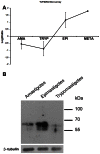
Figure 2. Expression of TcPARG throughout the Trypanosoma cruzi life-cycle.
(A) Microarray expression data for TcPARG over the course of T. cruzi life-cycle. TcPARG mRNA relative abundance was evaluated by using the transcriptome analysis of different T. cruzi stages available at Gene Expression Omnibus database (www.ncbi.nlm.nih.gov/geo, DataSets: GSE14641). Shown are mean microarray log2 ratios (stage/reference) for TS significantly regulated in Trypanosoma cruzi amastigotes (AMA), trypomastigotes (TRYP), epimastigotes (EPI), and metacyclic trypomastigotes (META). (B) Western blot analysis of the three life-cycle stages of T. cruzi. Protein extracts (35 µg) of amastigote, epimastigote or trypomastigote stages of T. cruzi were solved in a 10% polyacrylamide gel, transfer to a nitrocellulose membrane and revealed with an anti-TcPARG (1:10000) specific antiserum. β-tubulin was used as loading control.
Figure 3
Figure 3. Immunolocalization of PARG on Trypanosoma cruzi, CL Brener epimastigotes.
The parasites were fixed for 25 min with 3.8% (W/V) formaldehyde in PBS at 4°C, permeabilized with fresh PBS - 0,1% Triton X-100 and blocked for 1 h at room temperature with 5% (W/V) BSA in PBS. (A) Differential interference contrast (DIC). (B) Cells were counterstained with DAPI to identify nuclear DNA and kinetoplastid DNA. (C) PARG was detected with 1:500 mouse polyclonal TcPARG antibody followed by 1:600 Alexa Fluor 488 goat anti-mouse IgG antibody. (D) Merge of PARG and DNA signals show the nuclear localization of this enzyme. Bar: 10 µm. (E–F) For electron microscopy, epimastigotes were fixed in PBS 2.5% glutaraldehyde, 4% formaldehyde, embedded in epoxy resin and PARG detected with 1:50 mouse polyclonal TcPARG antibody followed by 1:100 anti-mouse antibody conjugated with 10-nm gold particle. N: nucleus; K: kinetoplast. Bar: 0.2 µm.
Figure 4
Figure 4. Inhibition of PARG activity in Trypanosoma cruzi.
(A) Dot Blot analysis of poly(ADP-ribose) accumulation during epimastigote growth in cultures untreated (Control) or treated with 1 µM of the PARG inhibitor DEA (DEA) for four days. (B) Dot Blot analysis of poly(ADP-ribose) formation and degradation after a genotoxic stimulus in untreated epimastigotes (Control) or in parasites preincubated with 1 µM of the PARG inhibitor ADP-HPD (ADP-HPD) for 30 min. After preincubation, hydrogen peroxide 300 µM was added (0 min), incubated for 10 minutes at 28°C (10 min) and the oxidizing agent was removed. Samples were obtained at 30 and 60 minutes post agent removal (indicated as 40 min and 70 min respectively). Five µg of total protein extract were dotted onto nitrocellulose membrane and an anti-pADPr polyclonal antibody was used to detect the formed polymers (upper panels). Ponceau Red was used to detect the whole protein sample in each dot (lower panels). (C–D) Data were normalized to protein content and are shown as the ratio of pADPr to loading control signals. Representative experiment of three independent assays.
Figure 5
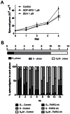
Figure 5. Role of TcPARG in Trypanosoma cruzi epimastigotes proliferation and cell cycle progression.
A) Effect of the PARG inhibitors ADP-HPD and DEA on T. cruzi growth and survival was determined by incubating epimastigotes at an initial density of 107 parasites/ml in the continuous presence of inhibitors at 1 µM. The number of epimastigotes was determined daily by counting formaldehyde-fixed parasites in a Neubauer chamber. All data points were determined in triplicates and shown as means with standard deviation. The significance of the results versus the control at day 4 was analyzed with t test and indicated in the figure (* p0.05). B) Effect of ADP-HPD at 1 µM concentration on cell cycle progression of epimastigotes was determined by adding the inhibitor at the indicated concentration to the culture media of hydroxyurea synchronized parasites after digitonin permeabilization. Samples were drawn every 2 hours for 14 hours and DNA content was determined by propidium iodide staining followed by flow cytometry analysis. The percentage of epimastigotes in each cell cycle phase was determined by setting gates according to the DNA content in the 0 hs of the control sample and maintained for all other samples. The data were analyzed using the Cyflogic software.
Figure 6
Figure 6. Effect of PARG inhibitors on T. cruzi infection on Vero or A549 host cells.
T. cruzi trypomastigotes were purified from the supernatant of previously infected cells and preincubated for 30 min in the respective culture medium in the absence (Control) or presence of 1 µM PARG inhibitor (DEA). Twenty-four hours Vero, A549 wild type or shPARG (hPARG silenced) cell monolayers were infected with 50 trypomastigotes/cell. The infection process was followed by microscopic direct visualization. At the indicated days (A and C) or at day 6 post-infection (B and D), percentage of infected cells and number of amastigotes intracellular were determined on May-Grünwald Giemsa stained samples. Amastigotes and cells were counted using the ImageJ software in at least 7 fields. The number of trypomastigotes/ml in the supernatant of infected cell cultures was determined by counting unfixed trypomastigotes in a Neubauer chamber at the indicated days (E) or at day 9 post-infection (F). All points were determined in triplicates and shown as means with standard deviation. Significance of the result versus the Control (***p0.001; two way ANOVA) or Wild Type Control (***p0.001; **, p0.01; two way ANOVA) is indicated.

Similar articles

Cited by

References

    1. Brochu G, Duchaine C, Thibeault L, Lagueux J, Shah GM et al. (1994) Mode of action of poly(ADP-ribose) glycohydrolase. Biochim Biophys Acta 1219: 342-350. doi:10.1016/0167-4781(94)90058-2. PubMed: 7918631. - PubMed
    1. D’Amours D, Desnoyers S, D’Silva I, Poirier GG (1999) Poly(ADP-ribosyl)ation reactions in the regulation of nuclear functions. Biochem J 342(2): 249-268. doi:10.1042/0264-6021:3420249. - PMC - PubMed
    1. Bonicalzi ME, Haince JF, Droit A, Poirier GG (2005) Regulation of poly(ADP-ribose) metabolism by poly(ADP-ribose) glycohydrolase: where and when? Cell Mol Life Sci 62: 739-750. doi:10.1007/s00018-004-4505-1. PubMed: 15868399. - PMC - PubMed
    1. Diefenbach J, Bürkle A (2005) Introduction to poly(ADP-ribose) metabolism. Cell Mol Life Sci 62: 721-730. doi:10.1007/s00018-004-4503-3. PubMed: 15868397. - PMC - PubMed
    1. Malanga M, Althaus FR (2005) The role of poly(ADP-ribose) in the DNA damage signaling network. Biochem Cell Biol 83: 354-364. doi:10.1139/o05-038. PubMed: 15959561. - PubMed

Publication types

MeSH terms