An increase of granulosa cell apoptosis mediates aqueous neem (Azadirachta indica) leaf extract-induced oocyte apoptosis in rat
- PMID: 23776837
- PMCID: PMC3678678
- DOI: 10.4103/2229-516X.112238
An increase of granulosa cell apoptosis mediates aqueous neem (Azadirachta indica) leaf extract-induced oocyte apoptosis in rat
Abstract
Objective: Neem plant (Azadirachta indica) has been extensively used in Ayurvedic system of medicine for female fertility regulation for a long time, but its mechanism of action remains poorly understood. Hence, the present study was aimed to determine whether an increase of granulosa cell apoptosis is associated with aqueous neem leaf extract (NLE)-induced oocyte apoptosis.
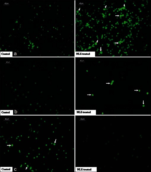
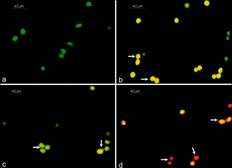
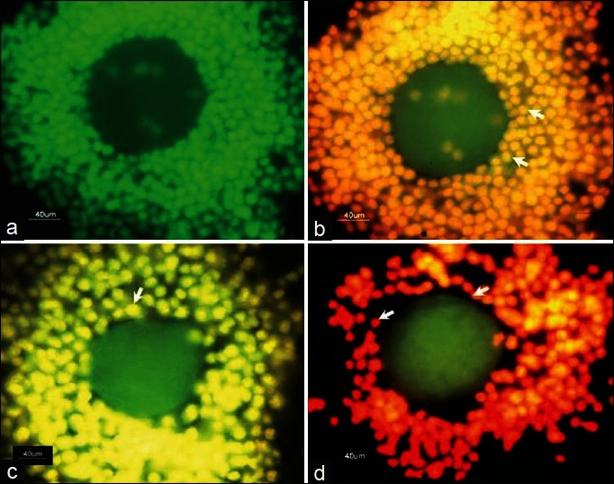
Materials and methods: Sexually immature female rats of 20 days old were fed NLE (50 mg/day) for 10 days and then subjected to superovulation induction protocol. The morphological changes in cumulus oocyte complexes (COCs), rate of oocyte apoptosis, hydrogen peroxide (H2O2), total nitrite, and cytochrome c concentrations, inducible nitric oxide synthase (iNOS), cytochrome c, p53, Bcl2 and Bax expressions, deoxyribonucleic acid (DNA) fragmentation, and estradiol 17β level in granulosa cells collected from preovulatory COCs were analyzed.
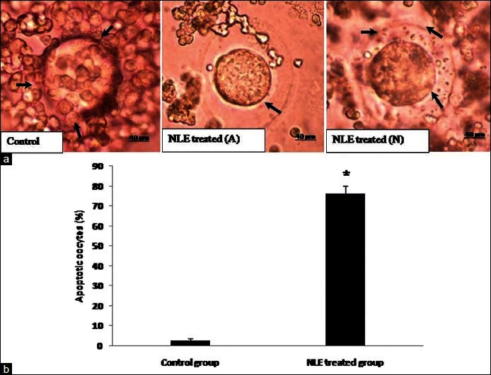
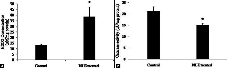
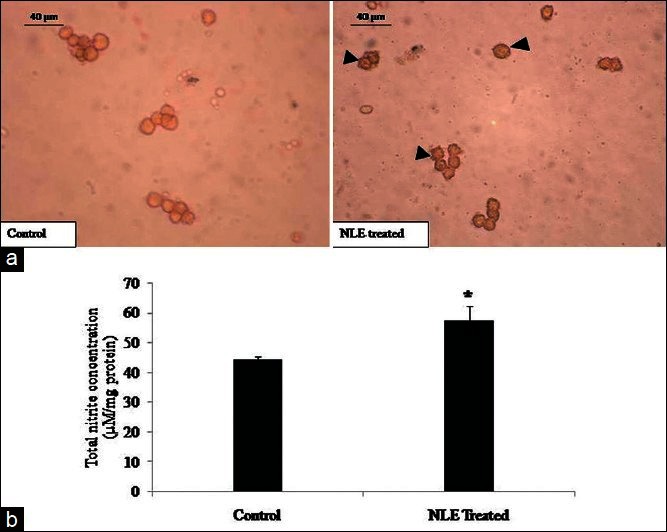
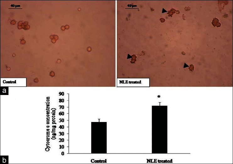
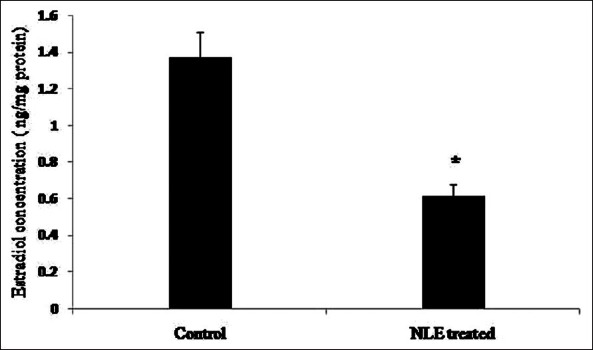
Results: Aqueous NLE increased H2O2 concentration and decreased catalase activity, increased iNOS expression and total nitrite concentration, increased p53, Bax, and p53 expressions but decreased Bcl2 expression, increased cytochrome c concentration and induced DNA fragmentation in granulosa cells. An increased granulosa cell apoptosis resulted in reduced estradiol 17β concentration and induced apoptosis in ovulated oocytes.
Conclusion: We conclude that aqueous NLE-induced granulosa cell apoptosis through the mitochondria-mediated pathway, reduced estradiol 17β concentration and induced apoptosis in ovulated oocytes. Thus, granulosa cell apoptosis mediates NLE-induced oocyte apoptosis during female fertility regulation in rat.
Keywords: Aqueous neem leaf extract; Bax/Bcl2; deoxyribonucleic acid fragmentation; granulosa cell apoptosis; rat oocytes; reactive oxygen species.
Conflict of interest statement
Figures
Similar articles
-
Neem (Azadirachta indica L.) leaf extract deteriorates oocyte quality by inducing ROS-mediated apoptosis in mammals.Springerplus. 2014 Aug 26;3:464. doi: 10.1186/2193-1801-3-464. eCollection 2014. Springerplus. 2014. PMID: 25197620 Free PMC article. Review.
-
Aqueous extract of Azadirachta indica (neem) leaf induces generation of reactive oxygen species and mitochondria-mediated apoptosis in rat oocytes.J Assist Reprod Genet. 2012 Jan;29(1):15-23. doi: 10.1007/s10815-011-9671-0. Epub 2011 Nov 17. J Assist Reprod Genet. 2012. PMID: 22089262 Free PMC article.
-
Extract of Azadirachta indica (neem) leaf induces apoptosis in rat oocytes cultured in vitro.Fertil Steril. 2006 Apr;85 Suppl 1:1223-31. doi: 10.1016/j.fertnstert.2005.11.034. Fertil Steril. 2006. PMID: 16616096
-
Presence of encircling granulosa cells protects against oxidative stress-induced apoptosis in rat eggs cultured in vitro.Apoptosis. 2017 Jan;22(1):98-107. doi: 10.1007/s10495-016-1324-4. Apoptosis. 2017. PMID: 27817140
-
Estradiol protects clomiphene citrate-induced apoptosis in ovarian follicular cells and ovulated cumulus-oocyte complexes.Fertil Steril. 2005 Oct;84 Suppl 2:1163-72. doi: 10.1016/j.fertnstert.2005.03.073. Fertil Steril. 2005. PMID: 16210008
Cited by
-
Plant-Derived Natural Compounds for Tick Pest Control in Livestock and Wildlife: Pragmatism or Utopia?Insects. 2020 Aug 1;11(8):490. doi: 10.3390/insects11080490. Insects. 2020. PMID: 32752256 Free PMC article. Review.
-
Autophagy in hypoxic ovary.Cell Mol Life Sci. 2019 Sep;76(17):3311-3322. doi: 10.1007/s00018-019-03122-4. Epub 2019 May 6. Cell Mol Life Sci. 2019. PMID: 31062072 Free PMC article. Review.
-
Morphological, cellular and molecular changes during postovulatory egg aging in mammals.J Biomed Sci. 2015 May 22;22(1):36. doi: 10.1186/s12929-015-0143-1. J Biomed Sci. 2015. PMID: 25994054 Free PMC article. Review.
-
Germ cell depletion from mammalian ovary: possible involvement of apoptosis and autophagy.J Biomed Sci. 2018 Apr 23;25(1):36. doi: 10.1186/s12929-018-0438-0. J Biomed Sci. 2018. PMID: 29681242 Free PMC article. Review.
-
An Overview on the Anticancer Activity of Azadirachta indica (Neem) in Gynecological Cancers.Int J Mol Sci. 2018 Dec 5;19(12):3898. doi: 10.3390/ijms19123898. Int J Mol Sci. 2018. PMID: 30563141 Free PMC article. Review.
References
-
- Allworth AE, Albertini DF. Meiotic maturation in cultured bovine oocytes is accompanied by remodeling of the cumulus cell cytoskeleton. Dev Biol. 1993;158:101–12. - PubMed
-
- Gilchrist RB, Lane M, Thompson JG. Oocyte-secreted factors: Regulators of cumulus cell function and oocyte quality. Hum Reprod Update. 2008;14:159–77. - PubMed
-
- Assidi M, Dieleman SJ, Sirard MA. Cumulus cell gene expression following the LH surge in bovine preovulatory follicles: Potential early markers of oocyte competence. Reproduction. 2010;140:835–52. - PubMed
LinkOut - more resources
Full Text Sources
Other Literature Sources
Research Materials
Miscellaneous