. 2013 Mar;17(2):219-27.
doi: 10.4103/2230-8210.109667.

Thyroid ultrasound

Affiliations

Thyroid ultrasound

Vikas Chaudhary et al. Indian J Endocrinol Metab. 2013 Mar.

Abstract

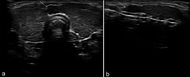
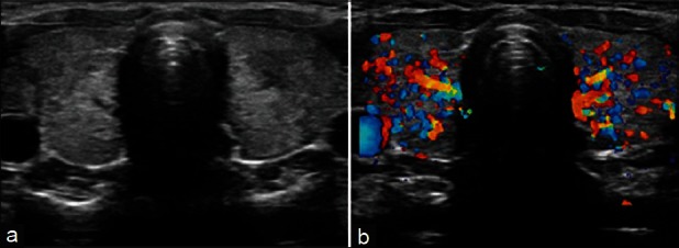
Thyroid ultrasonography has established itself as a popular and useful tool in the evaluation and management of thyroid disorders. Advanced ultrasound techniques in thyroid imaging have not only fascinated the radiologists but also attracted the surgeons and endocrinologists who are using these techniques in their daily clinical and operative practice. This review provides an overview of indications for ultrasound in various thyroid diseases, describes characteristic ultrasound findings in these diseases, and illustrates major diagnostic pitfalls of thyroid ultrasound.

Keywords: Color doppler; high resolution ultrasonography; thyroid; ultrasound; ultrasound elastography.


Conflict of interest statement

Conflict of Interest: None declared

Figures

Figure 1
Figure 1
Normal thyroid gland. (a) Gray scale ultrasound, transverse scan showing normal thyroid anatomy, (b) Arterial vascularization of the thyroid gland. On color Doppler, the inferior thyroid artery (arrow) is seen, (c) Blood flow pattern in normal thyroid gland. On spectral display, a low resistance flow with a high peak systolic velocity is obtained
Figure 2
Figure 2
Thyroglossal cyst in a patient who presented with midline neck swelling. Ultrasound neck (a) shows a well-defined anechoic cystic lesion with multiple low level internal echoes (asterisk) and posterior acoustic enhancement. Multiple low level internal echoes within the cyst may be due to hemorrhage or infection. X-ray neck lateral view (b) of the same patient shows large, soft tissue/cystic midline swelling (white arrow)
Figure 3
Figure 3
Benign thyroid adenoma in a 42-year-old female patient. Transverse gray-scale ultrasound neck (a) shows a large well circumscribed oval shaped (width>length), hyperechoic nodule in a thyroid lobe. The lesion has slight heterogeneous appearance due to presence of few tiny cystic spaces/clefts. A thin, hypoechoic capsule (arrow) is noted peripherally. Color Doppler image (b) demonstrates both central and peripheral vascularity with characteristic “spoke-and-wheel-like” appearance
Figure 4
Figure 4
Follicular adenoma thyroid in a 40-year-old female patient. Transverse gray-scale ultrasound neck (a) shows a large well circumscribed heterogeneous thyroid nodule with multiple internal cystic spaces giving characteristic “spongiform appearance” to the lesion. Color Doppler sonogram (b) demonstrates both central and peripheral vascularity. [Reproduced with permission from Indian Journal of Endocrinology and Metabolism.]
Figure 5
Figure 5
Benign cystic colloid nodule in a 55-year-old female patient. Transverse sonogram neck (a) reveals a well-circumscribed right-sided cystic nodule with solid peripheral component and ‘ring down’ artifact (thin arrow). The lesion demonstrates a thin hypoechoic rim (thick arrows) and posterior acoustic enhancement. Color Doppler image (b) demonstrates vascularity in the solid peripheral component. (Reproduced with permission from Indian Journal of Endocrinology and Metabolism)
Figure 6
Figure 6
Malignant thyroid nodule. Hürthle cell (follicular) carcinoma in a 60-year-old woman. Transverse sonogram (a) of right lobe of the thyroid shows a partially cystic tumor with solid internal nodule (arrow) and thick walls. Color Doppler sonogram (b) depicts increased vascularity in solid parts of the tumor (arrow)
Figure 7
Figure 7
Colloid multinodular goitre in a 50-year-old female patient. Transverse (a) and longitudinal (b) gray-scale ultrasound neck images reveal enlarged thyroid gland having multiple hyperechoic colloid nodules with internal cystic areas (arrows) showing ‘ring down’ sign. Color Doppler image (c) shows increased peripheral vascularity, with some intra goitrous vascularity
Figure 8
Figure 8
Adenomatous multinodular goitre in a 48-year-old patient with thyrotoxicosis. Transverse gray-scale ultrasound neck (a) shows diffuse enlargement of thyroid gland with multiple small (~5 mm size) echogenic nodules involving both the lobes and isthmus (arrows), with no normal-appearing intervening parenchyma. Color Doppler sonogram (b) demonstrates diffusely increased parenchymal vascularity. FNA biopsy confirmed the diagnosis of adenomatous multinodular goitre. However, it should be noted that FNA biopsy is not likely to be necessary in a diffusely enlarged thyroid gland with multiple nodules of similar US appearance and no normal intervening parenchyma
Figure 9
Figure 9
Focal Hashimoto's thyroiditis in a 35-year-old female patient, who presented with features of hypothyroidism and had anti-thyroid antibodies positive for the disease. Transverse gray-scale ultrasound neck (a) demonstrates ill-defined heterogeneous hypoechoic areas localized to postero-inferior aspect of thyroid lobes bilaterally (arrows). Longitudinal scan (b) left lobe thyroid (of same patient) clearly depicts the abnormal area which also shows increased vascularity on color Doppler sonogram (c)
Figure 10
Figure 10
Diffuse Hashimoto's thyroiditis in a 35-year-old female patient, who presented with features of hypothyroidism and had anti-thyroid antibodies positive for the disease. Transverse gray-scale ultrasound neck (a) demonstrates diffuse enlargement of thyroid gland with heterogeneous echotexture. Multiple tiny and discrete hypoechoic nodules (micronodules, arrows) and few linear echogenic septae (arrowhead) are also noted. Color Doppler sonogram (b) demonstrates mildly increased parenchymal vascularity
Figure 11
Figure 11
Nodular Hashimoto's thyroiditis (multiple echogenic nodules in sonographic background of diffuse Hashimoto's thyroiditis) in a 40-year-old female patient, who presented with hypothyroidism and had anti-thyroid antibodies positive for the disease. Transverse (a) and longitudinal (b) ultrasound neck images show diffusely enlarged and hypoechoic thyroid gland, with linear echogenic fibrous septae (arrowheads), and multiple small and discrete echogenic parenchymal nodules (arrows). Note, perinodular hypoechoic halo associated with few nodules in image b. Color Doppler imaging demonstrated increased parenchymal vascularity (image not shown). FNAC proved Hashimoto's thyroiditis
Figure 12
Figure 12
Nodular Hashimoto's thyroiditis (multiple hypoechoic nodules in sonographic background of diffuse Hashimoto's thyroiditis) in a 37-year-old male patient who presented with hypothyroidism and had anti-thyroid antibodies positive for the disease. Transverse gray-scale ultrasound neck (a) shows diffusely enlarged thyroid gland with multiple (>5) small and discrete hypoechoic nodules involving both the lobes and isthmus (arrows). Color Doppler image (b) demonstrates diffusely increased parenchymal vascularity. It should be noted that both benign and malignant nodules can co-exist within Hashimoto's thyroiditis; hence, FNAC may be required to differentiate them
Figure 13
Figure 13
Diffuse Hashimoto's thyroiditis (with cervical lymphadenopathy) in a 37-year-old female patient, who presented with features of hypothyroidism and anti-thyroid antibodies positive for the disease. Transverse (a) and longitudinal (b) gray-scale ultrasound neck images demonstrate the sonographic features of diffuse Hashimoto's thyroiditis, associated with a cervical lymph node just cephalad to the isthmus (the “Delphian” lymph node) (arrow). FNAC of the node revealed reactionary/inflammatory changes. The lymph node may be confused with thyroid isthmus nodule
Figure 14
Figure 14
Acute thyroiditis in a 12-year-old female patient, who presented with acute onset fever, neck pain and swelling. Transverse gray-scale ultrasound neck (a) shows bilaterally enlarged thyroid lobes with heterogeneous echo pattern. Color Doppler sonogram (b) demonstrates increased parenchymal vascularity in both lobes of the thyroid

References

    1. Solbiati L, Charboneau JW, Osti V, James EM, Hay ID. The thyroid gland. In: Rumack CM, Wilson SR, Charboneau JW, editors. Diagnostic Ultrasound. 3rd ed. Vol. 1. St. Louis, Missouri: Elsevier Mosby; 2005. pp. 735–70.
    1. Chaudhary V, Bano S. Imaging of the thyroid: Recent advances. Indian J Endocrinol Metab. 2012;16:371–6. - PMC - PubMed
    1. Gharib H, Papini E, Valcavi R, Baskin HJ, Crescenzi A, Dottorini ME, et al. American Association of Clinical Endocrinologists and Associazione Medici Endocrinologi medical guidelines for clinical practice for the diagnosis and management of thyroid nodules. Endocr Pract. 2006;12:63–102. - PubMed
    1. Baskin HJ. Ultrasound of thyroid nodules. In: Baskin HJ, editor. Thyroid Ultrasound and Ultrasound-guided FNA Biopsy. Boston: Kluwer Academic Publisher; 2007. pp. 71–86.
    1. Moon WJ, Jung SL, Lee JH, Na DG, Baek JH, Lee YH, et al. Benign and malignant thyroid nodules: US differentiation–multicenter retrospective study. Radiology. 2008;247:762–70. - PubMed