. 2013 Jun 18;6(1):182.
doi: 10.1186/1756-3305-6-182.

Interactions between Asaia, Plasmodium and Anopheles: new insights into mosquito symbiosis and implications in malaria symbiotic control

Affiliations

Interactions between Asaia, Plasmodium and Anopheles: new insights into mosquito symbiosis and implications in malaria symbiotic control

Aida Capone et al. Parasit Vectors. .

Abstract

Background: Malaria represents one of the most devastating infectious diseases. The lack of an effective vaccine and the emergence of drug resistance make necessary the development of new effective control methods. The recent identification of bacteria of the genus Asaia, associated with larvae and adults of malaria vectors, designates them as suitable candidates for malaria paratransgenic control.To better characterize the interactions between Asaia, Plasmodium and the mosquito immune system we performed an integrated experimental approach.

Methods: Quantitative PCR analysis of the amount of native Asaia was performed on individual Anopheles stephensi specimens. Mosquito infection was carried out with the strain PbGFPCON and the number of parasites in the midgut was counted by fluorescent microscopy.The colonisation of infected mosquitoes was achieved using GFP or DsRed tagged-Asaia strains.Reverse transcriptase-PCR analysis, growth and phagocytosis tests were performed using An. stephensi and Drosophila melanogaster haemocyte cultures and DsRed tagged-Asaia and Escherichia coli strains.

Results: Using quantitative PCR we have quantified the relative amount of Asaia in infected and uninfected mosquitoes, showing that the parasite does not interfere with bacterial blooming. The correlation curves have confirmed the active replication of Asaia, while at the same time, the intense decrease of the parasite.The 'in vitro' immunological studies have shown that Asaia induces the expression of antimicrobial peptides, however, the growth curves in conditioned medium as well as a phagocytosis test, indicated that the bacterium is not an immune-target.Using fluorescent strains of Asaia and Plasmodium we defined their co-localisation in the mosquito midgut and salivary glands.

Conclusions: We have provided important information about the relationship of Asaia with both Plasmodium and Anopheles. First, physiological changes in the midgut following an infected or uninfected blood meal do not negatively affect the residing Asaia population that seems to benefit from this condition. Second, Asaia can act as an immune-modulator activating antimicrobial peptide expression and seems to be adapted to the host immune response. Last, the co-localization of Asaia and Plasmodium highlights the possibility of reducing vectorial competence using bacterial recombinant strains capable of releasing anti-parasite molecules.


Figures
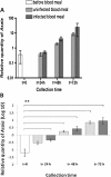
Figure 1
Figure 1
Quantitative analysis by qPCR of the amount of native Asaia in An. stephensi midgut. Mosquito collection time, after infected (dark grey bars) or uninfected (light grey bars) blood meal, has been fixed before (t=0) and 24, 48, 72 h after blood meals. The relative amount of Asaia was expressed as a ratio of bacterial 16S rRNA and mosquito rps7 gene copies in logarithmic scale (panel A) and Log10 values (panel B); in both panels amounts were mean±SEM of ten individuals. Two asterisks in panel B denoted p<0.01 in the samples indicated by horizontal lines, compared by both ANOVA and Post-hoc test ‘Bonferroni’.
Figure 2
Figure 2
Correlation curves between Asaia and P. berghei amounts in An. stephensi midguts. Relative quantities of Asaia obtained from ten individuals presented on a logarithmic scale and the corresponding parasite numbers were reported at 24, 48 and 72 h after the infected blood meal (A, B and C respectively). The average numbers of parasites (Anp) and the curve slopes (Y) were shown. Analysis of linear regression revealed p values <0.05 obtaining a similar correlation curve (R2) at 48 h and 72 h.
Figure 3
Figure 3
Semi-quantitative analysis by RT-PCR of bacterial induction of AMPs in immunocyte cultures. AMPs expression was evaluated in An. stephensi (panel A) and D. melanogaster (panel B). Defensin (a), cecropin (b), gambicin (c, panel A) and drosomycin (c, panel B) expression at 0h, 4 h, 8 h and 12 h after Asaia or E. coli bacterial challenge. Actin (d) has been used as a constitutive control gene. The expression of AMPs has been evaluated after electrophoresis in 1% agarose gel, documentation was collected using a “Gel Doc XR” and digitally evaluated with “Quantity One” as schematized below each panel. One representative set of data out of 7 replicates is shown.
Figure 4
Figure 4
Bacterial growth curves. Asaia (A) and E. coli (B) were cultured in the presence of control Schneider’s medium (.....), or half-diluted (− − −) and undiluted mosquito haemocytes conditioned medium (___).
Figure 5
Figure 5
Phagocytosis test analysis. Phagocytic activity was evaluated in vitro using cultured An. stephensi (A) and D. melanogaster (B) haemocytes, and DsRed-labelled bacterial strains of Asaia and E. coli. The percentage of haemocytes showing fluorescent phagocytised bacteria, E. coli (black bars) or Asaia (grey bars), has been evaluated after 1 h and 2 h. Values are expressed as mean±SD of ten replicates.
Figure 6
Figure 6
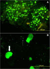
Co-localisation of GFP-Asaia and P. berghei PbGFPCON oocysts in the midgut of An. stephensi. Microscopic fluorescence analysis was carried out 11 days after the infected blood meal and at the 15th day after bacterial administration. A massive presence of oocysts is evident in the midgut (A), parasite and bacterial co-localisation is appreciable in the magnified image showing the bacterium (thin arrow) as able to surround and overlap the mature oocysts (thick arrow) (B).
Figure 7
Figure 7
Co-localization of DsRed-Asaia and P berghei PbGFPCON sporozoites in the proximity of mature oocysts and within the salivary glands of An. stephensi. Microscopic fluorescence analysis was carried out on the 17th day after infection and at the 21st day after bacterial administration. The presence of red fluorescent Asaia (thin arrow) in the proximity of mature oocysts and GFP-tagged sporozoites (thick arrow) (A and B) as well as the co-localisation of the two microrganisms in the salivary gland lobes (C and D) can be detected.

Similar articles

Cited by

References

    1. Shiff C. Integrated approach to malaria control. Clin Microbiol Rev. 2002;15:278–293. - PMC - PubMed
    1. Sinden RE. Plasmodium differentiation in the mosquito. Parassitologia. 1999;41:139–148. - PubMed
    1. Sinden RE, Alavi Y, Raine JD. Mosquito-malaria interactions: a reappraisal of the concepts of susceptibility and refractoriness. Insect Biochem Mol Biol. 2004;34:625–629. - PubMed
    1. Vaughan JA. Population dynamics of Plasmodium sporogony. Trends Parasitol. 2007;23:63–70. - PubMed
    1. Taylor LH. Infection rates in, and the number of Plasmodiumfalciparum genotypes carried by Anopheles mosquitoes in Tanzania. Ann Trop Med Parasitol. 1999;93:659–662. - PubMed

Publication types

MeSH terms