

Novel patterns for the growing main bronchi in the human fetus: an anatomical, digital and statistical study
- PMID: 23778946
- PMCID: PMC3890071
- DOI: 10.1007/s00276-013-1145-x
Novel patterns for the growing main bronchi in the human fetus: an anatomical, digital and statistical study
Abstract
Purpose: Intensive progress in prenatal medicine results in performing airway management in the fetus affected by life-threatening congenital malformations. This study aimed to examine age-specific reference intervals and growth dynamics for length, proximal and distal external transverse diameters, and projection surface areas of the two main bronchi at varying gestational ages, including their relative growth in length and projection surface area.
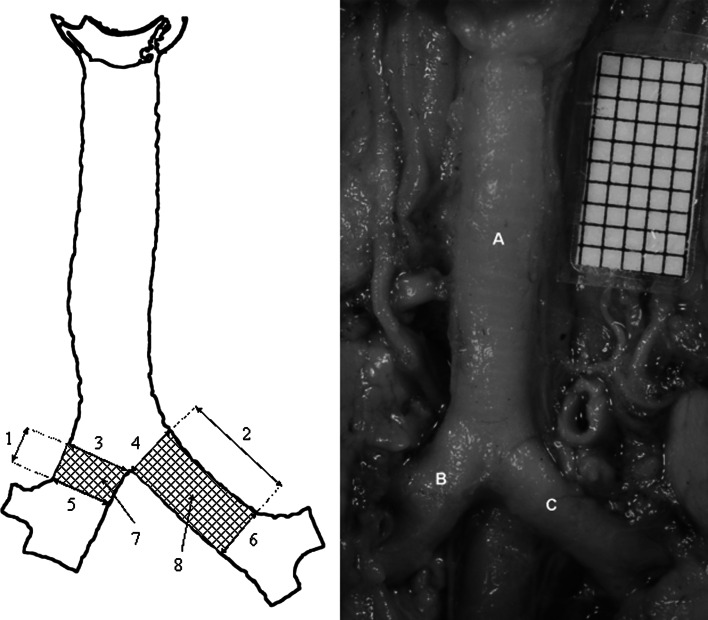
Materials and methods: Using anatomical dissection, digital image analysis and statistics, length, proximal and distal external transverse diameters, and projection surface areas of the right and left main bronchi were examined in 73 human fetuses (39 males, 34 females) aged 14-25 weeks, derived from spontaneous abortions and stillbirths.
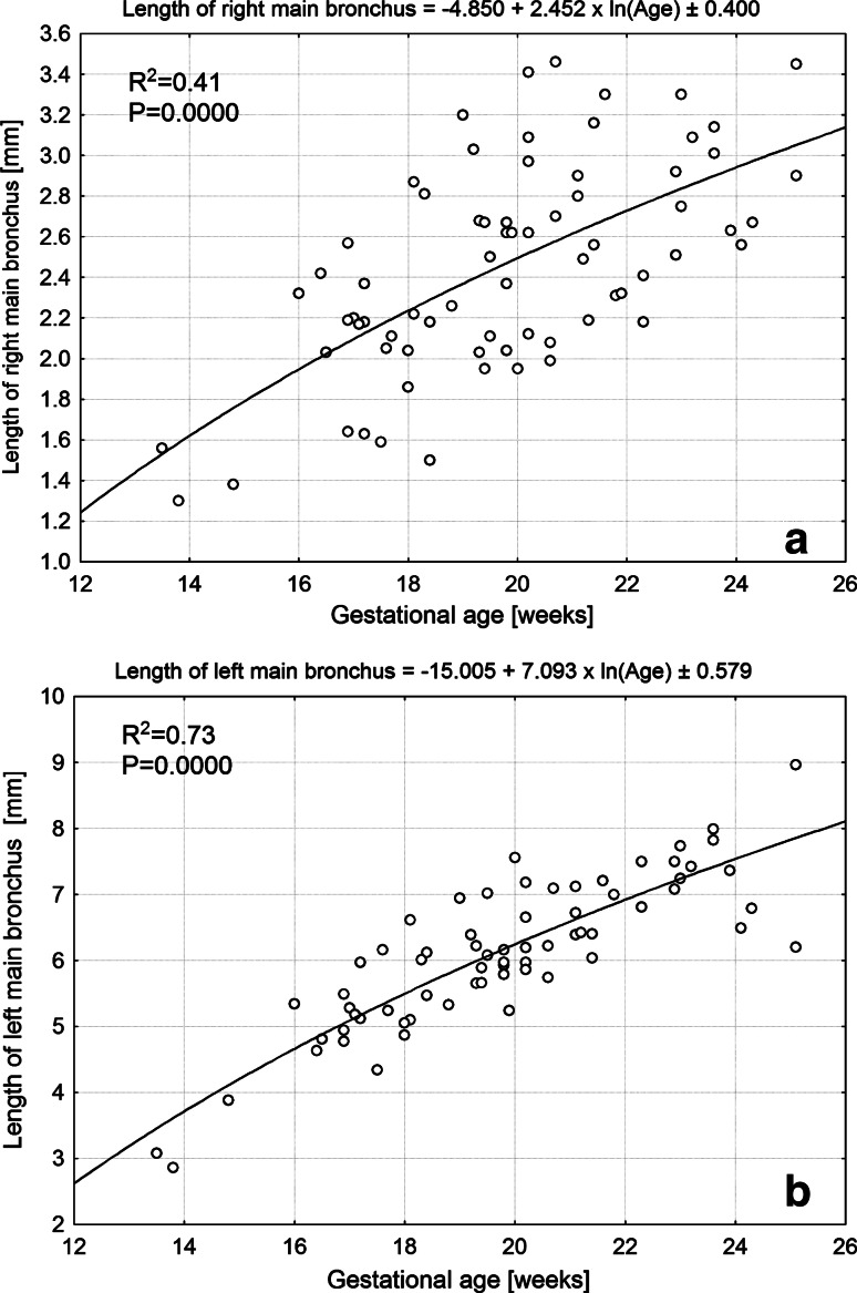
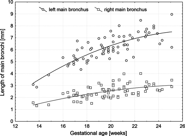
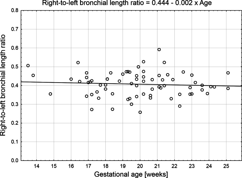
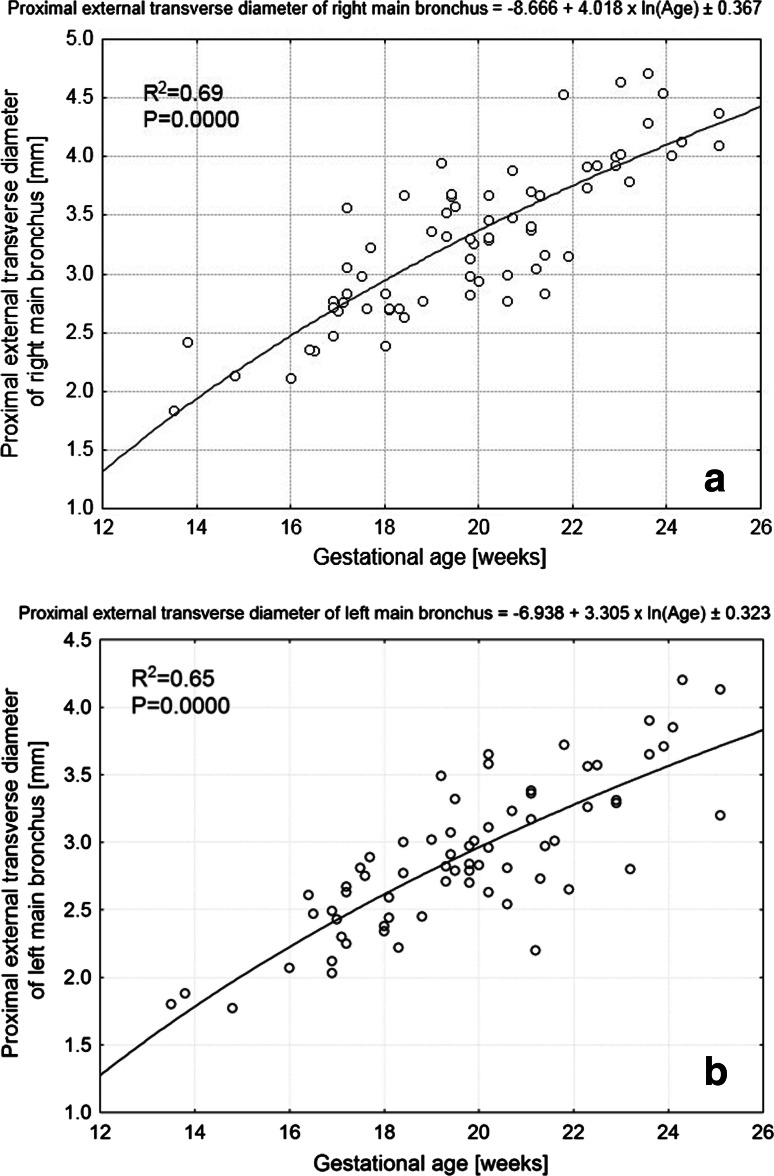
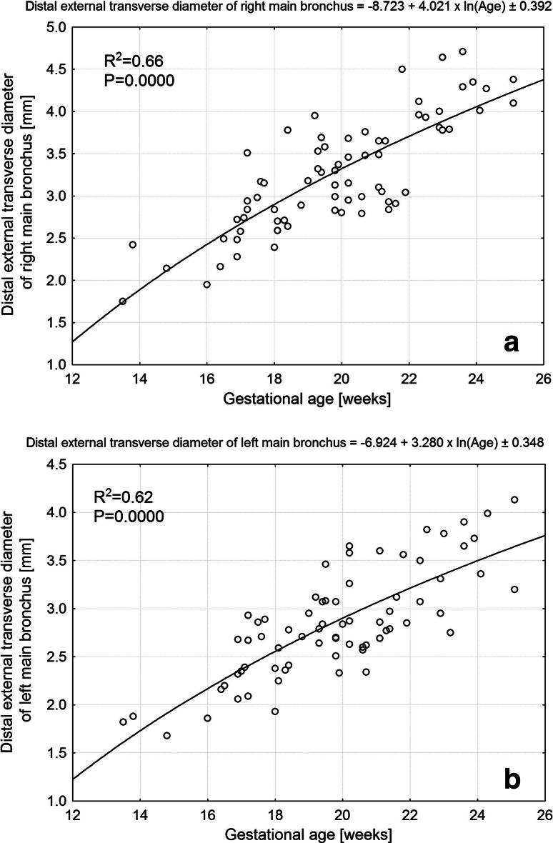
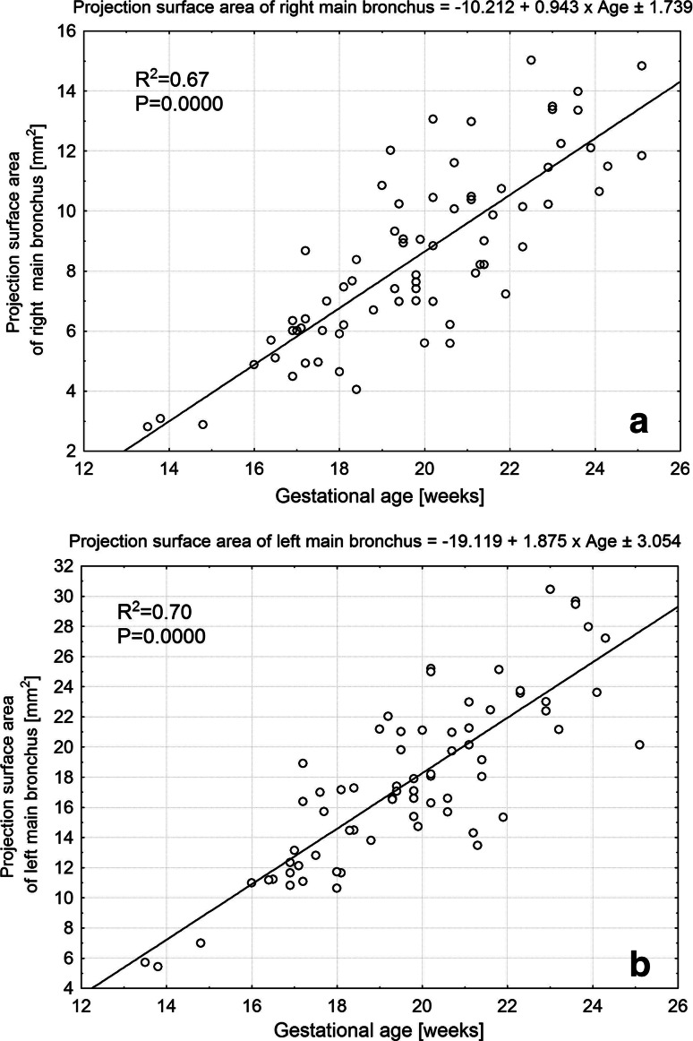
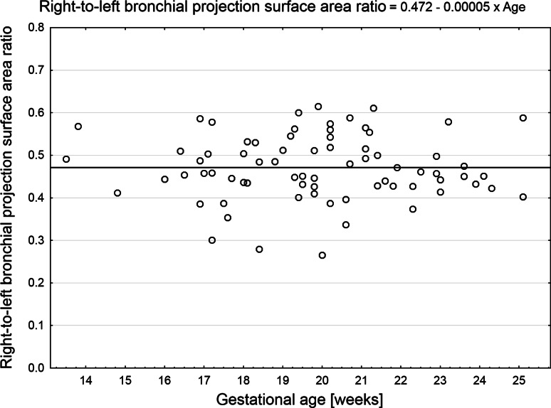
Results: Statistical analysis showed no sex differences. Between the 14 and 25th week of gestation, the lengths of the right and left main bronchi increased from 1.43 ± 0.18 to 3.18 ± 0.39 mm, and from 2.97 ± 0.16 to 7.58 ± 1.95 mm, in accordance with the functions: [Formula: see text], respectively. The proximal external transverse diameters of the right and left main bronchi varied from 2.13 ± 0.41 to 4.24 ± 0.20 mm, and from 1.84 ± 0.06 to 3.67 ± 0.66 mm, following the logarithmic models: [Formula: see text], respectively. The distal external transverse diameter rose from 2.09 ± 0.47 to 4.24 ± 0.20 mm, as [Formula: see text] for the right main bronchus, and from 1.85 ± 0.04 to 3.67 ± 0.66 mm, like [Formula: see text] for the left one. On either side, there were no statistically significant differences between values of the proximal and distal transverse diameters of the main bronchus. The projection surface areas of the right and left main bronchi ranged from 2.95 ± 0.19 to 13.34 ± 2.12 mm(2), and from 5.57 ± 0.21 to 28.52 ± 5.24 mm(2), as [Formula: see text] and [Formula: see text]. The two main bronchi revealed a proportionate increase in both length and projection surface area, since the right-to-left bronchial length ratio and the right-to-left bronchial projection surface area ratio were stable, 0.41 ± 0.07 and 0.47 ± 0.08, respectively, throughout the analyzed period.
Conclusions: The main bronchi show no sex differences. The right and left main bronchi grow logarithmically in length and external transverse diameter, and linearly in projection surface area. The right and left main bronchi evolve proportionately, with the right-to-left bronchial ratios of 0.41 ± 0.07 for length, and 0.47 ± 0.08 for projection surface area.
Figures
References
-
- Chow MYH, Liam BL, Thng CH, Chong BK. Predicting the size of a double-lumen endobronchial tube using computed tomographic scan measurements of the left main bronchus diameter. Anesth Analg. 1999;88:302–305. - PubMed
MeSH terms
LinkOut - more resources
Full Text Sources
Other Literature Sources
