. 2013 Aug;9(8):1214-27.
doi: 10.4161/auto.24870. Epub 2013 May 8.

IκB kinase complex (IKK) triggers detachment-induced autophagy in mammary epithelial cells independently of the PI3K-AKT-MTORC1 pathway

Affiliations

IκB kinase complex (IKK) triggers detachment-induced autophagy in mammary epithelial cells independently of the PI3K-AKT-MTORC1 pathway

Nan Chen et al. Autophagy. 2013 Aug.

Abstract

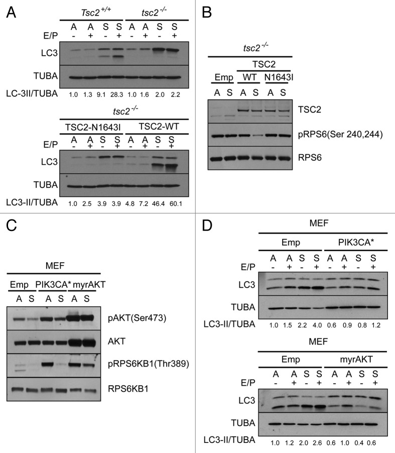
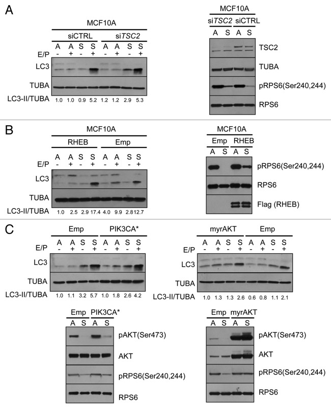
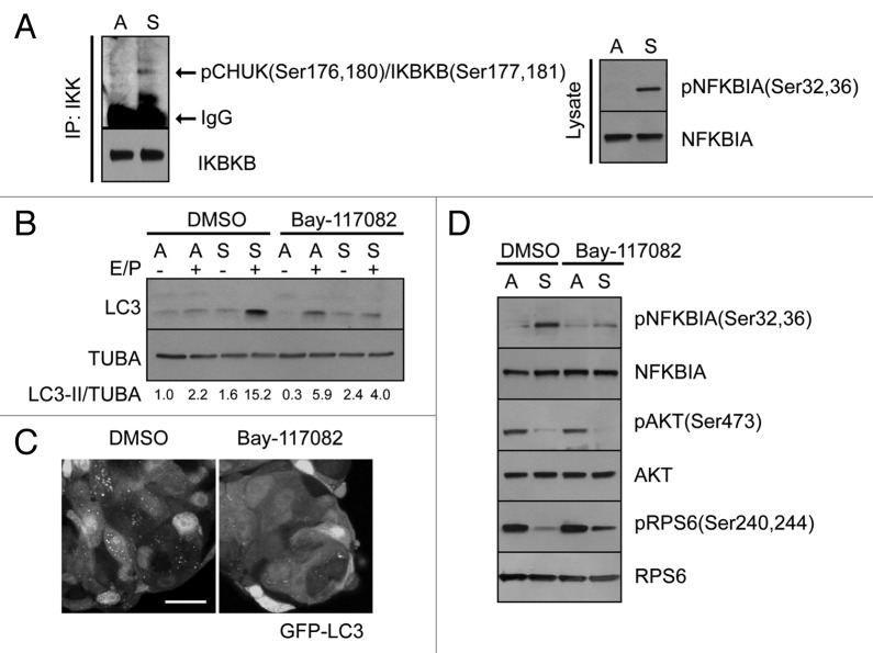
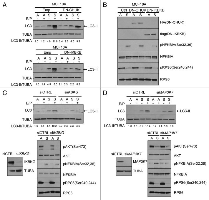
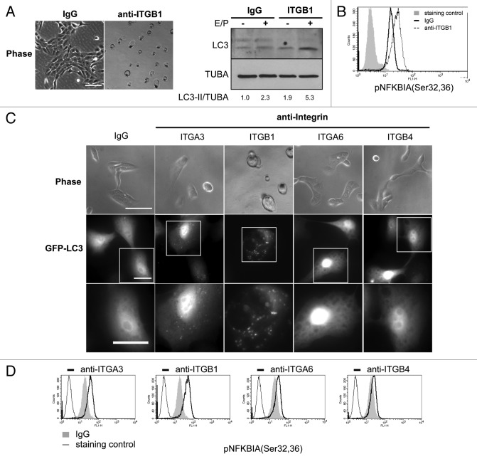
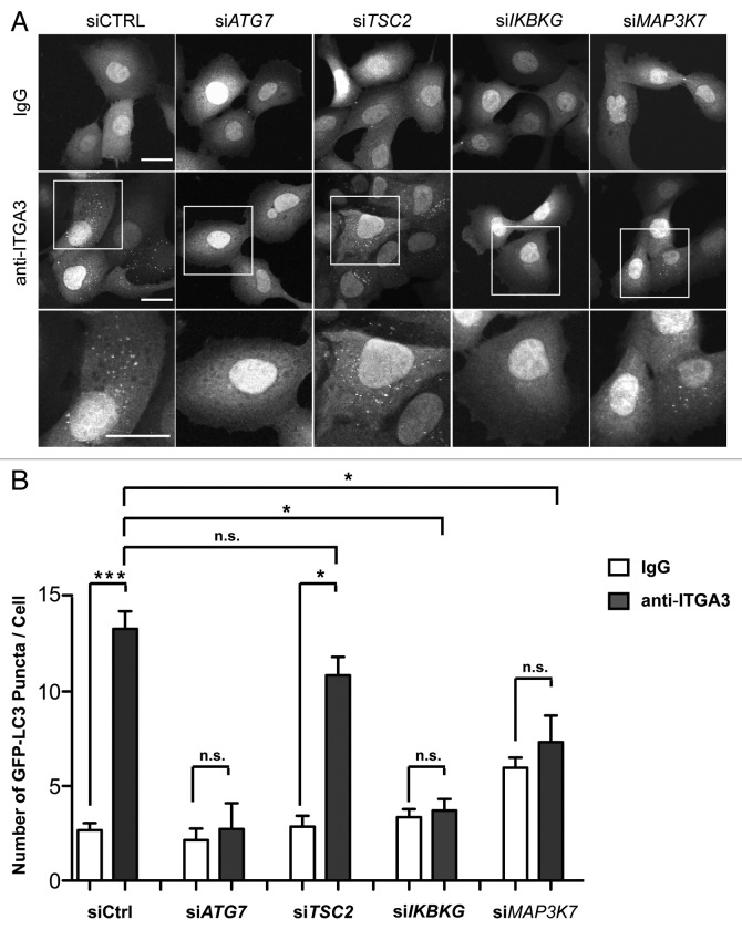
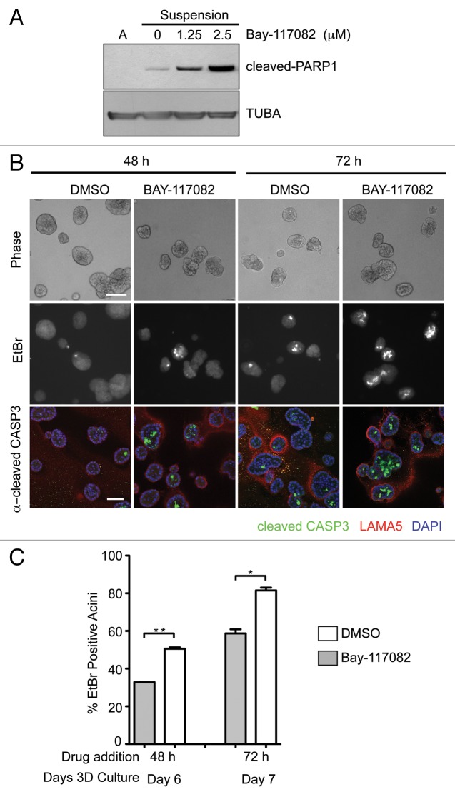
Adherent cells require proper integrin-mediated extracellular matrix (ECM) engagement for growth and survival; normal cells deprived of proper ECM contact undergo anoikis. At the same time, autophagy is induced as a survival pathway in both fibroblasts and epithelial cells upon ECM detachment. Here, we further define the intracellular signals that mediate detachment-induced autophagy and uncover an important role for the IκB kinase (IKK) complex in the induction of autophagy in mammary epithelial cells (MECs) deprived of ECM contact. Whereas the PI3K-AKT-MTORC1 pathway activation potently inhibits autophagy in ECM-detached fibroblasts, enforced activation of this pathway is not sufficient to suppress detachment-induced autophagy in MECs. Instead, inhibition of IKK, as well as its upstream regulator, MAP3K7/TAK1, significantly attenuates detachment-induced autophagy in MECs. Furthermore, function-blocking experiments corroborate that both IKK activation and autophagy induction result from decreased ITGA3-ITGB1 (α3β1 integrin) function. Finally, we demonstrate that pharmacological IKK inhibition enhances anoikis and accelerates luminal apoptosis during acinar morphogenesis in three-dimensional culture. Based on these results, we propose that the IKK complex functions as a key mediator of detachment-induced autophagy and anoikis resistance in epithelial cells.

Keywords: anoikis; autophagy; extracellular matrix; integrin; mammary epithelial cells.


Figures

None
Figure 1. Activation of PI3K-AKT-MTORC1 pathway suppresses ECM detachment-induced autophagy in mouse embryonic fibroblasts (MEFs). (A) Top: Lysates from Tsc2+/+ or tsc2−/− MEFs grown attached (A) or suspended (S) for 24 h were immunoblotted with anti-LC3 and anti-tubulin (TUBA) antibodies. Where indicated, E64d and pepstain A (E/P) were added 6 h prior to harvesting. Bottom: tsc2−/− MEFs stably expressing TSC2N1643I or wild-type TSC2 (TSC2-WT) were grown attached or suspended for 24 h were immunoblotted with anti-LC3 and anti-TUBA antibodies. Where indicated, E64d and pepstain A (E/P) were added 6 h prior to harvesting. (B) Lysates from tsc2−/− MEFs stably expressing empty vector (Emp), TSC2-WT or TSC2N1643I were grown attached (A) or in suspended (S) for 24 h were subject to immunoblotting with antibodies against TSC2, phospho-RPS6 ribosomal protein (Ser240,244) [pRPS6(Ser240,244)] and RPS6 ribosomal protein (RPS6). (C) Wild-type MEFs stably expressing empty vector (Emp), activated PIK3CA (PIK3CA*), or myristoylated AKT (myrAKT) were grown attached (A) or suspended (S) for 24 h. Cell lysates were subject to immunoblotting with antibodies against phospho-AKT(Ser473) [pAKT(Ser473)], AKT, phospho-p70 RPS6 Kinase(Thr389) [pRPS6KB1(Thr389)] or RPS6KB1. (D) Wild-type MEFs stably expressing empty vector (Emp) and PIK3CA* (top) or myrAKT (bottom) were grown attached (A) or suspended (S) for 24 h were immunoblotted with anti-LC3 and anti-TUBA antibodies. Where indicated, E64d and pepstain A (E/P) were added 6 h prior to harvesting.
None
Figure 2. Activation of PI3K-AKT-MTORC1 pathway is not sufficient to suppress autophagy upon ECM detachment in mammary epithelial cells (MECs). (A) Left: MCF10A cells transfected with pooled nontargeting control (siCTRL) or siRNA against TSC2 (siTSC2) were grown attached (A) or suspended (S) for 24 h were lysed and immunoblotted with anti-LC3 and anti-TUBA antibodies. Where indicated, E64d and pepstain A (E/P) were added 6 h prior to harvesting. Right: Lysates from MCF10A cells transfected with siCTRL or siTSC2 and then grown attached (A) or suspended (S) for 24 h were subject to immunoblotting with antibodies against TSC2, TUBA, pRPS6(Ser240,244) and RPS6. (B) Left: MCF10A cells stably expressing empty vector (Emp) or flag-tagged RHEB (RHEB) were grown attached (A) or suspended (S) for 24 h were lysed and immunoblotted with anti-LC3 and anti-TUBA antibodies. Where indicated, E64d and pepstain A (E/P) were added 6 h prior to harvesting. Right: Lysates from MCF10A cells stably expressing empty vector (Emp) or flag-tagged RHEB (RHEB) grown attached (A) or suspended (S) for 24 h were subject to immunoblotting with indicated antibodies. (C) Top: MCF10A cells stably expressing empty vector (Emp), PIK3CA* (left), or myrAKT (right) were grown attached (A) or suspended (S) for 24 h were lysed and immunoblotted with anti-LC3 and anti-TUBA antibodies. Where indicated, E64d and pepstain A (E/P) were added 6 h prior to harvesting. Bottom: MCF10A cells stably expressing empty vector (Emp), PIK3CA*, or myrAKT were grown attached or suspended for 24 h, lysed and immunoblotted with indicated antibodies.
None
Figure 3. Inhibition of IKK activity suppresses autophagy induction in ECM deprived MECs. (A) Left: Lysates from MCF10A cells grown in attached (A) or suspended (S) for 24 h were subject to immunoprecipitation with antibodies against CHUK and IKBKB. Immunoprecipitates were subject to immunoblotting with antibodies against phospho-CHUK(Ser176,180)/IKBKB(Ser177,181). Right: Lysates from MCF10A cells grown in attached (A) or suspended (S) for 24 h were subject to immunoblotting with antibodies against phospho-NFKBIA(Ser32,36) [pIkBa(Ser32,36)] and NFKBIA (IκBα). (B) MCF10A cells treated with DMSO or 10 μM Bay-117082 were grown attached (A) or suspended (S) for 24 h, lysed and immunoblotted with anti-LC3 and anti-TUBA antibodies. Where indicated, E64d and pepstain A (E/P) were added 6 h prior to harvesting. (C) MCF10A cells stably expressing GFP-LC3 were grown suspended for 24 h in the presence of DMSO (left) or 10 μM Bay-117082 (right). Scale bar: 25 μm. (D) MCF10A cells treated with DMSO or 10 μM Bay-117082 were grown attached (A) or suspended (S) for 24 h, lysed and immunoblotted with the indicated antibodies.
None
Figure 4. MAP3K7/IKK pathway promotes ECM detachment-induced autophagy in MECs. (A) MCF10A cells transiently transected with empty vector (Emp), vector expressing dominant negative CHUK (DN-CHUK) (top), or dominant negative IKBKB (DN-IKBKB) (bottom) were grown attached (A) or suspended (S) for 24 h, lysed and immunoblotted with anti-LC3 and anti-TUBA antibodies. Where indicated, E64d and pepstain A (E/P) were added 6 h prior to harvesting. LC3-I detection was minimal in this experiment. (B) MCF10A cells transiently transected with empty vector (Emp), vector expressing DN-CHUK or DN-IKBKB were grown attached (A) or suspended (S) for 24 h, lysed and immunoblotted with the indicated antibodies. (C) Top: MCF10A cells transfected with siCTRL or siIKBKG were grown attached or suspended for 24 h. Where indicated, E64d and pepstain A (E/P) were added 6 h prior to harvesting. Cell lysates were subject to immunoblotting with antibodies against LC3 and TUBA. LC3-I detection was minimal in this experiment. Bottom left: IKBKG protein levels in MCF10A cells transfected with siCTRL or siIKBKG. Bottom right: siCTRL or siIKBKG expressing MECs were grown attached or suspended for 24 h, lysed and immunoblotted with indicated antibodies. (D) Top: MCF10A cells transfected with siCTRL or siMAP3K7 were grown attached or suspended for 24 h. Where indicated, E64d and pepstain A (E/P) were added 6 h prior to harvesting. Cell lysates were subject to immunoblotting with antibodies against LC3 and TUBA. LC3-I detection was minimal in this experiment. Bottom left: MAP3K7 protein levels in MCF10A cells transfected with siCTRL or siMAP3K7. siCTRL or siMAP3K7-expressing MECs were grown attached or suspended for 24 h, lysed and immunoblotted with the indicated antibodies.
None
Figure 5. Antibody-mediated blockade of ITGA3-ITGB1 function induces IKK activation and autophagy in MECs. (A) Left: Phase images of MCF10A cells incubated with 20 μg/ml IgG or anti-ITGB1 prior to seeding to rBM (Matrigel)-coated culture plates for 24 h. Scale bar: 100 μm. Right: MCF10A cells preincubated with 20 μg/ml IgG or anti-ITGB1 were cultured on rBM-coated culture plates for 24 h. Cell lysates were subject to immunoblotting against LC3 and TUBA. Where indicated, E64d and pepstain A (E/P) were added 6 h prior to harvesting. (B) MCF10A cells were preincubated with 20 μg/ml IgG or anti-ITGB1 prior to seeding to rBM-coated culture plates for 24 h. pNFKBIA(Ser32,36) levels were detected by flow cytometry in permeabilized cells. (C) MCF10A cells expressing GFP-LC3 were incubated with the indicated antibodies at 20 μg/ml before seeding to rBM-coated culture plates for 16 h. Top: Phase images. Scale bar: 100 μm. Middle: Fluorescent images of GFP-LC3. Scale bar: 20 μm. Bottom: enlarged images of indicated area in middle panel. Scale bar: 40 μm. (D) Flow cytometry detection of intracellular levels of pNFKBIA(Ser 32,36) in cells preincubated with the indicated antibodies before seeding to rBM-coated culture plates for 16 h.
None
Figure 6. MAP3K7-IKK pathway promotes autophagy in MECs induced upon function blockade of ITGA3. (A) Confocal images of MCF10A cells expressing GFP-LC3 transfected with the indicated siRNAs, preincubated with IgG (top) or anti-ITGA3 antibody (middle), and seeded to rBM-coated culture plates for 16 h. Scale bar: 25 μm. Bottom: Enlarged images of the indicated area in middle panel. Scale bar: 50 μm. (B) Quantification of number of GFP-LC3 puncta per cell (mean ± SEM). In total, 300 cells from three independent experiments were quantified.
None
Figure 7. Inhibition of IKK enhances anoikis and luminal apoptosis in 3D culture. (A) MCF10A cells were cultured attached (A) or in suspension in the presence of DMSO control or Bay-117082 at the indicated doses. Cell lysates were subject to immunoblotting with indicated antibodies. (B) Top: Phase images of MCF10A cell in 3D culture treated with DMSO or 10 μM Bay-117082 for indicated period of time. Scale bar: 100 μm. Middle: Fluorescent images of EtBr staining of MCF10A cell 3D culture under indicated conditions. Bottom: Confocal images of cleaved-CASP3 staining of MCF10A cell 3D culture treated with indicated conditions. Scale bar: 50 μm. (C) Quantification of percentage of EtBr positive acini in MCF10A 3D culture under the indicated conditions (mean ± SEM). In total, 300 acini from three independent experiments were quantified.

Similar articles

Cited by

References

    1. Gilmore AP. Anoikis. Cell Death Differ. 2005;12(Suppl 2):1473–7. doi: 10.1038/sj.cdd.4401723. - DOI - PubMed
    1. Frisch SM, Francis H. Disruption of epithelial cell-matrix interactions induces apoptosis. J Cell Biol. 1994;124:619–26. doi: 10.1083/jcb.124.4.619. - DOI - PMC - PubMed
    1. Debnath J, Brugge JS. Modelling glandular epithelial cancers in three-dimensional cultures. Nat Rev Cancer. 2005;5:675–88. doi: 10.1038/nrc1695. - DOI - PubMed
    1. Fung C, Lock R, Gao S, Salas E, Debnath J. Induction of autophagy during extracellular matrix detachment promotes cell survival. Mol Biol Cell. 2008;19:797–806. doi: 10.1091/mbc.E07-10-1092. - DOI - PMC - PubMed
    1. He C, Klionsky DJ. Regulation mechanisms and signaling pathways of autophagy. Annu Rev Genet. 2009;43:67–93. doi: 10.1146/annurev-genet-102808-114910. - DOI - PMC - PubMed

Publication types

MeSH terms