

Fibromuscular dysplasia affecting a two-branched renal artery in a patient with a solitary kidney: case presentation
- PMID: 23780717
- PMCID: PMC6649647
- DOI: 10.1002/clc.22149
Fibromuscular dysplasia affecting a two-branched renal artery in a patient with a solitary kidney: case presentation
Abstract
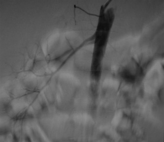
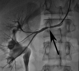
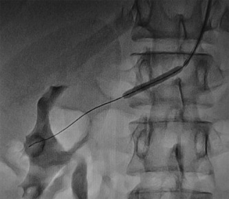
Fibromuscular dysplasia (FMD) is a nonatherosclerotic, noninflammatory arterial disease, commonly involving the renal arteries. Here we report a case of a 16-year-old Chinese male who was found to have severe hypertension with proteinuria for 2 years. Computed tomography showed absence of the left kidney and enlargement of the right kidney. Subsequent angiography confirmed the above findings and revealed narrowing of both the upper and lower branches of the right renal artery caused by FMD. These combined lesions are very rare, and individuals affected are at increased risk of renal dysfunction if left untreated. Treatment with percutaneous balloon angioplasty is the first choice in such a patient and usually results in optimal outcomes.
© 2013 Wiley Periodicals, Inc.
Figures


Comment in
-
Response to 'Fibromuscular dysplasia affecting a two-branched renal artery in a patient with a solitary kidney: case presentation'.Clin Cardiol. 2013 Dec;36(12):E50. doi: 10.1002/clc.22221. Epub 2013 Oct 17. Clin Cardiol. 2013. PMID: 24136776 Free PMC article.
References
-
- Olin JW, Sealove BA. Diagnosis, management, and future developments of fibromuscular dysplasia. J Vasc Surg. 2011;53:3. - PubMed
-
- Safian RD, Textor SC. Renal‐artery stenosis. N Engl J Med. 2001;344:431–442. - PubMed
-
- Blondin D, Lanzman R, Schellhammer F, et al. Fibromuscular dysplasia in living renal donors: still a challenge to computed tomographic angiography. Eur J Radiol. 2010;75:67–71. - PubMed
Publication types
MeSH terms
LinkOut - more resources
Full Text Sources
Other Literature Sources
