. 2013:2013:916348.
doi: 10.1155/2013/916348. Epub 2013 May 23.

A Case of Primary T-Cell Central Nervous System Lymphoma: MR Imaging and MR Spectroscopy Assessment

Affiliations

A Case of Primary T-Cell Central Nervous System Lymphoma: MR Imaging and MR Spectroscopy Assessment

G Manenti et al. Case Rep Radiol. 2013.

Abstract

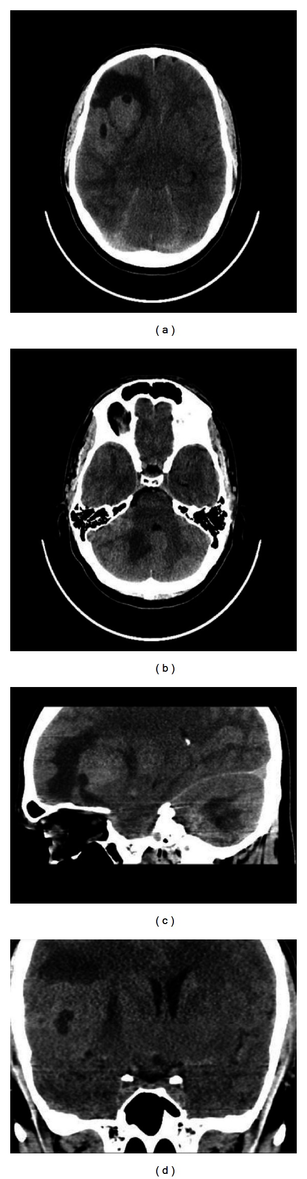
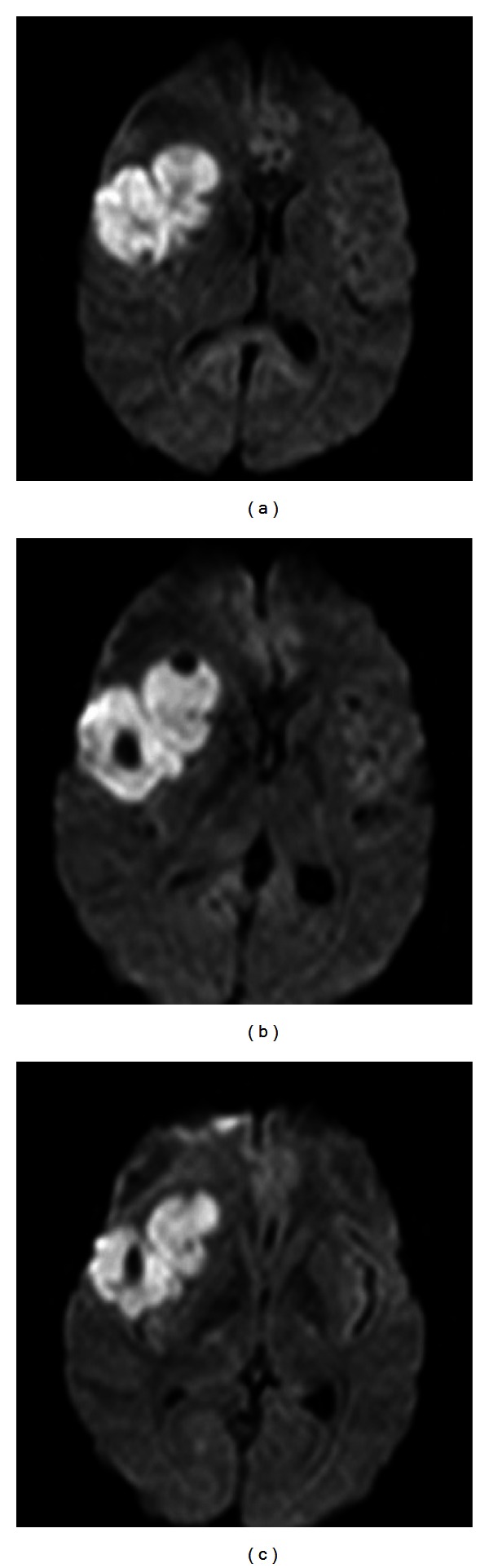
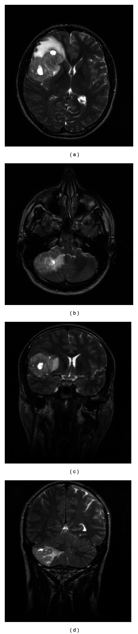
Primary central nervous system lymphomas (PCNSLs) are mainly B-cells lymphomas. A risk factor for the development of PCNSL is immunodeficiency, which includes congenital disorders, iatrogenic immunosuppression, and HIV. The clinical course is rapidly fatal; these patients usually present signs of increased intracranial pressure, nausea, papilledema, vomiting, and neurological and neuropsychiatric symptoms. PCNSL may have a characteristic appearance on CT and MR imaging. DWI sequences and MR spectroscopy may help to differentiate CNS lymphomas from other brain lesions. In this paper, we report a case of a 23-year-old man with T-primary central nervous system lymphoma presenting with a mass in the right frontotemporal lobe. We describe clinical, CT, and MRI findings. Diagnosis was confirmed by stereotactic biopsy of the lesion.


Figures

Figure 1
Figure 1
Axial CT acquisition (a)-(b) with sagittal (c) and coronal (d) multiplanar reformatted images showing a mild hyperdense complex lobulated mass in the right frontotemporal region associated with oedema and a similar smaller lesion in the right cerebellar lobe.
Figure 2
Figure 2
Isotropic diffusion weighted images (DWI) showing signal restriction associated with the fronto-temporal lesion.
Figure 3
Figure 3
Axial and coronal T2-weighted images showing inhomogeneous hyperintensity of the cerebellar and fronto-temporal right lobe lesions associated with ovalar hyperintensities and perilesional oedema.
Figure 4
Figure 4
Gadolinium-DTPA enhanced T1-weighted multiplanar images (a), (b), and (c) reveal heterogenous enhancement throughout the fronto-temporal mass; single-voxel H MR spectroscopy (d) shows a lactate/lipid peak, increase of Choline/creatine ratio, and depression of N-acetylaspartate/creatine ratio in the fronto-temporal lesion.

References

    1. Shibamoto Y, Ogino H, Suzuki G, et al. Primary central nervous system lymphoma in Japan: changes in clinical features, treatment, and prognosis during 1985–2004. Neuro-Oncology. 2008;10(4):560–568. - PMC - PubMed
    1. Zhang D, Hu LB, Henning TD, et al. MRI findings of primary CNS lymphoma in 26 immunocompetent patients. Korean Journal of Radiology. 2010;11(3):269–277. - PMC - PubMed
    1. DeAngelis LM. Primary central nervous system lymphoma. Recent Results in Cancer Research. 1994;135:155–169. - PubMed
    1. Ferreri AJM, Reni M, Villa E. Primary central nervous system lymphoma in immunocompetent patients. Cancer Treatment Reviews. 1995;21(5):415–446. - PubMed
    1. Villano JL, Koshy M, Shaikh H, Dolecek TA, McCarthy BJ. Age, gender, and racial differences in incidence and survival in primary CNS lymphoma. British Journal of Cancer. 2011;105(9):1414–1418. - PMC - PubMed

LinkOut - more resources