. 2013;54(4-5):296-304.
doi: 10.3109/03008207.2013.814646. Epub 2013 Aug 26.

Utilization of transgenic models in the evaluation of osteogenic differentiation of embryonic stem cells

Affiliations

Utilization of transgenic models in the evaluation of osteogenic differentiation of embryonic stem cells

Dario Repic et al. Connect Tissue Res. 2013.

Abstract

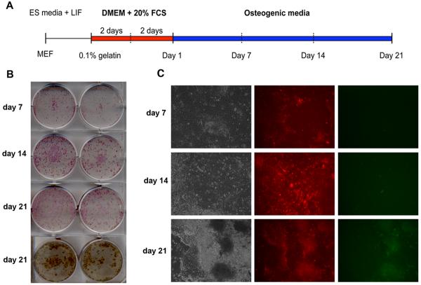
Previous studies reported that embryonic stem cells (ESCs) can be induced to differentiate into cells showing a mature osteoblastic phenotype by culturing them under osteo-inductive conditions. It is probable that osteogenic differentiation requires that ESCs undergo differentiation through an intermediary step involving a mesenchymal lineage precursor. Based on our previous studies indicating that adult mesenchymal progenitor cells express α-smooth muscle actin (αSMA), we have generated ESCs from transgenic mice in which an αSMA promoter directs the expression of red fluorescent protein (RFP) to mesenchymal progenitor cells. To track the transition of ESC-derived MSCs into mature osteoblasts, we have utilized a bone-specific fragment of rat type I collagen promoter driving green fluorescent protein (Col2.3GFP). Following osteogenic induction in ESCs, we have observed expression of alkaline phosphatase (ALP) and subsequent mineralization as detected by von Kossa staining. After 1 week of osteogenic induction, ESCs begin to express αSMARFP. This expression was localized to the peripheral area encircling a typical ESC colony. Nevertheless, these αSMARFP positive cells did not show activation of the Col2.3GFP promoter, even after 7 weeks of osteogenic differentiation in vitro. In contrast, Col2.3GFP expression was detected in vivo, in mineralized areas following teratoma formation. Our results indicate that detection of ALP activity and mineralization of ESCs cultured under osteogenic conditions is not sufficient to demonstrate osteogenic maturation. Our study indicates the utility of the promoter-visual transgene approach to assess the commitment and differentiation of ESCs into the osteoblast lineage.


Figures

Figure 1
Figure 1. Osteogenic potential of primary bone marrow stromal cells
(A) Diagram of αSMARFP and Col2.3GFP transgenic constructs. (B) Images of day 7 primary BMSC cultures derived from dual αSMARFP/Col2.3GFP transgenic mice (brightfield, left panel; RFP, middle panel; GFP, right panel). C) FACS analysis confirming the activity of αSMARFP and absence of Col2.3GFP before sorting. D) Images obtained one week after sorting, prior to osteogenic induction, and E) nine days after osteogenesis was induced (brightfield images red (αSMARFP) and green fluorescence (Col2.3GFP) and overlayed images are shown). Mineralization was assessed with von Kossa method (F) and expression levels of mature osteoblast markers osteocalcin and bone sialoprotein were evaluated by real-time PCR (Figure 1G). Transgene activation and osteogenic differentiation was evaluated in three independent biological experiments and results from a representative experiment are presented.
Figure 2
Figure 2. Generation of murine ESCs lines
A) By crossing αSMARFP × Col2.3GFP mice, we have generated dual transgenic ESC lines. Genotyping was done using a set of primers that distinguishes GFP from RFP sequences. B) Following induction of differentiation the αSMA expression was detected in cells surrounding ESC colonies, with no expression of αSMARFP within ESC colony. Morphologically αSMARFP+ cells exhibited fibroblastic shape. Edges between the two distinct populations are indicated using arrows.
Figure 3
Figure 3. Evaluation of osteogenic differentiation of ESCs using monolayer conditions
(A) Description of the cell culture protocol and timeline for analysis. (B) Histochemical detection of alkaline phosphatase. Mineralization was detected using von Kossa method on day 21. (C) Images of mouse ESCs following osteogenic induction. Phase contrast, left panel; RFP, middle panel; GFP, right panel. Epifluorescence imaging for GFP shows only a background signal from mineralized tissue. Experiments were completed as three independent biological replicates.
Figure 4
Figure 4. Evaluation of osteogenic differentiation of ESCs derived through EB step
(A) Description of the cell culture protocol and timeline for analysis. (B) Detection of ALP on day 7–21 and mineralization on day 21. (C) Phase contrast image of mouse ESCs after osteogenic induction (left panel). αSMARFP expression is shown in middle panel, while expression of Col2.3GFP is shown on right panel. Experiments were completed as four independent biological replicates.
Figure 5
Figure 5. Analysis of gene expression during osteogenic induction of ESCs
Time course of bone marker expression in ESCs differentiated as monolayered cultures (A) or through formation of EBs (B). RNA was extracted at various time points during osteogenic differentiation and assayed for Runx2, Osterix, Col1a2, Bsp and Oc. Undifferentiated ESCs were used to normalize the expression, and RNA from day 21 BMSCs was used as a positive control. Results presented are from one of three independent experiments.
Figure 6
Figure 6. In vivo analysis of Col2.3GFP expression using teratoma formation assay
ESCs from αSMARFP/Col2.3GFP transgenic mice were transplanted into the femoral muscle of immunodeficient mice. (A) Epifluorescence and (B) von Kossa staining of the corresponding section confirmed the presence of mineralized tissues in the areas in which Col2.3GFP activates. Four mice were utilized and ESCs were injected in femoral muscle on both sides. (Bar= 100μm).

References

    1. Keller GM. In vitro differentiation of embryonic stem cells. Curr Opin Cell Biol. 1995;7:862–9. - PubMed
    1. Granero-Molto F, Weis JA, Longobardi L, Spagnoli A. Role of mesenchymal stem cells in regenerative medicine: application to bone and cartilage repair. Expert Opin Biol Ther. 2008;8:255–68. - PubMed
    1. Caplan AI. Adult mesenchymal stem cells for tissue engineering versus regenerative medicine. J Cell Physiol. 2007;213:341–7. - PubMed
    1. Gao C, Seuntjens J, Kaufman GN, Tran-Khanh N, Butler A, Li A, Wang H, Buschmann MD, Harvey EJ, Henderson JE. Mesenchymal stem cell transplantation to promote bone healing. J Orthop Res. 2012;30:1183–9. - PubMed
    1. Krebsbach PH, Kuznetsov SA, Satomura K, Emmons RV, Rowe DW, Robey PG. Bone formation in vivo: comparison of osteogenesis by transplanted mouse and human marrow stromal fibroblasts. Transplantation. 1997;63:1059–69. - PubMed

Publication types

Substances

LinkOut - more resources