Review
. 2013 Oct 26;382(9902):1445-57.
doi: 10.1016/S0140-6736(13)60178-6. Epub 2013 Jun 17.

Retinopathy of prematurity

Affiliations
Review

Retinopathy of prematurity

Ann Hellström et al. Lancet. .

Abstract

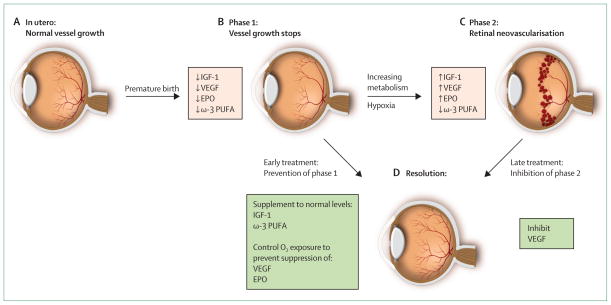
The immature retinas of preterm neonates are susceptible to insults that disrupt neurovascular growth, leading to retinopathy of prematurity. Suppression of growth factors due to hyperoxia and loss of the maternal-fetal interaction result in an arrest of retinal vascularisation (phase 1). Subsequently, the increasingly metabolically active, yet poorly vascularised, retina becomes hypoxic, stimulating growth factor-induced vasoproliferation (phase 2), which can cause retinal detachment. In very premature infants, controlled oxygen administration reduces but does not eliminate retinopathy of prematurity. Identification and control of factors that contribute to development of retinopathy of prematurity is essential to prevent progression to severe sight-threatening disease and to limit comorbidities with which the disease shares modifiable risk factors. Strategies to prevent retinopathy of prematurity will depend on optimisation of oxygen saturation, nutrition, and normalisation of concentrations of essential factors such as insulin-like growth factor 1 and ω-3 polyunsaturated fatty acids, as well as curbing of the effects of infection and inflammation to promote normal growth and limit suppression of neurovascular development.


Conflict of interest statement

Conflicts of interest

AH previously owned shares in PremaCure Holding, which controlled PremaCure (Uppsala, Sweden), a company that had rights to the WINROP system and held patents and patent applications that covered prevention of retinopathy of prematurity with insulin-like growth factor 1. LEHS and OD declare that they have no conflicts of interest.

Figures

Figure 1
Figure 1. Progression of retinopathy of prematurity
(A) Oxygen tension is low in utero and vascular growth is normal. (B) Phase 1: after birth until roughly 30 weeks postmenstrual age, retinal vascularisation is inhibited because of hyperoxia and loss of the nutrients and growth factors provided at the maternal–fetal interface. Blood-vessel growth stops and as the retina matures and metabolic demand increases, hypoxia results. (C) Phase 2: the hypoxic retina stimulates expression of the oxygen-regulated factors such as erythropoietin (EPO) and vascular endothelial growth factor (VEGF), which stimulate retinal neovascularisation. Insulin-like growth factor 1 (IGF-1) concentrations increase slowly from low concentrations after preterm birth to concentrations high enough to allow activation of VEGF pathways. (D) Resolution of retinopathy might be achieved through prevention of phase 1 by increasing IGF-1 to in-utero concentrations and by limiting oxygen to prevent suppression of VEGF; alternatively, VEGF can be suppressed in phase 2 after neovascularisation with laser therapy or an antibody. EPO=erythropoietin. ω-3 PUFA=ω-3 polyunsaturated fatty acids. Adapted from reference , by permission of the Association for Research in Vision and Ophthalmology.
Figure 2
Figure 2. Infection, inflammation, and retinopathy of prematurity
Exposure to infection and inflammation seems to modify risk of retinopathy of prematurity, especially before (prephase) and a few weeks after birth (phase 2), when oxygen concentrations are relatively low compared with phase 1 (immediately after birth). Whereas prenatal inflammation seems to exert a sensitising effect without directly increasing risk, postnatal infection and inflammation are associated with an increased risk, perhaps most prominently in phase 2. Adapted from reference , by permission of Elsevier.
Figure 3
Figure 3. Zones and stages of retinopathy of prematurity
The retina is divided into three zones (A, diagram shows right eye) and the extent or severity of retinopathy in these zones is classified as stages (B). Stage 1 is characterised by a thin demarcation line between vascularised and non-vascularised retina, stage 2 by a ridge, stage 3 by extraretinal fibrovascular proliferation, stage 4 by part retinal detachment, and stage 5 by total retinal detachment. In stage 3, extraretinal neovascularisation can become severe enough to cause retinal detachment (stages 4–5), which usually leads to blindness. Part B is courtesy of Lisa Hård.
Figure 4
Figure 4. Plus disease
Compared with a normal retina (A), plus disease (B) is characterised by venous dilation and increased arterial tortuosity of posterior vessels. Reproduced from reference , by permission of the American Medical Association.

References

    1. Silverman WA. Retrolental fibroplasia: a modern parable. New York: Grune & Stratton; 1980.
    1. Campbell K. Intensive oxygen therapy as a possible cause of retrolental fibroplasias: a clinical approach. Med J Aust. 1951;2:48–50. - PubMed
    1. Carlo WA, Finer NN, Walsh MC, et al. Target ranges of oxygen saturation in extremely preterm infants. N Engl J Med. 2010;362:1959–69. - PMC - PubMed
    1. Stenson B, Brocklehurst P, Tarnow-Mordi W. Increased 36-week survival with high oxygen saturation target in extremely preterm infants. N Engl J Med. 2011;364:1680–82. - PubMed
    1. Chen J, Smith LE. Retinopathy of prematurity. Angiogenesis. 2007;10:133–40. - PubMed

Publication types