. 2013 Jun 19:10:62.
doi: 10.1186/1742-4690-10-62.

Genistein interferes with SDF-1- and HIV-mediated actin dynamics and inhibits HIV infection of resting CD4 T cells

Affiliations

Genistein interferes with SDF-1- and HIV-mediated actin dynamics and inhibits HIV infection of resting CD4 T cells

Jia Guo et al. Retrovirology. .

Abstract

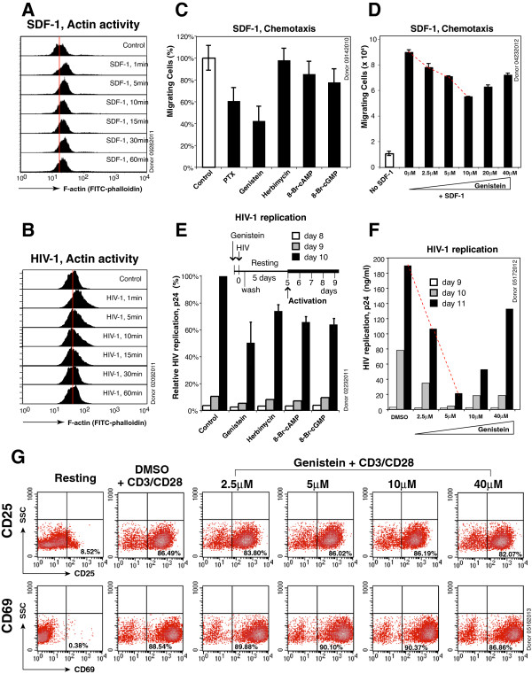
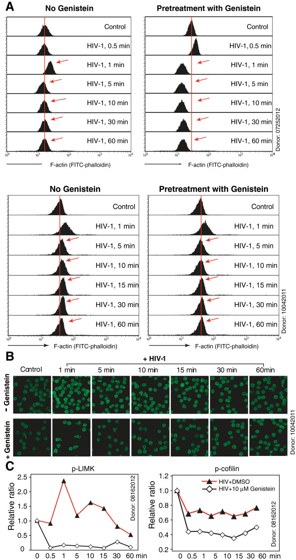
Background: Binding of HIV to the chemokine coreceptor CXCR4 mediates viral fusion and signal transduction that promotes actin dynamics critical for HIV infection of blood resting CD4 T cells. It has been suggested that this gp120-mediated actin activity resembles the chemotactic actin dynamics mediated by chemokines such as SDF-1. To determine whether inhibiting SDF-1-mediated chemotactic activity can also inhibit HIV infection, we screened several inhibitors known to reduce SDF-1-mediated chemotaxis of T cells.

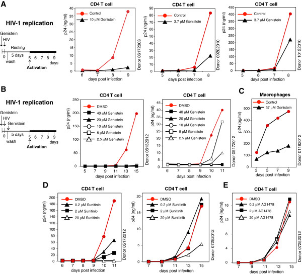
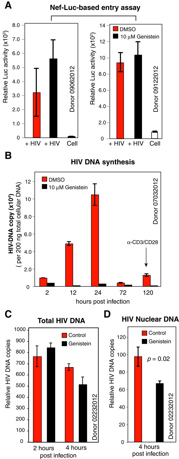
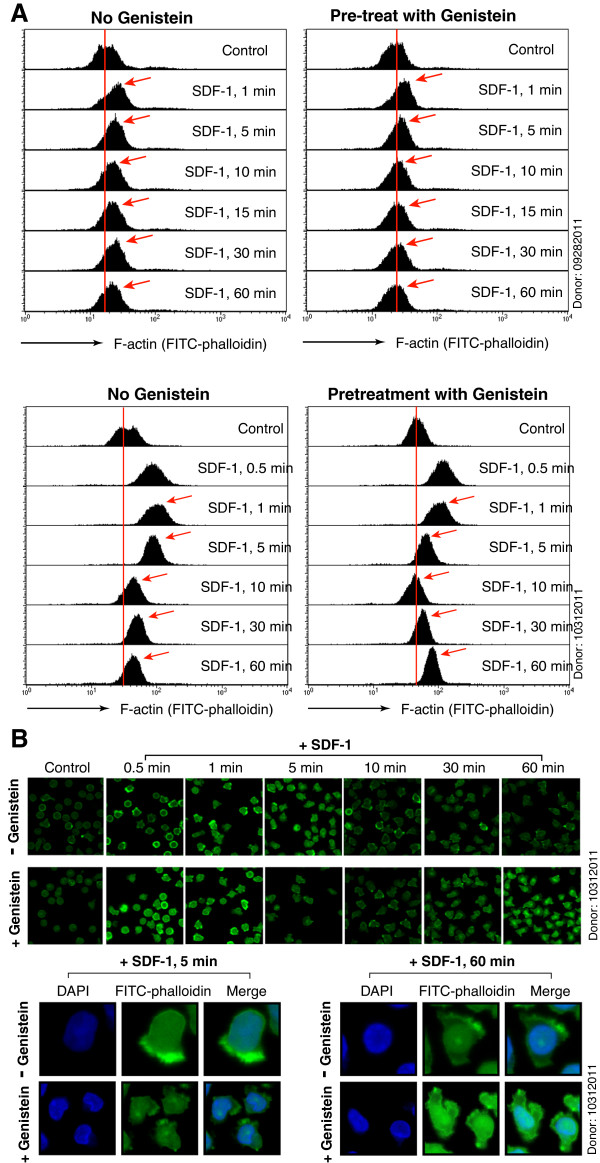
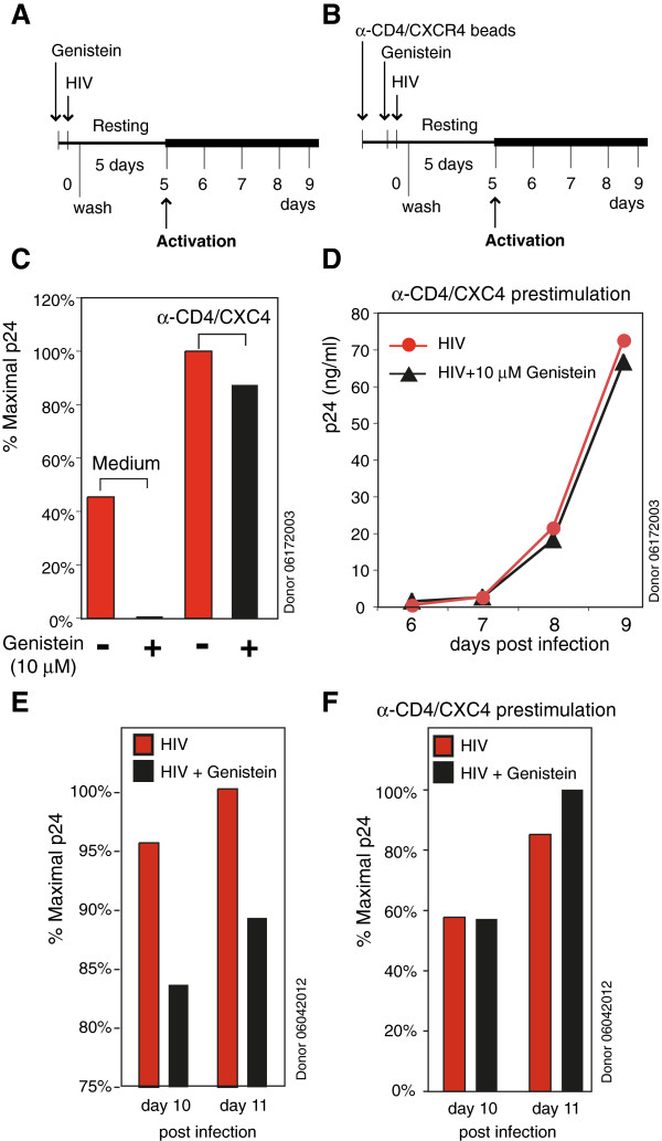
Results: We found that a tyrosine kinase inhibitor, genistein, inhibited both SDF-1-mediated chemotaxis and HIV infection of resting CD4 T cells. Genistein was also found to interfere with SDF-1- and HIV-mediated actin dynamics in CD4 T cells. This reduction in actin activity correlates with genistein-mediated inhibition of viral DNA accumulation in resting CD4 T cells. In addition, we also tested two other tyrosine kinase inhibitors, sunitinib and AG1478. Sunitinib, but not AG1478, inhibited HIV infection of resting CD4 T cells. We further tested the safety of genistein in 3 Chinese rhesus macaques (Macaca mulatta), and each animal was given a monotherapy of genistein at 10 mg/kg orally for 12 weeks. No adverse drug effects were observed in these animals.

Conclusions: Our results suggest that novel therapeutic strategies can be developed based on targeting cellular proteins involved in HIV-dependent signaling. This approach can interfere with HIV-mediated actin dynamics and inhibit HIV infection.


Figures

Figure 1
Figure 1
Genistein inhibits SDF-1-mediated chemotaxis and HIV infection of resting CD4 T cells. (A) Resting memory CD4 T cells were stimulated with SDF-1 (12.5 nM) for various times, stained with FITC-phalloidin, and then analyzed with flow cytometer. (B) Resting memory CD4 T cells were stimulated with HIV-1NL4-3 for various times and similarly stained with FITC-phalloidin and analyzed. (C) Resting memory CD4 T ells were pretreated with pertussis toxin (PTX, 100 ng/ml), genistein (3.7 μM), herbimycin (1 μM), 8-Br-cAMP (100 μM), 8-Br-cGMP (100 μM), or DMSO (1%, control) for 15–60 minutes, and then assayed for migration towards SDF-1 (12.5 nM). Results are expressed as the relative percentage of migrating cells. (D) Dosage-dependent inhibition of SDF-1-mediated chemotaxis by genistein. Experiments were carried out as described in (C) with memory T cells pretreated with different dosages of genistein. (E) Resting CD4 T cells were similarly pretreated with genistein, herbimycin, 8-Br-cAMP, 8-Br-cGMP, or DMSO, and infected with HIV-1NL4-3 for 2 hours in the continuous presence of these inhibitors. Following infection, cells were washed twice and then cultured in the absence of the inhibitors for 5 days. Cells were activated at day 5 with anti-CD3/CD28 magnetic beads (4 beads per cell), and viral replication was measured by p24 release. (F) Dosage-dependent inhibition of HIV-1 infection by genistein. Experiments were carried out as described in (E) with resting T cells pretreated with different dosages of genistein. (G) Effects of genistein on T cell activation. Resting CD4 T cells were treated with genistein at different dosages for 3 hours, washed twice, and then cultured in the absence of genistein as described in (E). Cells were similarly activated with anti-CD3/CD28 beads. At 24 hours after stimulation, cells were stained with PE-labeled monoclonal antibody against human CD25 or CD69, and then analyzed with flow cytometry.
Figure 2
Figure 2
Genistein inhibits HIV infection of resting CD4 T cells and macrophages. (A) Genistein inhibits HIV infection of resting CD4 T cells in additional three donors. Experimental procedures are described in Figure 1E. (B) Dosage-dependent inhibition of HIV infection of resting CD4 T cells. Resting CD4 T cells from two additional donors were pretreated with genistein at different dosages for 1 hour, and then infected with HIV-1NL4-3 for 2 hours at 37°C in the continuous presence of these inhibitors. Following infection, cells were washed twice and a single dose of genistein was added to the culture. Cells were culture for 5 days and activated at day 5 with anti-CD3/CD28 magnetic beads (4 beads per cell). Viral replication was measured by p24 release. (C) Genistein inhibits HIV infection of primary macrophages. Human peripheral blood monocyte-derived macrophages were pretreated with genistein or DMSO (1%, control) for 1 hour, and then infected with HIV (THRO.c/2626) for 2 hours. Following infection, cells were washed to remove HIV and the inhibitor, and then continuously cultured in the absence of genistein to monitor viral replication. (D) Sunitinib inhibits HIV infection of resting CD4 T cells. Cells from two donors were pretreated with different dosages of sunitinib for 1 hour, and then infected with HIV-1NL4-3 for 2 hours in the continuous presence of the inhibitor. Following infection, cells were washed twice and then cultured in the absence of the inhibitor for 5 days. Cells were activated at day 5 with anti-CD3/CD28 magnetic beads, and viral replication was measured by p24 release. (E) AG1478 does not inhibit HIV infection of resting CD4 T cells. Resting CD4 T cells were similarly pretreated with AG1478, and infected with HIV-1, as described in (D).
Figure 3
Figure 3
Genistein inhibits HIV DNA synthesis and viral DNA nuclear localization. (A) Genistein does not inhibit viral entry into resting CD4 T cells. Resting CD4 T cells from two donors were pretreated with genistein for 1 hour, and then infected with Nef-luciferase tagged HIV-1 for 2 hours. Cells were washed and luciferase activity was measured in live cells. (B) Genistein inhibits viral DNA synthesis in CD4 T cells. Resting memory CD4 T cells were pretreated with genistein (10 μM) or DMSO (1%, control) for 1 hour, and then infected with HIV-1NL4-3 for 2 hour in the presence of genistein. Cells were washed twice to remove HIV and genistein, cultured for 5 days, and then activated at day 5 with anti-CD3/CD28 magnetic beads. Infected cells were harvested and lysed at different time points following infection to extract total cellular DNA. HIV DNA was measured by using real time PCR using equal amount of total DNA. (C and D) Resting memory CD4 T cells were similarly pretreated with genistein (10 μM) and infected with HIV. Viral DNA was measured at 2 and 4 hours post infection. Infected cells were also used for fractionation and purification of nuclear DNA. HIV nuclear DNA was measured using real time PCR using equal amount of total DNA (D).
Figure 4
Figure 4
Genistein interferes with SDF-1-mediated actin dynamics in resting memory CD4 T cells. (A) Resting memory CD4 T cells from two donors were pretreated with genistein (3.7 μM) or mock-treated for 1 hour at 37°C, and then stimulated with SDF-1 (12.5 nM) for various times, from 0.5 minute to 60 minutes. Cells were fixed, stained with FITC-phalloidin for F-actin, and then analyzed with flow cytometer. (B) Confocal images of cells stained in (A), showing F-actin staining. Images were acquired and analyzed at identical conditions for all samples. Some cells were also stained with DAPI (4′, 6-diamidino-2-phenylindole) for nuclear DNA.
Figure 5
Figure 5
Genistein interferes with HIV-mediated actin dynamics in resting memory CD4 T cells. (A) Resting memory CD4 T cells from two donors were pretreated with genistein (3.7 μM) or mock-treated for 1 hour at 37°C, and then stimulated with HIV-1NL4-3 (100 ng) for various times. Cells were fixed, stained with FITC-phalloidin for F-actin, and then analyzed with a flow cytometer. (B) Confocal images of cells stained in (A), showing F-actin staining. Images were acquired and analyzed at identical conditions for all samples. (C) Resting memory CD4 T cells were pretreated with 10 μM genistein for 1 hour, and then treated with HIV-1NL4-3 for a time course. Cells were then analyzed by Western blot for LIMK and cofilin activation, using an anti-phospho-LIMK antibody or an anti-cofilin antibody. The same blots were also probed with an anti-human GAPDH antibody for loading control and normalization.
Figure 6
Figure 6
Anti-CD4/CXCR4 bead pre-stimulation abolishes genistein inhibition of HIV infection of resting CD4 T cells. (A and B) Purified resting CD4 T cells were cultured in medium overnight (A) or pre-stimulated with anti-CD4/CXCR4 magnetic beads (2 beads per cell) overnight (B), and then treated with genistein (10 μM) for 1 hour. Cells were infected with HIV-1NL4-3 for 2 hours, washed, incubated for 5 days in the absence of genistein, and then activated by anti-CD3/CD28 magnetic beads (2 beads per cell). Viral replication was measured by p24 release into the supernatant. (C) Viral replication measured at day 8 post infection. (D) The full viral replication course in cells pre-stimulated with anti-CD4/CXCR4 beads. (E and F) A repeat of the experiment in another donor. Viral replication at day 10 and 11 is shown.
Figure 7
Figure 7
Genistein monotherapy on Chinese rhesus macaques with chronic SIV infectionc. (A and B) Three Chinese rhesus macaques were chronically infected with SIVmac251. Each animal received 10 mg/kg of genistein orally for 12 weeks. The dynamics of the absolute CD4+ T cell count (A) and the percentages of CD4+ T cells (B) in peripheral blood were monitored. The percentage of CD4+ T cells were calculated by gating on total CD3+ T lymphocytes analyzed by flow cytometry. The absolute CD4+ T cell counts were generated from the calculation of percentages of CD4+ T cells, CD3+ T cells and lymphocytes from the complete blood count (CBC). (C) Plasma viral load before and after genistein monotherapy was also measured.

Similar articles

Cited by

References

    1. Klatzmann D, Champagne E, Chamaret S, Gruest J, Guetard D, Hercend T, Gluckman JC, Montagnier L. T-lymphocyte T4 molecule behaves as the receptor for human retrovirus LAV. Nature. 1984;312:767–768. doi: 10.1038/312767a0. - DOI - PubMed
    1. Dalgleish AG, Beverley PC, Clapham PR, Crawford DH, Greaves MF, Weiss RA. The CD4 (T4) antigen is an essential component of the receptor for the AIDS retrovirus. Nature. 1984;312:763–767. doi: 10.1038/312763a0. - DOI - PubMed
    1. Feng Y, Broder CC, Kennedy PE, Berger EA. HIV-1 entry cofactor: functional cDNA cloning of a seven-transmembrane, G protein-coupled receptor. Science. 1996;272:872–877. doi: 10.1126/science.272.5263.872. - DOI - PubMed
    1. Alkhatib G, Combadiere C, Broder CC, Feng Y, Kennedy PE, Murphy PM, Berger EA. CC CKR5: a RANTES, MIP-1alpha, MIP-1beta receptor as a fusion cofactor for macrophage-tropic HIV-1. Science. 1996;272:1955–1958. doi: 10.1126/science.272.5270.1955. - DOI - PubMed
    1. Choe H, Farzan M, Sun Y, Sullivan N, Rollins B, Ponath PD, Wu L, Mackay CR, LaRosa G, Newman W. et al.The beta-chemokine receptors CCR3 and CCR5 facilitate infection by primary HIV-1 isolates. Cell. 1996;85:1135–1148. doi: 10.1016/S0092-8674(00)81313-6. - DOI - PubMed

Publication types

MeSH terms

LinkOut - more resources