. 2013 Aug;24(16):2506-14.
doi: 10.1091/mbc.E13-03-0150. Epub 2013 Jun 19.

RanGTP and CLASP1 cooperate to position the mitotic spindle

Affiliations

RanGTP and CLASP1 cooperate to position the mitotic spindle

Stephen L Bird et al. Mol Biol Cell. 2013 Aug.

Abstract

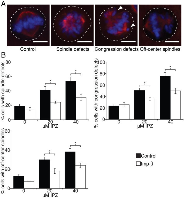
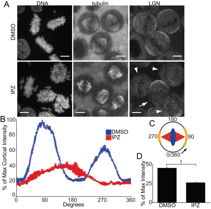
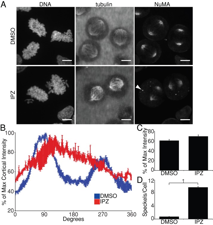
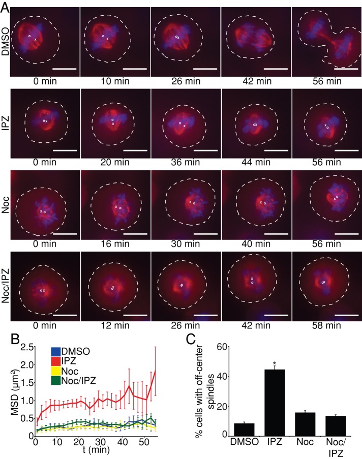
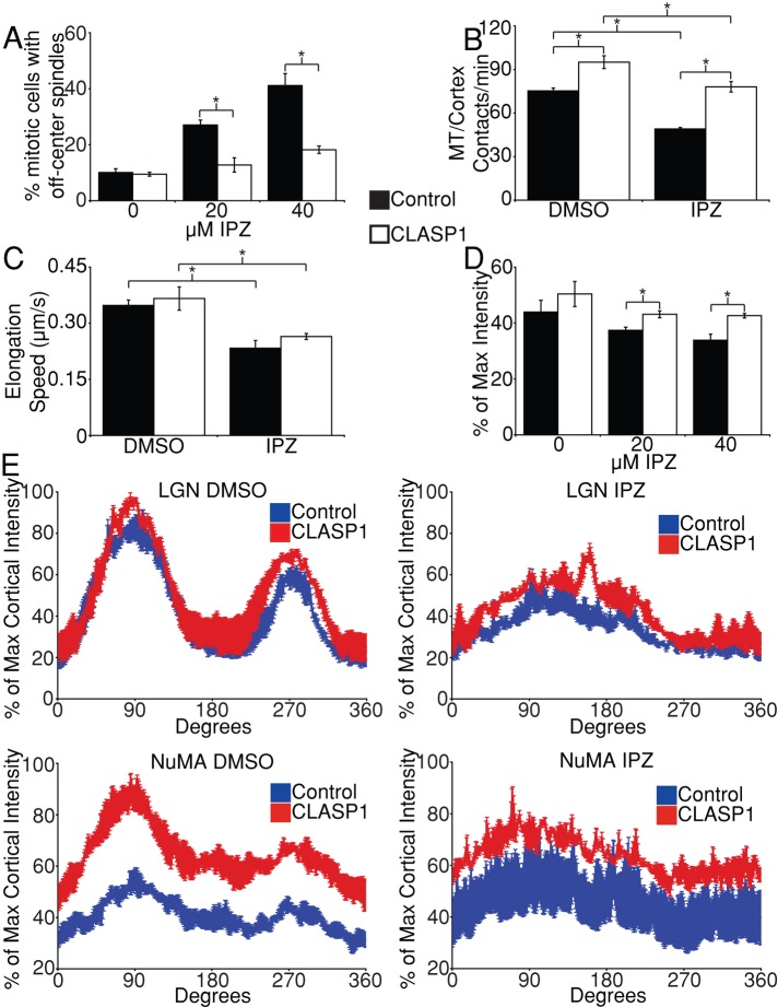
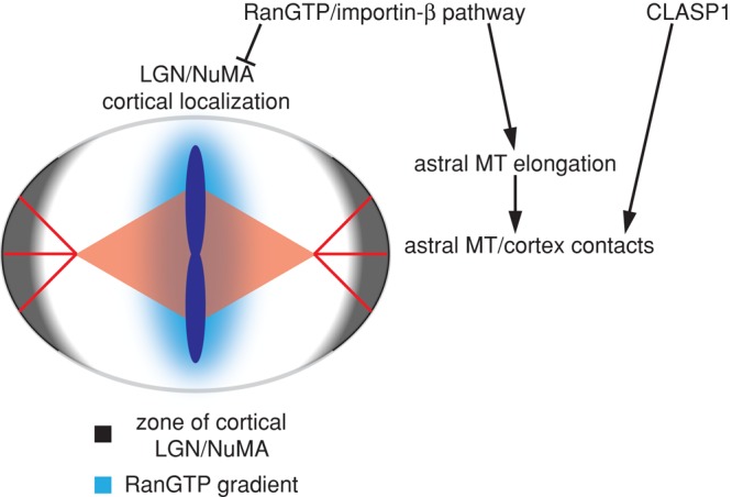
Accurate positioning of the mitotic spindle is critical to ensure proper distribution of chromosomes during cell division. The small GTPase Ran, which regulates a variety of processes throughout the cell cycle, including interphase nucleocytoplasmic transport and mitotic spindle assembly, was recently shown to also control spindle alignment. Ran is required for the correct cortical localization of LGN and nuclear-mitotic apparatus protein (NuMA), proteins that generate pulling forces on astral microtubules (MTs) through cytoplasmic dynein. Here we use importazole, a small-molecule inhibitor of RanGTP/importin-β function, to study the role of Ran in spindle positioning in human cells. We find that importazole treatment results in defects in astral MT dynamics, as well as in mislocalization of LGN and NuMA, leading to misoriented spindles. Of interest, importazole-induced spindle-centering defects can be rescued by nocodazole treatment, which depolymerizes astral MTs, or by overexpression of CLASP1, which does not restore proper LGN and NuMA localization but stabilizes astral MT interactions with the cortex. Together our data suggest a model for mitotic spindle positioning in which RanGTP and CLASP1 cooperate to align the spindle along the long axis of the dividing cell.


Figures

FIGURE 1:
FIGURE 1:
Importazole specifically disrupts importin-β–mediated spindle positioning. (A) Representative images of DMSO-treated (control) and 40 μM importazole-treated cells displaying importazole-associated mitotic phenotypes in spindle assembly, chromosome congression, and spindle centering. Dashed white lines indicate the cell cortex, and arrowheads point to lagging chromosomes. Note that multiple mitotic phenotypes often occur in the same cell. (B) Asynchronously growing HeLa cells expressing YFP–importin-β or YFP alone (control) were treated with DMSO, 20 μM, or 40 μM importazole for 1 h, and their mitotic defects were quantified as a percentage of total mitotic cells. Note that importin-β overexpression partially rescues importazole-induced defects. Scale bars, 10 μm. In B, n = 5, and 100 metaphase cells were counted per condition. Bars, SE. Asterisks denote statistical significance (p < 0.05).
FIGURE 2:
FIGURE 2:
Importazole impairs localization of cortical LGN. (A) DNA, tubulin, and LGN localization in synchronized mitotic HeLa cells treated with DMSO or 40 μM importazole for 1 h. LGN in DMSO-treated cells localized to two arcs along the cortex in line with the axis of the mitotic spindle. The cortical staining pattern of LGN was disrupted in importazole-treated cells, with individual cells displaying more severe importazole phenotypes showing a more complete disruption of LGN staining (arrowheads) than cells displaying relatively minor importazole phenotypes (arrow). (B) Quantification of cortical LGN staining in synchronized metaphase HeLa cells treated with DMSO or 40 μM importazole for 1 h. LGN fluorescence intensity was measured as percentage of maximum intensity starting at the cortex in line with the metaphase plate and proceeding in the direction of the more prominent arc of cortical staining, as diagrammed (C). DMSO-treated cells displayed two arcs of cortical LGN staining at 90 and 270°, but importazole-treated cells displayed a single, smaller arc of LGN staining at ∼180°. (D) Mean cortical LGN fluorescence as a percentage of maximum intensity in DMSO- and importazole-treated metaphase cells. Scale bars, 10 μm. For B and D, n = 3, and 40 cells were measured per condition. Bars, SE. Asterisk denotes statistical significance (p < 0.001).
FIGURE 3:
FIGURE 3:
Importazole impairs localization of cortical NuMA. (A) DNA, tubulin, and NuMA localization in synchronized mitotic HeLa cells treated with DMSO or 40 μM importazole for 1 h. NuMA in DMSO-treated cells localized to spindle poles, as well as to two arcs along the cortex in line with the long axis of the mitotic spindle, similar to the cortical staining pattern of LGN. NuMA's cortical localization was disrupted in importazole-treated cells. Cytoplasmic NuMA foci also appeared in importazole-treated cells (arrowhead). (B) Quantification of cortical NuMA staining in synchronized metaphase HeLa cells treated with DMSO or 40 μM importazole was performed as described in Figure 2B. (C) Mean cortical NuMA fluorescence as percentage of maximum intensity in DMSO and importazole-treated metaphase cells. (D) Quantification of the mean number of cytoplasmic NuMA foci observed per cell in DMSO- and importazole-treated cells. Scale bars, 10 μm. For B and C, n = 3, and 40 cells were measured per condition; for D, n = 3, and 20 cells were measured per condition. Bars, SE. Asterisk denotes statistical significance (p < 0.001).
FIGURE 4:
FIGURE 4:
Importazole causes astral MT-dependent spindle movement. (A) Selected frames from 60-min movies of HeLa cells stably expressing GFP-tubulin and mCherry-H2B treated with DMSO, 40 μM importazole, 20 nM nocodazole, or 20 nM nocodazole and 40 μM importazole. Spindles in DMSO-treated cells move to the cell center before anaphase, whereas spindles in importazole-treated cells move about the cytoplasm. Spindles in nocodazole- and nocodazole/importazole-treated cells remained close to the center of the cell. (B) Mean square displacement as a function of time measured for mitotic spindles from GFP-tubulin– and mCherry-H2B–expressing HeLa cells under the indicated treatment conditions. (C) Asynchronously growing HeLa cells were treated with DMSO, 40 μM importazole, 20 nM nocodazole, or 20 nM nocodazole and 40 μM importazole for 1 h, and cells displaying spindle-centering defects were quantified as a percentage of total mitotic cells. Dashed white lines indicate the cell cortex, white Xs indicate the cell center, and white Os indicate the spindle center. Scale bars, 10 μm. Spindles from six cells were analyzed for each condition in B. For C, n = 3, and 100 cells were counted per condition. Bars, SE. Asterisk denotes statistical significance from all other conditions (p < 0.001).
FIGURE 5:
FIGURE 5:
RanGTP and CLASP1 independently promote spindle positioning through astral MTs. (A) Quantification of spindle-centering defects in asynchronously growing HeLa cells expressing either GFP-CLASP1 or YFP alone (control) treated with DMSO, 20 μM, or 40 μM importazole for 1 h. (B) The mean number of astral MT/cortex contacts per minute measured in asynchronous HeLa cells stably expressing GFP-EB3 transfected with BFP-CLASP1 or BFP alone (control) and treated with DMSO or 40 μM importazole 10 min before live-cell imaging for 2 min. (C) Mean astral MT elongation speed determined in EB3 HeLa cells transfected with BFP-CLASP1 and treated with DMSO or 40 μM importazole as in B. (D) Mean cortical LGN fluorescence as a percentage of maximum intensity in synchronized metaphase HeLa cells expressing GFP-CLASP1 or YFP alone (control) treated with DMSO, 20 μM, or 40 μM importazole for 1 h before fixation. (E) Quantification of cortical LGN and NuMA staining in synchronized metaphase HeLa cells expressing GFP-CLASP1 or YFP treated with DMSO or 40 μM importazole for 1 h before fixation. For A–E, n = 3, with 100 metaphase cells counted/condition in A, 5 cells analyzed/condition in B, 50 MTs from 5 cells measured/condition in C, and 20 cells measured/condition in D and E. Bars, SE. Asterisks denote statistical significance (p < 0.05).
FIGURE 6:
FIGURE 6:
Model for RanGTP and CLASP1 regulation of mitotic spindle positioning. The RanGTP gradient negatively regulates the cortical localization of LGN and NuMA in proximity to the chromosomes along the metaphase plate axis. At the same time, the RanGTP gradient elicits release of importin-β cargo factors that promote astral MT elongation along the long axis of the spindle. Growing astral MTs contact the cortex and are stabilized by CLASP1. The cortical activity of NuMA and LGN is promoted in the vicinity of astral MTs, creating a zone of cortex-bound dynein/dynactin complexes that elicit pulling forces on the astral MTs in line with the long axis of the spindle.

References

    1. Al-Bassam J, Chang F. Regulation of microtubule dynamics by TOG-domain proteins XMAP215/Dis1 and CLASP. Trends Cell Biol. 2011;21:604–614. - PMC - PubMed
    1. Albertson DG. Formation of the first cleavage spindle in nematode embryos. Dev Biol. 1984;101:61–72. - PubMed
    1. Clarke PR, Zhang C. Spatial and temporal control of nuclear envelope assembly by Ran GTPase. Symp Soc Exp Biol. 2004;56:193–204. - PubMed
    1. Dishinger JF, Kee HL, Jenkins PM, Fan S, Hurd TW, Hammond JW, Truong YN, Margolis B, Martens JR, Verhey KJ. Ciliary entry of the kinesin-2 motor KIF17 is regulated by importin-beta2 and RanGTP. Nat Cell Biol. 2010;12:703–710. - PMC - PubMed
    1. Du Q, Macara IG. Mammalian Pins is a conformational switch that links NuMA to heterotrimeric G proteins. Cell. 2004;119:503–516. - PubMed

Publication types

MeSH terms