. 2013 Aug;24(15):2340-9.
doi: 10.1091/mbc.E12-10-0728. Epub 2013 Jun 19.

Motion of variable-length MreB filaments at the bacterial cell membrane influences cell morphology

Affiliations

Motion of variable-length MreB filaments at the bacterial cell membrane influences cell morphology

Christian Reimold et al. Mol Biol Cell. 2013 Aug.

Abstract

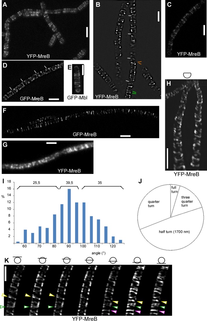
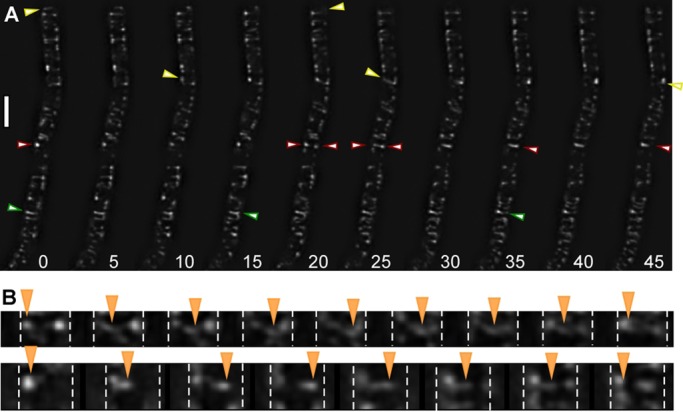
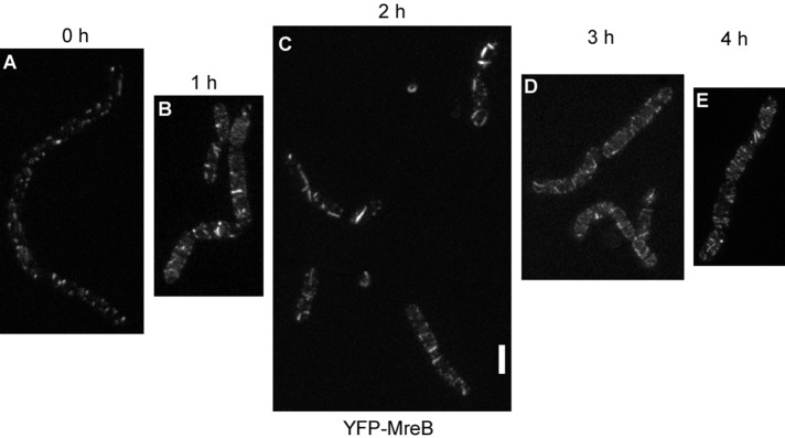
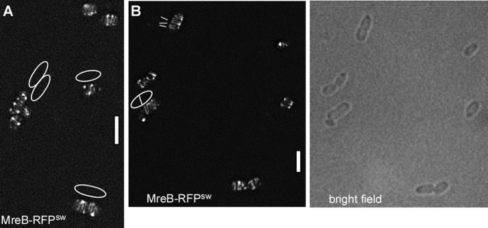
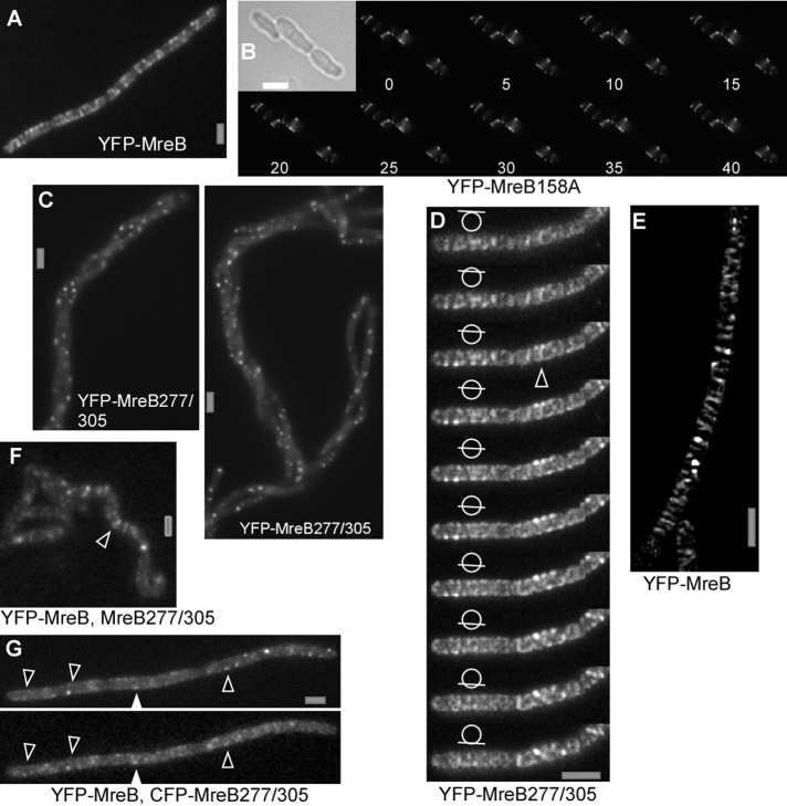
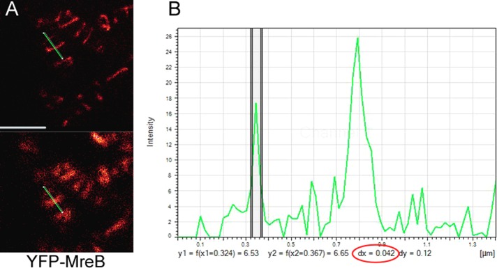
The maintenance of rod-cell shape in many bacteria depends on actin-like MreB proteins and several membrane proteins that interact with MreB. Using superresolution microscopy, we show that at 50-nm resolution, Bacillus subtilis MreB forms filamentous structures of length up to 3.4 μm underneath the cell membrane, which run at angles diverging up to 40° relative to the cell circumference. MreB from Escherichia coli forms at least 1.4-μm-long filaments. MreB filaments move along various tracks with a maximal speed of 85 nm/s, and the loss of ATPase activity leads to the formation of extended and static filaments. Suboptimal growth conditions lead to formation of patch-like structures rather than extended filaments. Coexpression of wild-type MreB with MreB mutated in the subunit interface leads to formation of shorter MreB filaments and a strong effect on cell shape, revealing a link between filament length and cell morphology. Thus MreB has an extended-filament architecture with the potential to position membrane proteins over long distances, whose localization in turn may affect the shape of the cell wall.


Figures

FIGURE 1:
FIGURE 1:
Superresolution microscopy of MreB in exponentially growing bacterial cells. (A) STED image of YFP-MreB (80-nm resolution). (B) N-SIM image of YFP-MreB (125-nm resolution). Green bars show neighboring filaments with different angles; orange bars show opposite angles relative to 90°. (C) G-STED image of YFP-MreB expressed at very low concentration of inducer (0.005% xylose). (D) SIM (Zeiss) image of GFP-MreB expressed from original locus. (E) STED image of GFP-Mbl (original promoter). (F) STED image of GFP-MreB expressed from an ectopic site on the chromosome as sole source of the protein (Formstone and Errington, 2005; Dominguez-Escobar et al., 2011). (G) TIRF image of YFP-MreB (corresponding to A, B, and D). (H) N-SIM 3D reconstruction of the upper two-thirds of a chain of cells. (I) Angular distribution of GFP-MreB filaments in B. subtilis. Numbers above the bars show percentage of filaments with 90° (±5°) relative to the longitudinal axis or with angles with >10° difference relative to the diametral axis of the cell. (J) Relative contributions of filaments of different sizes. (K) SIM (Zeiss) Z-stack with planes taken every 150 nm. Bars show the position of focal planes relative to the cell body. Green triangles indicate filament making a full helical turn; yellow triangle, filament making a quarter turn (not completed full turn); and purple triangle, filament making a half-turn. White bars, 2 μm.
FIGURE 2:
FIGURE 2:
Dynamics of YFP-MreB filaments. (A) Time-lapse experiments with images captured every 5 s, using N-SIM. Yellow triangles indicate filaments moving from left to right; green triangle, filament reversing its direction; red triangles, filaments moving in from opposite directions and fusing, with the left-moving filament prevailing in its direction. (B) Two examples of filaments reversing their direction of movement, indicated by orange triangles; images captured every 5 s. Dashed white lines mark the lateral sides of the cells. The filament in the top moves right at an angle, whereas the filament in the bottom moves close to 90°. Both filaments move back at the same angle as they enter the field of view. White bar, 2 μm.
FIGURE 3:
FIGURE 3:
STED microscopy of cells grown in culture in the absence of shaking (A) and successive changes in the pattern of localization of YFP-MreB after commencement of shaking (B–E). White bar, 2 μm; images are scaled equally.
FIGURE 4:
FIGURE 4:
E. coli MreB forms filaments. (A, B) 3D SIM (Zeiss) images of E. coli cells expressing a sandwich MreB-RFP fusion (the RFP is integrated into a loop within the MreB molecule), EcMreB-RFPsw. (A) White lines indicate the orientation of filaments; outlines of cells are shown that are difficult to deduce from the fluorescence signal. (B) Outlines of cells seen by bright-field illumination. White bars, 2 μm.
FIGURE 5:
FIGURE 5:
Fluorescence microscopy of exponentially growing chains of B. subtilis cells. (A) Epifluorescence of YFP-MreB. (B) Time-lapse SIM (Zeiss; 5-s intervals) of cells expressing D158A mutant YFP-MreB; inset, outlines of cells by bright-field light. (C) Expression of double mutant YFP-MreB277/305 for 8 h, epifluorescence. (D) YFP-MreB277/305 expressed for 3 h; SIM (Zeiss) stack with planes indicated by bars in circle; triangle shows an example of an extended (<750 nm) filament. (E) SIM (Zeiss) image of cells expressing YFP-MreB under identical conditions as in D. (F) Localization of YFP-MreB 4 h after expression of double mutant MreB. Note that the helical phenotype has not fully arisen at this time, but the localization of proteins is easier to see because they are still mostly flat. White triangle indicates clear, extended filament. (G) Colocalization of CFP-MreB and YFP-MreB277/305 (examples indicated by triangles) 4 h after expression of double-mutant MreB (epifluorescence). White bars, 2 μm.
FIGURE 6:
FIGURE 6:
Measurement of the thickness of YFP-MreB filaments in exponentially growing B. subtilis cells. (A) G-STED image (top) and conventional confocal image (bottom). The green bar indicates the line scan through a thin and a much thicker filament. (B) Intensity plot of the line scan. The y axis shows fluorescence intensity, and the x axis shows size in micrometers. The two vertical lines indicate the half-maximum intensity of the filament, which has a diameter of 42 nm (number circled in red); the thicker filament is on the right and has a size of 85 nm.

References

    1. Adams DW, Errington J. Bacterial cell division: assembly, maintenance and disassembly of the Z ring. Nat Rev Microbiol. 2009;7:642–653. - PubMed
    1. Barry RM, Gitai Z. Self-assembling enzymes and the origins of the cytoskeleton. Curr Opin Microbiol. 2011;14:704–711. - PMC - PubMed
    1. Bendezu FO, de Boer PA. Conditional lethality, division defects, membrane involution, and endocytosis in mre and mrd shape mutants of Escherichia coli. J Bacteriol. 2008;190:1792–1811. - PMC - PubMed
    1. Bendezu FO, Hale CA, Bernhardt TG, de Boer PA. RodZ (YfgA) is required for proper assembly of the MreB actin cytoskeleton and cell shape in E. coli. EMBO J. 2009;28:193–204. - PMC - PubMed
    1. Cabeen MT, Jacobs-Wagner C. Bacterial cell shape. Nat Rev Microbiol. 2005;3:601–610. - PubMed

Publication types

MeSH terms