. 2013 Sep;41(16):7861-74.
doi: 10.1093/nar/gkt543. Epub 2013 Jun 19.

Hepatitis C virus 3'UTR regulates viral translation through direct interactions with the host translation machinery

Affiliations

Hepatitis C virus 3'UTR regulates viral translation through direct interactions with the host translation machinery

Yun Bai et al. Nucleic Acids Res. 2013 Sep.

Abstract

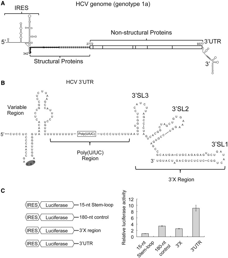
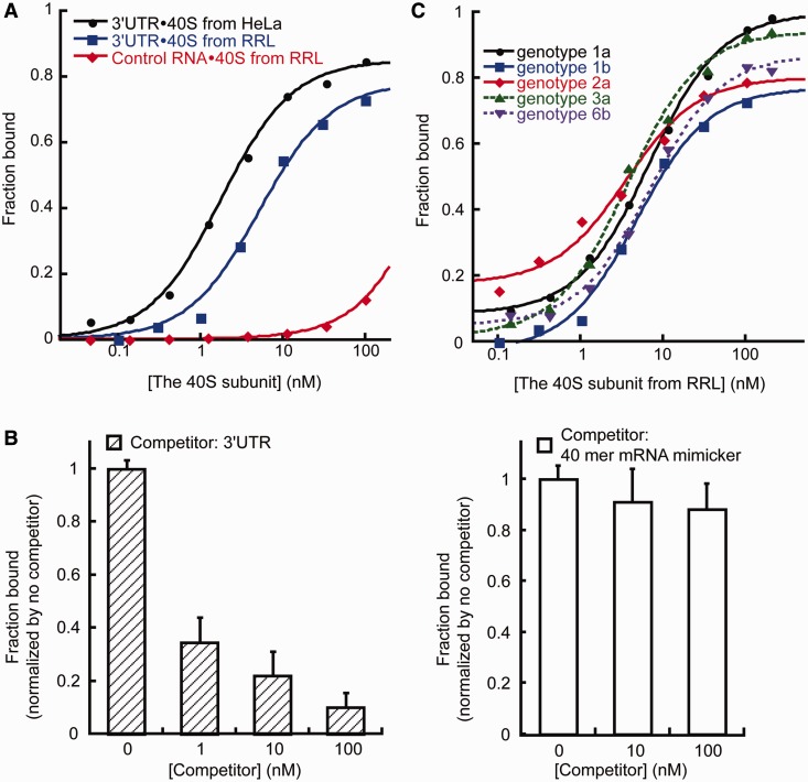
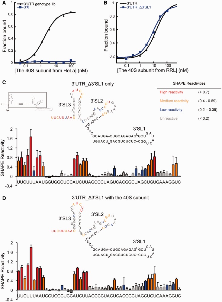
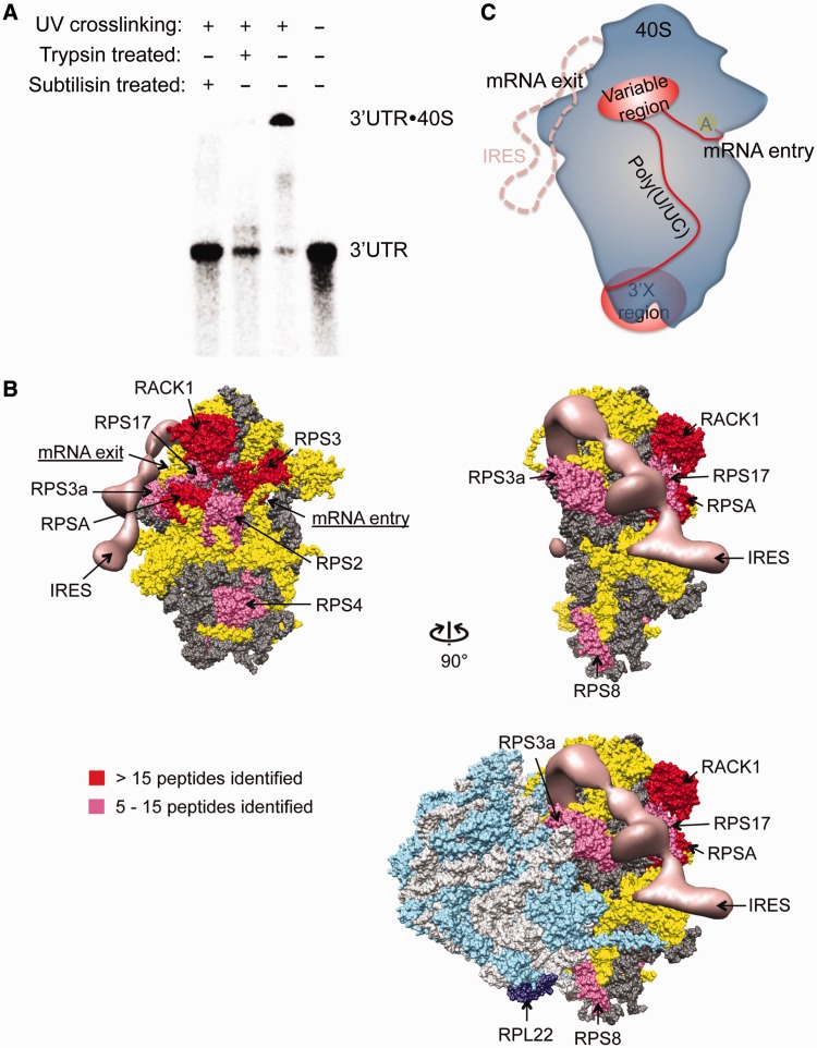
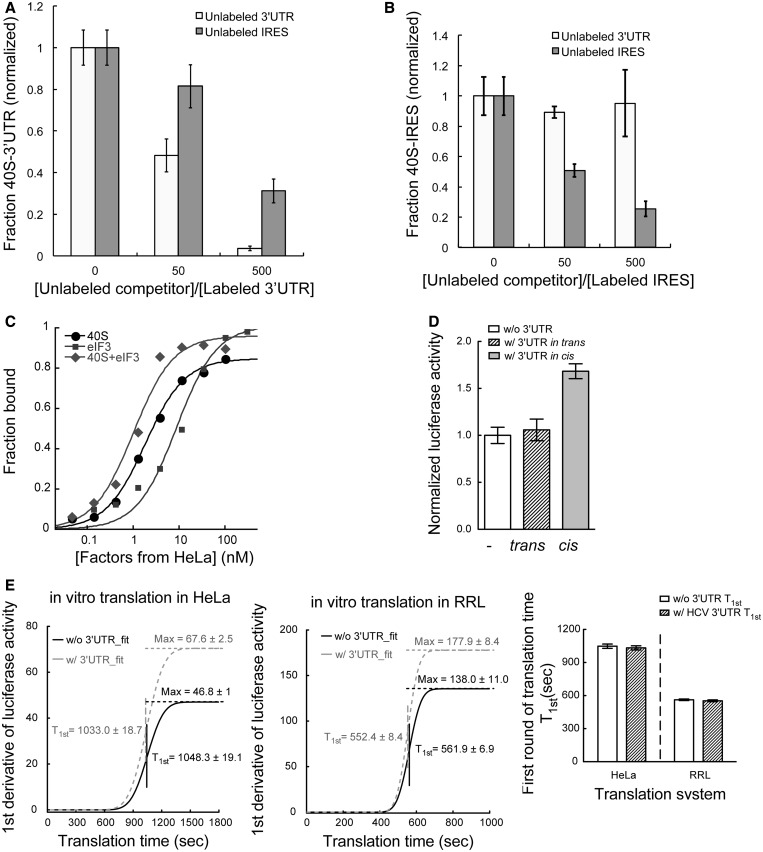
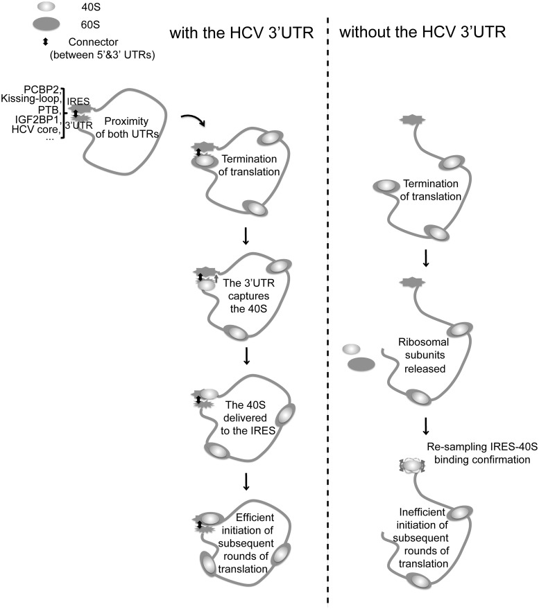
The 3' untranslated region (3'UTR) of hepatitis C virus (HCV) messenger RNA stimulates viral translation by an undetermined mechanism. We identified a high affinity interaction, conserved among different HCV genotypes, between the HCV 3'UTR and the host ribosome. The 3'UTR interacts with 40S ribosomal subunit proteins residing primarily in a localized region on the 40S solvent-accessible surface near the messenger RNA entry and exit sites. This region partially overlaps with the site where the HCV internal ribosome entry site was found to bind, with the internal ribosome entry site-40S subunit interaction being dominant. Despite its ability to bind to 40S subunits independently, the HCV 3'UTR only stimulates translation in cis, without affecting the first round translation rate. These observations support a model in which the HCV 3'UTR retains ribosome complexes during translation termination to facilitate efficient initiation of subsequent rounds of translation.


Figures

Figure 1.
Figure 1.
The HCV 3′UTR stimulates IRES-dependent translation in cell culture. (A). Schematic drawing of the HCV genome. Secondary structures of both UTRs are indicated. The coding region is shown in thick lines for structural proteins and in boxes for non-structural proteins. Numbers are labeled according to genotype 1a strain H77 (B). Secondary structure of the 3′UTR of HCV genotype 1a. The variable region, poly(U/UC) region and three-stem loops in the 3′X region are labeled (C). Schematic drawing of different constructs used in translation assays are shown on the left. In the control construct, a 15-nt stem-loop (CUGCCGUAUAGGCAG) was attached to the 3′end the luciferase mRNA via a 5-nt linker (GUUCA) to ensure mRNA stability. The 180-nt control sequence is adopted from the pUC19 vector (447–630). On the right shows luciferase activities from cell-based translation assays using different RNA constructs. Luciferase activities in all experiments are normalized against that of the construct with a 15-nt stem loop downstream of the luciferase mRNA.
Figure 2.
Figure 2.
The HCV 3′UTR exhibits strong, specific and conserved interaction with the 40S subunit. (A). Binding isotherms for the 3′UTR-40S interaction. The HCV 3′UTR interacts with the 40S subunits isolated from both HeLa extract and RRL, whereas an RNA of similar size, corresponding to part of the HCV NS5B protein-coding region directly upstream of the 3′UTR (nucleotides 9184–9386 from HCV strain H77) showed no obvious interaction. (B). The 3′UTR-40S interaction is subject to competition by unlabeled 3′UTR competitor (left panel). The 3′UTR-40S interaction is not subject to competition by a random 40-nt mRNA mimicker (right panel). The amount of competitor used is indicated. The bound fraction shown has been normalized. (C). Binding isotherm of the HCV 3′UTR from different genotypes binding to the 40S subunit. The Kd values are 6.7 ± 0.7 nM (genotype 1a), 4.7 ± 0.9 nM (genotype 1b), 3.5 ± 1 nM (genotype 2a), 3.7 ± 0.4 nM (genotype 3a), 6.7 ± 0.7 nM (genotype 6b).
Figure 3.
Figure 3.
The contribution of the 3′X region in the HCV 3′UTR-40S interaction (A). Binding isotherms showing the 3′X region alone cannot interact with the 40S subunit. (B). Binding isotherms showing full-length 3′UTR and 3′UTR_Δ3′SL1 interact with the 40S subunit similarly. (C and D). SHAPE analysis of the 3′UTR either alone or in complex with the 40S subunit. The gray box shows the construct used in these experiments. In red are highly reactive nucleotides, in orange are medium reactive nucleotides, in blue are nucleotides with low reactivity, and in gray are unreactive nucleotides. Nucleotides either with no SHAPE data or not included in the construct are shown in black.
Figure 4.
Figure 4.
Mapping of the 3′UTR interacting region on the 40S subunit. (A). Non-specifically cross-linked 3′UTR-40S complex can be degraded to the 3′UTR RNA alone by both trypsin and subtilisin. (B). Mapping result from 4-thiouridine mediated cross-linking of the 3′UTR to the 40S subunit. The hits were categorized based on the total number of spectra observed. Proteins with >15 spectra identified are assigned as strong binders (red), whereas those with between 5 and 15 spectra are assigned as moderate binders (magenta). Entries with four or less spectra are considered background noise from non-specific cross-linking. The HCV IRES is shown in salmon. In dark and light gray are ribosomal RNAs of the 40S and the 60S subunit, respectively. In yellow are the 40S ribosomal proteins not interacting with the 3′UTR. In cyan and dark purple are the 60S ribosomal proteins with RPL22 labeled in dark purple, which was indicated to interact with the 3′X region (34). All the 3′UTR interacting ribosomal/ribosome associated proteins are labeled. (C). Binding model for the HCV 3′UTR and the 40S ribosome. In blue is the 40S subunit, with position of the A site indicated by the yellow oval. In red is the 3′UTR with the variable and the 3′X region shown in oval and the poly(U/UC) tract as well as the linker between the stop codon and the beginning of the variable region stem-loop shown as curved lines. The HCV IRES-binding position on the 40S ribosome is shown in salmon dash line.
Figure 5.
Figure 5.
The HCV 3′UTR stimulates translation only in cis, without affecting the rate of translation (A). Competition assaying showing the 3′UTR-40S interaction is subject to partial competition by unlabeled IRES (dark gray) with the competition less efficient than that from unlabeled 3′UTR (light gray). The competition by 50 times of unlabeled 3′UTR seems modest because the 40S concentration (10 nM) used in these experiment is well above Kd. (B). The IRES-40S interaction is subject to competition by unlabeled IRES (dark gray) but not unlabeled 3′UTR (light gray). The x-axis shows the ratio between unlabeled competitor and radiolabeled probe. (C). Binding isotherms for the interactions between the HCV 3′UTR and the 40S subunit (Kd = 1.8 ± 0.1 nM), eIF3 (Kd = 8.9 ± 2 nM), as well as the 40S-eIF3 complex (Kd = 1.0 ± 0.2 nM) (D). In vitro translation assays showing the 3′UTR can only stimulate translation when in cis with the IRES and reporter mRNA. Addition of free 3′UTR in trans showed no stimulation on IRES-dependent translation. (E). First derivative of the real-time recorded luciferase activity was fitted to a cumulative function of normal distribution. Values for both T1st and Max are labeled. For both RRL (left) and Hela lysate (middle) system, presence of the 3′UTR does not affect the T1st but leads to a higher Max value. The bar graph (right) shows the comparison of the T1st for transcripts with and without the HCV 3′UTR in both translation systems.
Figure 6.
Figure 6.
Model of the HCV 3′UTR function in IRES-dependent translation. The two UTRs of HCV are brought to proximity by either long-range RNA–RNA kissing interactions or protein factors (16,17,40,45,46). At the termination stage of translation, when the stop codon is recognized by the A site, the variable region is presented to the 3′UTR binding region on the 40S subunit, promoting the 3′UTR-40S interaction. This interaction retains the 40S subunit after ribosome recycling and transfers it to the IRES in a favorable conformation for effective interactions, which can lead to efficient initiation for subsequence rounds of translation. Without the 3′UTR, for each round of translation, the IRES needs to recruit translation factors from the environment and sample through a variety of binding conformations with the 40S subunit, leading to inefficient initiation of translation.

References

    1. Wells SE, Hillner PE, Vale RD, Sachs AB. Circularization of mRNA by eukaryotic translation initiation factors. Mol. Cell. 1998;2:135–140. - PubMed
    1. Komarova AV, Brocard M, Kean KM. The case for mRNA 5′ and 3′ end cross talk during translation in a eukaryotic cell. Prog. Nucleic Acid Res. Mol. Biol. 2006;81:331–367. - PubMed
    1. Treder K, Kneller EL, Allen EM, Wang Z, Browning KS, Miller WA. The 3′ cap-independent translation element of Barley yellow dwarf virus binds eIF4F via the eIF4G subunit to initiate translation. RNA. 2008;14:134–147. - PMC - PubMed
    1. Gazo BM, Murphy P, Gatchel JR, Browning KS. A novel interaction of Cap-binding protein complexes eukaryotic initiation factor (eIF) 4F and eIF(iso)4F with a region in the 3′-untranslated region of satellite tobacco necrosis virus. J. Biol. Chem. 2004;279:13584–13592. - PubMed
    1. Miller WA, Wang Z, Treder K. The amazing diversity of cap-independent translation elements in the 3′-untranslated regions of plant viral RNAs. Biochem. Soc. Trans. 2007;35:1629–1633. - PMC - PubMed

Publication types

MeSH terms