. 2013 Nov;99(11):860-9.
doi: 10.1002/bip.22321.

Protein folding and unfolding under force

Affiliations

Protein folding and unfolding under force

Bharat Jagannathan et al. Biopolymers. 2013 Nov.

Abstract

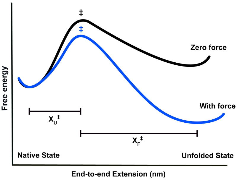
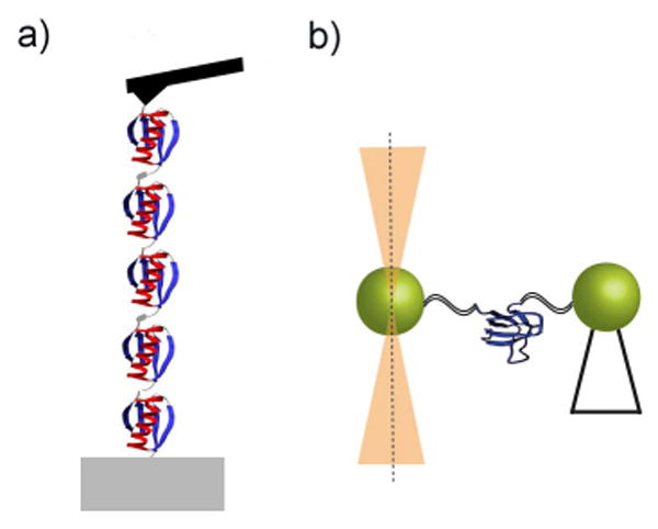
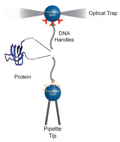
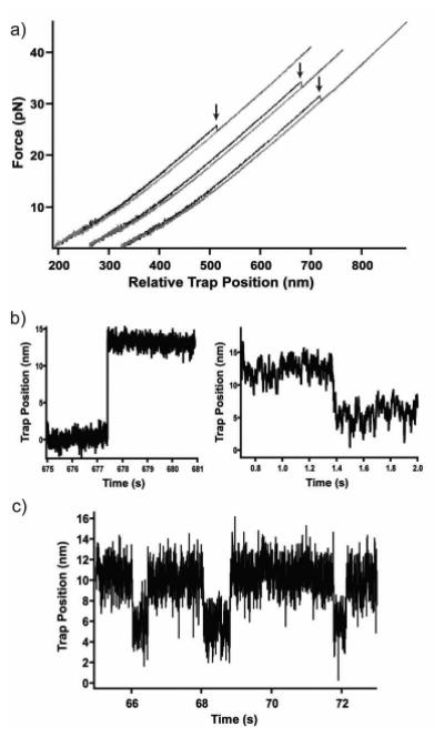
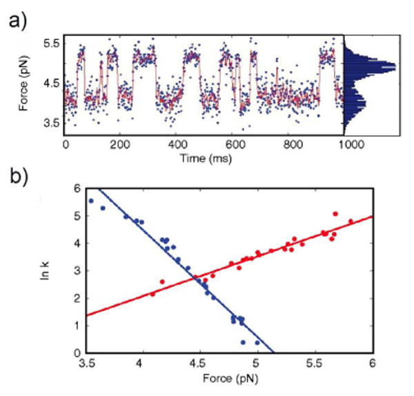
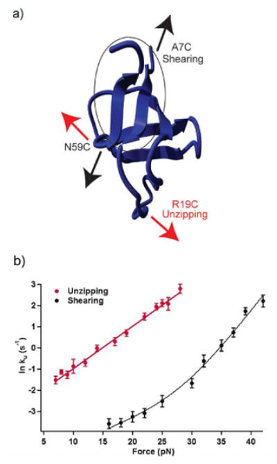
The recent revolution in optics and instrumentation has enabled the study of protein folding using extremely low mechanical forces as the denaturant. This exciting development has led to the observation of the protein folding process at single molecule resolution and its response to mechanical force. Here, we describe the principles and experimental details of force spectroscopy on proteins, with a focus on the optical tweezers instrument. Several recent results will be discussed to highlight the importance of this technique in addressing a variety of questions in the protein folding field.

Keywords: force spectroscopy; optical tweezers; protein folding.


Figures

Figure 1
Figure 1
The effect of force on the free energy landscape of a two-state system. In the absence of force (black curve), the native state is lower in free energy and the protein is predominantly folded. The application of force (blue curve) lowers the free energy of the transition state (‡) and the unfolded state relative to the native state. The force-dependent change in folding and unfolding rates scales with the distance to the transition state (XF and XU, respectively).
Figure 2
Figure 2
Schematic representation of (a) an atomic force microscopy setup, and (b) single-trap optical tweezers where one of the beads is held by suction on a pipette tip.
Figure 3
Figure 3
Schematic representation of the experimental setup used to apply force on single protein molecules with single-trap optical tweezers. Double stranded DNA molecules are linked to specific cysteine residues on the protein via disulfide bonds, and act as handles to apply force on the protein.
Figure 4
Figure 4
Typical traces obtained in (a) force-ramp experiments where unfolding events are observed as ‘rips’ (indicated by arrows), (b) force-jump experiments where protein unfolding (left) and refolding (right) are monitored after jumping to a set force that favors the transition, (c) constant-force experiments in which the protein ‘hops’ between two conformational states at a given force.
Figure 5
Figure 5
(a) Stretching (red) and relaxation (blue) force extension curves from RNase H identified the presence of a partially folded intermediate state. (b) Constant-force experiments revealed that the protein ‘hops’ between the unfolded and the intermediate states before folding to the native state, inset shows a longer time trace.
Figure 6
Figure 6
(a) Typical trace obtained from constant-trap position experiments on apomyoglobin during which the protein molecule spontaneously folds and unfolds. (b) The force-dependence of the folding (blue) and unfolding (red) rates obtained from these experiments were fit to the Bell model to estimate the distances to the transition state.
Figure 7
Figure 7
(a) Three-dimensional structure and schematic of T4 lysozyme showing the energetically coupled N- and C-domains (green and blue respectively). (b) Typical unfolding (red) and refolding (blue) work distributions used to estimate the equilibrium free energies by applying the Crooks fluctuation theorem.
Figure 8
Figure 8
(a) The effect of pulling geometry on mechanical unfolding was studied by applying a shearing and an unzipping force on the src SH3 domain. (b) The force dependence of unfolding rates in the shearing geometry (black) is biphasic, indicating the presence of parallel unfolding pathways.

References

    1. Bustamante C, Chemla YR, Forde NR, Izhaky D. Annu Rev Biochem. 2004;73:705–748. - PubMed
    1. Oberhauser AF, Carrion-Vazquez M. J Biol Chem. 2008;283:6617–6621. - PMC - PubMed
    1. Maillard RA, Chistol G, Sen M, Righini M, Tan J, Kaiser CM, Hodges C, Martin A, Bustamante C. Cell. 2011;145:459–469. - PMC - PubMed
    1. Aubin-Tam ME, Olivares AO, Sauer RT, Baker TA, Lang M. J Cell. 2011;145:257–267. - PMC - PubMed
    1. Street TO, Courtemanche N, Barrick D. Methods Cell Biol. 2008;84:295–325. - PubMed

Publication types