. 2014 Jan 1;134(1):32-42.
doi: 10.1002/ijc.28335. Epub 2013 Jul 19.

Mixed-polarization phenotype of ascites-associated macrophages in human ovarian carcinoma: correlation of CD163 expression, cytokine levels and early relapse

Affiliations

Mixed-polarization phenotype of ascites-associated macrophages in human ovarian carcinoma: correlation of CD163 expression, cytokine levels and early relapse

Silke Reinartz et al. Int J Cancer. .

Abstract

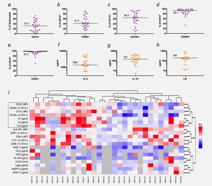
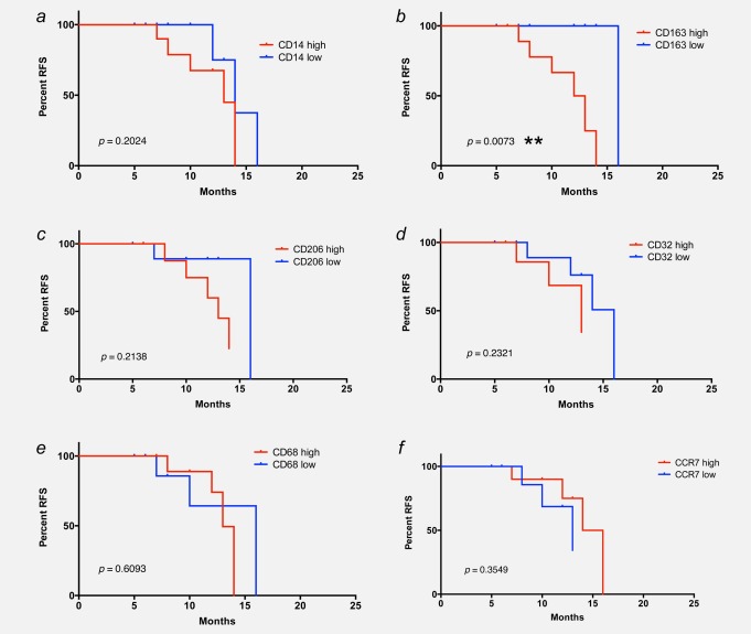
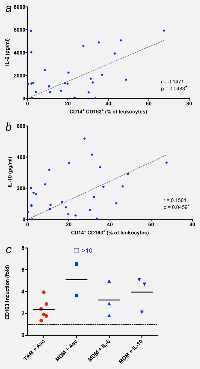
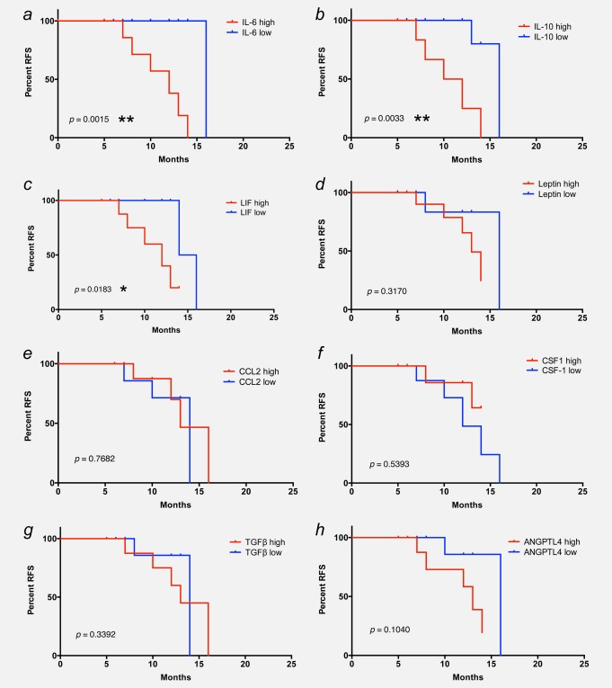
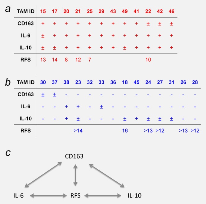
Ovarian cancer is typically accompanied by the occurrence of malignant ascites containing large number of macrophages. It has been suggested that these tumor-associated macrophages (TAMs) are skewed to alternative polarization (M2) and thereby play an essential role in therapy resistance and metastatic spread. In our study, we have investigated the nature, regulation and clinical correlations of TAM polarization in serous ovarian cancer. Macrophage polarization markers on TAMs and ascites cytokine levels were analyzed for 30 patients and associated with relapse-free survival (RFS) in a prospective study with 20 evaluable patients. Surface expression of the M2 marker CD163 on TAMs was inversely associated with RFS (p < 0.01). However, global gene expression profiles determined for 17 of these patients revealed a mixed-polarization phenotype unrelated to the M1/M2 classification. CD163 surface expression also correlated with the ascites levels of IL-6 and IL-10 (p < 0.05), both cytokines induced CD163 expression, and their ascites levels showed a clear inverse association with RFS (p < 0.01). These findings define a subgroup of patients with high CD163 expression, high IL-6 and/or IL-10 levels and poor clinical outcome.

Keywords: CD163; IL-10; IL-6; ovarian carcinoma; tumor-associated macrophages.


Figures

Figure 1
Figure 1
Expression patterns of surface markers on TAMs and cytokine ascites levels in ovarian cancer patients. (ae) Expression of surface markers associated with macrophage polarization and tumor progression (n = 30; FACS analyses). (fh) Distribution of IL-6, IL-10 and LIF levels (n = 30; ELISA). (i) Heat map generated by hierarchal clustering of expression data. Data were median centered and scaled from −1 to +1 for each marker or cytokine, with bright red indicating the highest expression levels and bright blue the lowest expression level. Missing values are indicated in gray. Samples with more than 25% of markers/cytokines missing were not considered for clustering of markers or cytokines and replaced by 0 when clustering samples.
Figure 2
Figure 2
Association of RFS with CD163 surface expression on ovarian carcinoma TAMs. Kaplan–Meier plots showing the RFS of patients with high or low levels (median centered) of CD14 (% of leukocytes; panel a), CD163 (% of CD14+; panel b), CD206 (% of CD14+; panel c), CD32 (MFI; panel d), CD68 (% of CD14+; panel e) and CCR7 (% of CD14+; panel f) expression on their TAMs. p-Values were determined by Mantel–Cox log-rank test.
Figure 3
Figure 3
Expression of polarization-associated genes in TAMs and MDMs. RNAs from 17 TAM and three MDM samples were analyzed by transcriptional profiling on Agilent 44K microarrays, and relative expression levels were plotted for a range of established M1 (a) and M2 (b) markers. Horizontal bars indicate the respective median; p-values were determined by t-test (*p < 0.05; **p < 0.01; ***p < 0.001).
Figure 4
Figure 4
CD163 surface expression is associated with IL-6 and IL-10. (a and b) Correlation of CD163 surface expression with ascites levels of IL-6 and IL-10. *p < 0.05; **p < 0.01 by Pearson’s r correlation. (c) Induction of CD163 in TAMs and MDMs by ascites and/or IL-10 and IL-10. TAMs were cultured for 2 days with or without cell-free ascites (10% of total culture medium). MDMs from healthy donors were differentiated in the presence of GM-CSF and either cell-free ascites (10% of total culture medium) or the indicated recombinant cytokines for 5 days. Expression of surface marker CD163 was determined by FACS and is expressed as fold induction, i.e., normalized to 1 for the respective untreated control (untreated TAMs and MMDs, respectively, are represented by the dotted line). Each data point within the same treatment group represents a different donor. Horizontal bars indicate averages. The induction value represented by the open box could not be precisely determined because of an extremely low expression value in untreated MDMs. [Color figure can be viewed in the online issue, which is available at wileyonlinelibrary.com.]
Figure 5
Figure 5
Association of RFS with cytokine levels in ovarian carcinoma ascites. Kaplan–Meier plots showing the RFS of patients with high or low ascites levels of IL-6 (a), IL-10 (b), LIF (c), leptin (d), CCL2 (e), CSF1 (f), TGF-β (g) and ANGPTL4 (h). Cutoff values were 1,500 ng/Ml for IL-6 and CSF-1, 200 ng/Ml for IL-10 and LIF, 100 ng/Ml for leptin and 150 ng/Ml for CCL2. p-Values were determined by Mantel–Cox log-rank test.
Figure 6
Figure 6
Summary of the correlations identified in our study. Patients were categorized according to the levels of CD163, IL-6 and IL-10 (high in panel a and low in panel b). The bottom row shows the RFS times after first-line surgery. Empty field represent patients that are not yet evaluable (relapse-free <12 months postsurgery). Panel c shows a schematic representations of the correlations between CD163, IL-6, IL-10 and RFS. [Color figure can be viewed in the online issue, which is available at wileyonlinelibrary.com.]

References

    1. Hanahan D, Weinberg RA. Hallmarks of cancer: the next generation. Cell. 2011;144:646–74. - PubMed
    1. Condeelis J, Pollard JW. Macrophages: obligate partners for tumor cell migration, invasion, and metastasis. Cell. 2006;124:263–6. - PubMed
    1. Sica A, Bronte V. Altered macrophage differentiation and immune dysfunction in tumor development. J Clin Invest. 2007;117:1155–66. - PMC - PubMed
    1. Qian BZ, Pollard JW. Macrophage diversity enhances tumor progression and metastasis. Cell. 2010;141:39–51. - PMC - PubMed
    1. Gabrilovich DI, Ostrand-Rosenberg S, Bronte V. Coordinated regulation of myeloid cells by tumours. Nat Rev Immunol. 2012;12:253–68. - PMC - PubMed

Publication types

MeSH terms