Review
. 2013 Jun 19;33(25):10195-208.
doi: 10.1523/JNEUROSCI.0822-13.2013.

Gangliosides and gangliosidoses: principles of molecular and metabolic pathogenesis

Affiliations
Review

Gangliosides and gangliosidoses: principles of molecular and metabolic pathogenesis

Konrad Sandhoff et al. J Neurosci. .

Abstract

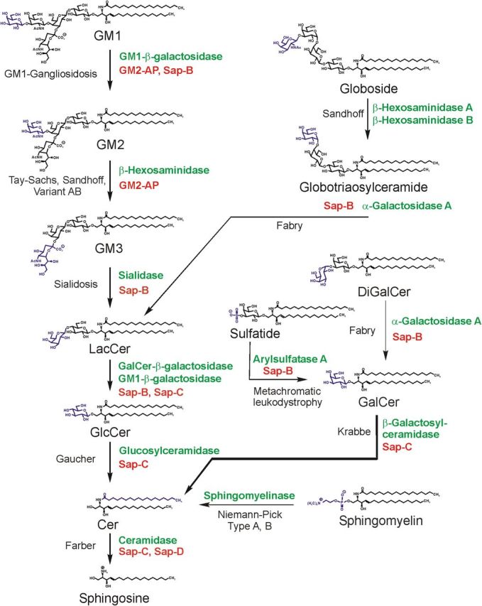
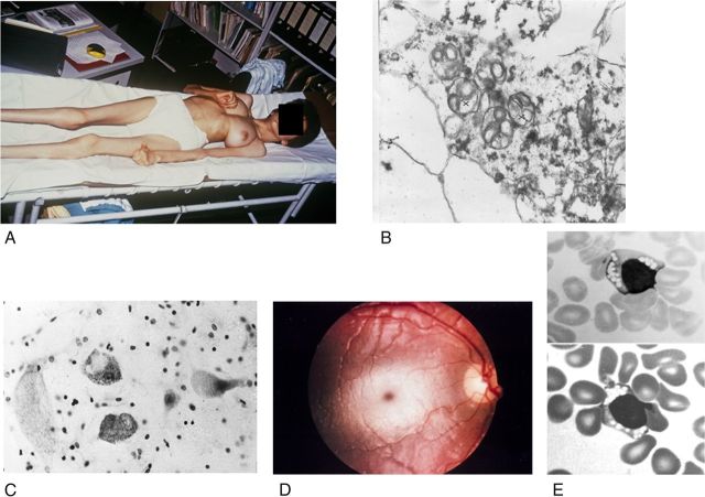
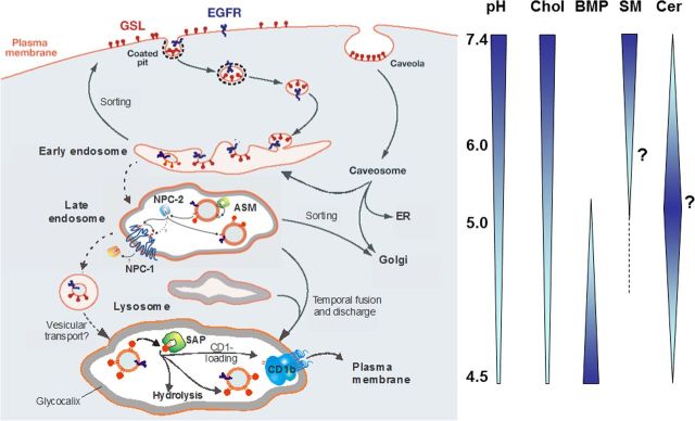
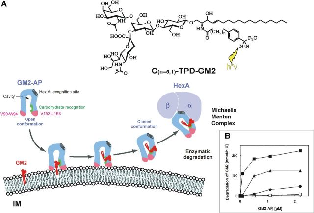
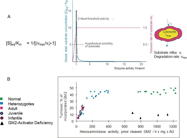
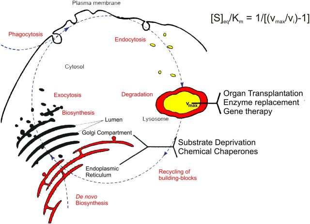
Gangliosides are the main glycolipids of neuronal plasma membranes. Their surface patterns are generated by coordinated processes, involving biosynthetic pathways of the secretory compartments, catabolic steps of the endolysosomal system, and intracellular trafficking. Inherited defects in ganglioside biosynthesis causing fatal neurodegenerative diseases have been described so far almost exclusively in mouse models, whereas inherited defects in ganglioside catabolism causing various clinical forms of GM1- and GM2-gangliosidoses have long been known. For digestion, gangliosides are endocytosed and reach intra-endosomal vesicles. At the level of late endosomes, they are depleted of membrane-stabilizing lipids like cholesterol and enriched with bis(monoacylglycero)phosphate (BMP). Lysosomal catabolism is catalyzed at acidic pH values by cationic sphingolipid activator proteins (SAPs), presenting lipids to their respective hydrolases, electrostatically attracted to the negatively charged surface of the luminal BMP-rich vesicles. Various inherited defects of ganglioside hydrolases, e.g., of β-galactosidase and β-hexosaminidases, and of GM2-activator protein, cause infantile (with tetraparesis, dementia, blindness) and different protracted clinical forms of GM1- and GM2-gangliosidoses. Mutations yielding proteins with small residual catabolic activities in the lysosome give rise to juvenile and adult clinical forms with a wide range of clinical symptomatology. Apart from patients' differences in their genetic background, clinical heterogeneity may be caused by rather diverse substrate specificities and functions of lysosomal hydrolases, multifunctional properties of SAPs, and the strong regulation of ganglioside catabolism by membrane lipids. Currently, there is no treatment available for neuronal ganglioside storage diseases. Therapeutic approaches in mouse models and patients with juvenile forms of gangliosidoses are discussed.


Figures

Figure 1.
Figure 1.
Pathway of lysosomal sphingolipid degradation. The eponyms of known metabolic diseases and those of SAPs (red) necessary for in vivo degradation are indicated. Hydrolases are given in green. Heterogeneity in the lipid part of the sphingolipids is not indicated. Variant AB, Variant AB of GM2-gangliosidosis [deficiency of GM2-activator protein (GM2-AP)]; Sap, sphingolipid activator protein (modified from Sandhoff and Kolter, 1995).
Figure 2.
Figure 2.
Clinical and morphological observations in gangliosidoses. A, Near end stage in a Spanish 13-year-old patient with GM2-gangliosidosis variant B1 (photograph courtesy of H.H. Goebel, Mainz). B, GM2-gangliosidosis variant 0 in a 23-week-old fetus; and part of a cortical neuron; early stage of membranous cytoplasmic bodies (two clusters marked by crosses), i.e., lipid storing secondary lysosomes, ×36,000 (courtesy W. Schlote, Tübingen). C, The large cells are neurons distended by lysosomal storage in the medulla oblongata of a 1.5-year-old patient with GM1-gangliosidosis. The small dark bodies are nuclei from increased numbers of glial cells (“reactive gliosis”). Cresyl violet stain ×300 (courtesy of H.U. Benz, Tübingen). D, Cherry red macular spot in an infantile patient with GM2-gangliosidosis variant B (Tay–Sachs disease). The dark red spot in the middle of the light central area is secondary to lipid storage in neuronal cells in this area; the storing cells have lost their processes that normally cover the fovea centralis. The fovea normally appears as yellow, but is changed to the red macular spot showing the color of the choroidea behind the retina. E, Lymphocytes with clusters of vacuoles (storage lysosomes) in blood smear of an early infantile patient with GM1-gangliosidosis, indicating the “generalized storage disorder.” May-Grünwald stain; ×1500 (courtesy of H.U. Benz, Tübingen).
Figure 3.
Figure 3.
Proposed topology of endocytosis and lysosomal lipid and membrane digestion (Gallala et al., 2011). “A section of the plasma membrane is internalized by way of coated pits or caveolae. These membrane patches include GSLs (red) and receptors such as epidermal growth factor receptor (EGFR; blue). These vesicles fuse with the early endosomes, which mature to late endosomes. Endosomal perimeter membranes form invaginations, controlled by ESCRT proteins (Wollert and Hurley, 2010), which bud off, forming intra-endosomal vesicles. Lipid sorting occurs at this stage. The pH of the lumen is ∼5. At this pH, ASM is active and degrades sphingomyelin of the intra-endosomal vesicles to ceramide, whereas the perimeter membrane is protected against the action of ASM by the glycocalyx facing the lumen. This decrease in sphingomyelin, coupled with the increase in ceramide, facilitates the binding of cholesterol to NPC2 and its transport to the perimeter membrane of the late endosome where it is transferred to NPC1 (Kwon et al., 2009). This protein mediates the export of cholesterol through the glycocalyx, eventually reaching cholesterol binding proteins in the cytosol. Ultimately, late endosomes fuse with lysosomes. The GSLs are harbored in intralysosomal vesicles that face the lumen of the lysosome, and are degraded by hydrolases with the assistance of SAPs. The products of this degradation are exported to the cytosol or loaded on CD1b immunoreceptors and exported to the plasma membrane for antigen presentation. Gradients of pH in the lysosol, and intra-endolysosomal vesicle content of cholesterol (Chol), BMP, sphingomyelin (SM, hypothetical), and ceramide (Cer, hypothetical) are shown” (modified from Kolter and Sandhoff, 2010).
Figure 4.
Figure 4.
Mechanism of GM2-AP-Liftase. A, Model for the interaction of GM2-activator protein with luminal lysosomal membranes (IM) in the degradation of ganglioside GM2 (modified from Wendeler et al., 2004a). “GM2-AP interacts with the membrane by dipping the two exposed hydrophobic loops, V90–W94 and V153–L163, into the apolar part of the membrane. Ganglioside GM2 is recognized by specific sites at the rim of the cavity. In the open protein conformation, the large hydrophobic area reaching from the apolar phase of the membrane to the activator's cavity lowers the energy barrier for lipids leaving the membrane in an upward direction. After the ceramide tail has arrived inside the activator's cavity, the inward-bending of the hydrophobic loop V153–L163 is favored, and the conformation changes to the closed form (Wright et al., 2003). This folding in of the hydrophobic loop leaves a more polar patch close to the membrane. The activator is then anchored only by the loop V90–W94. It may now rotate slightly upward to expose all polar patches more fully to the solvent, and it may also leave the membrane and interact with the degrading enzyme. The photoaffinity label analogs C(n = 5,1)-TPD-GM2 were photo-incorporated specifically into the region V153–L163” (Sandhoff, 2012). B, BMP and GM2-AP stimulate the hydrolysis of LUV-bound ganglioside GM2. The degradation of ganglioside GM2 inserted into LUVs doped with 0 mol % BMP (○), 5 mol % BMP(□), 10 mol % BMP (·), 20 mol % BMP (▴), or 25 mol % BMP (■) was measured in the presence of increasing concentrations of GM2-AP (0–2.2 μm) (Werth et al., 2001).
Figure 5.
Figure 5.
Residual catabolic activity correlates with clinical forms of GM2 gangliosidoses (modified from Sandhoff, 2012). A, Steady-state substrate concentration as a function of enzyme concentration and activity (Conzelmann and Sandhoff, 1983). The model underlying this theoretical calculation assumes influx of the substrate into the lysosomal compartment at a constant rate (vi) and its subsequent utilization by the catabolic enzyme. Blue line = [S]eq steady-state substrate concentration; ………… = theoretical threshold of enzyme activity; ——- = critical threshold value, taking limited solubility of substrate into account; red line = turnover rate of substrate (flux rate). B, To experimentally verify the basic assumptions for this model, studies were performed in cell culture (Leinekugel et al., 1992). The radiolabeled substrate ganglioside GM2 was added to cultures of skin fibroblasts with different activities of β-hexosaminidase A and its uptake and turnover measured. The correlation between residual enzyme activity and the turnover rate of the substrate was essentially as predicted: degradation rate of ganglioside GM2 increased steeply with residual activity, to reach the control level at a residual activity of ∼10–15% of normal. All cells with an activity above this critical threshold had a normal turnover. Comparison of the results of these feeding studies with the clinical status of the donor of each cell line basically confirmed our notions but also revealed the limitations of the cell culture approach.
Figure 6.
Figure 6.
Intracellular metabolic flux of sphingolipids. vi = the influx rate of the substrate into the lysosome; for further information see the text. Modified from Kolter and Sandhoff, 1999).

References

    1. Abdul-Hammed M, Breiden B, Adebayo MA, Babalola JO, Schwarzmann G, Sandhoff K. Role of endosomal membrane lipids and NPC2 in cholesterol transfer and membrane fusion. J Lipid Res. 2010;51:1747–1760. doi: 10.1194/jlr.M003822. - DOI - PMC - PubMed
    1. Andersson U, Smith D, Jeyakumar M, Butters TD, Borja MC, Dwek RA, Platt FM. Improved outcome of N-butyldeoxygalactonojirimycin-mediated substrate reduction therapy in a mouse model of Sandhoff disease. Neurobiol Dis. 2004;16:506–515. doi: 10.1016/j.nbd.2004.04.012. - DOI - PubMed
    1. Appelqvist H, Sandin L, Björnström K, Saftig P, Garner B, Ollinger K, Kågedal K. Sensitivity to lysosome-dependent cell death is directly regulated by lysosomal cholesterol content. PLoS One. 2012;7:e50262. doi: 10.1371/journal.pone.0050262. - DOI - PMC - PubMed
    1. Arfi A, Bourgoin C, Basso L, Emiliani C, Tancini B, Chigorno V, Li YT, Orlacchio A, Poenaru L, Sonnino S, Caillaud C. Bicistronic lentiviral vector corrects beta-hexosaminidase deficiency in transduced and cross-corrected human Sandhoff fibroblasts. Neurobiol Dis. 2005;20:583–593. doi: 10.1016/j.nbd.2005.04.017. - DOI - PubMed
    1. Assadi M, Baseman S, Janson C, Wang DJ, Bilaniuk L, Leone P. Serial 1H-MRS in GM2 gangliosidoses. Eur J Pediatr. 2008;167:347–352. doi: 10.1007/s00431-007-0469-0. - DOI - PubMed

Publication types