Point mutations in the paramyxovirus F protein that enhance fusion activity shift the mechanism of complement-mediated virus neutralization
- PMID: 23785199
- PMCID: PMC3754036
- DOI: 10.1128/JVI.01111-13
Point mutations in the paramyxovirus F protein that enhance fusion activity shift the mechanism of complement-mediated virus neutralization
Abstract
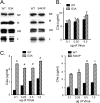
Parainfluenza virus 5 (PIV5) activates and is neutralized by the alternative pathway (AP) in normal human serum (NHS) but not by heat-inactivated (HI) serum. We have tested the relationship between the fusion activity within the PIV5 F protein, the activation of complement pathways, and subsequent complement-mediated virus neutralization. Recombinant PIV5 viruses with enhanced fusion activity were generated by introducing point mutations in the F fusogenic peptide (G3A) or at a distal site near the F transmembrane domain (S443P). In contrast to wild-type (WT) PIV5, the mutant G3A and S443P viruses were neutralized by both NHS and HI serum. Unlike WT PIV5, hyperfusogenic G3A and S443P viruses were potent C4 activators, C4 was deposited on NHS-treated mutant virions, and the mutants were neutralized by factor B-depleted serum but not by C4-depleted serum. Antibodies purified from HI human serum were sufficient to neutralize both G3A and S443P viruses in vitro but were ineffective against WT PIV5. Electron microscopy data showed greater deposition of purified human antibodies on G3A and S443P virions than on WT PIV5 particles. These data indicate that single amino acid changes that enhance the fusion activity of the PIV5 F protein shift the mechanism of complement activation in the context of viral particles or on the surface of virus-infected cells, due to enhanced binding of antibodies. We present general models for the relationship between enhanced fusion activity in the paramyxovirus F protein and increased susceptibility to antibody-mediated neutralization.
Figures


Similar articles
-
The paramyxovirus fusion protein C-terminal region: mutagenesis indicates an indivisible protein unit.J Virol. 2012 Mar;86(5):2600-9. doi: 10.1128/JVI.06546-11. Epub 2011 Dec 14. J Virol. 2012. PMID: 22171273 Free PMC article.
-
Bimolecular complementation of paramyxovirus fusion and hemagglutinin-neuraminidase proteins enhances fusion: implications for the mechanism of fusion triggering.J Virol. 2009 Nov;83(21):10857-68. doi: 10.1128/JVI.01191-09. Epub 2009 Aug 26. J Virol. 2009. PMID: 19710150 Free PMC article.
-
Interactions of human complement with virus particles containing the Nipah virus glycoproteins.J Virol. 2011 Jun;85(12):5940-8. doi: 10.1128/JVI.00193-11. Epub 2011 Mar 30. J Virol. 2011. PMID: 21450814 Free PMC article.
-
Paramyxovirus fusion and entry: multiple paths to a common end.Viruses. 2012 Apr;4(4):613-36. doi: 10.3390/v4040613. Epub 2012 Apr 19. Viruses. 2012. PMID: 22590688 Free PMC article. Review.
-
[Significance of paramyxovirinae protein F in physiopathology and immunity].Bull Acad Natl Med. 2000;184(6):1255-64; discussion 1265. Bull Acad Natl Med. 2000. PMID: 11268674 Review. French.
Cited by
-
In silico prediction of B and T cell epitopes based on NDV fusion protein for vaccine development against Newcastle disease virus.Vet Res Forum. 2021 Spring;12(2):157-165. doi: 10.30466/vrf.2019.98625.2351. Epub 2021 Jun 15. Vet Res Forum. 2021. PMID: 34345381 Free PMC article.
-
Paramyxovirus activation and inhibition of innate immune responses.J Mol Biol. 2013 Dec 13;425(24):4872-92. doi: 10.1016/j.jmb.2013.09.015. Epub 2013 Sep 20. J Mol Biol. 2013. PMID: 24056173 Free PMC article. Review.
-
In the Crosshairs: RNA Viruses OR Complement?Front Immunol. 2020 Sep 29;11:573583. doi: 10.3389/fimmu.2020.573583. eCollection 2020. Front Immunol. 2020. PMID: 33133089 Free PMC article. Review.
-
Complement Inhibitors Vitronectin and Clusterin Are Recruited from Human Serum to the Surface of Coronavirus OC43-Infected Lung Cells through Antibody-Dependent Mechanisms.Viruses. 2021 Dec 24;14(1):29. doi: 10.3390/v14010029. Viruses. 2021. PMID: 35062233 Free PMC article.
-
Role of Defective Interfering Particles in Complement-Mediated Lysis of Parainfluenza Virus-Infected Cells.Viruses. 2025 Mar 28;17(4):488. doi: 10.3390/v17040488. Viruses. 2025. PMID: 40284931 Free PMC article.
References
-
- McSharry JJ, Pickering RJ, Caliguiri LA. 1981. Activation of the alternative complement pathway by enveloped viruses containing limited amounts of sialic acid. Virology 114:507–515 - PubMed
-
- Devaux P, Christiansen D, Plumet S, Gerlier D. 2004. Cell surface activation of the alternative complement pathway by the fusion protein of measles virus. J. Gen. Virol. 85:1665–1673 - PubMed
-
- Gasque P. 2004. Complement: a unique innate immune sensor for danger signals. Mol. Immunol. 41:1089–1098 - PubMed
Publication types
MeSH terms
Substances
Grants and funding
LinkOut - more resources
Full Text Sources
Other Literature Sources
Miscellaneous

