. 2013;9(6):e1003411.
doi: 10.1371/journal.ppat.1003411. Epub 2013 Jun 13.

Viral retasking of hBre1/RNF20 to recruit hPaf1 for transcriptional activation

Affiliations

Viral retasking of hBre1/RNF20 to recruit hPaf1 for transcriptional activation

Gregory J Fonseca et al. PLoS Pathog. 2013.

Abstract

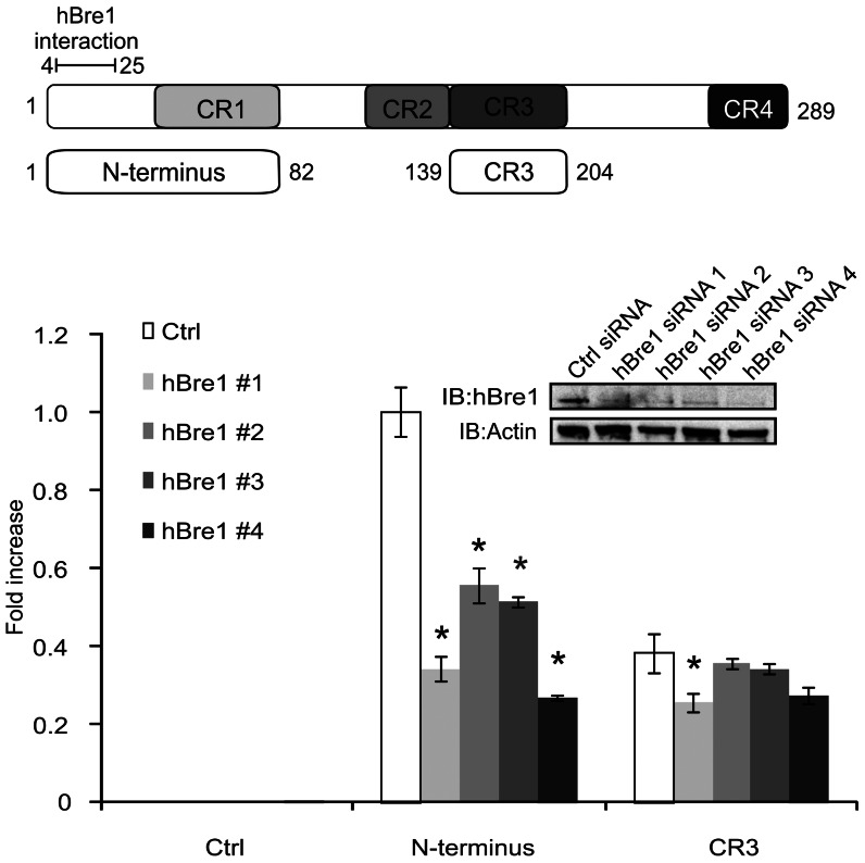
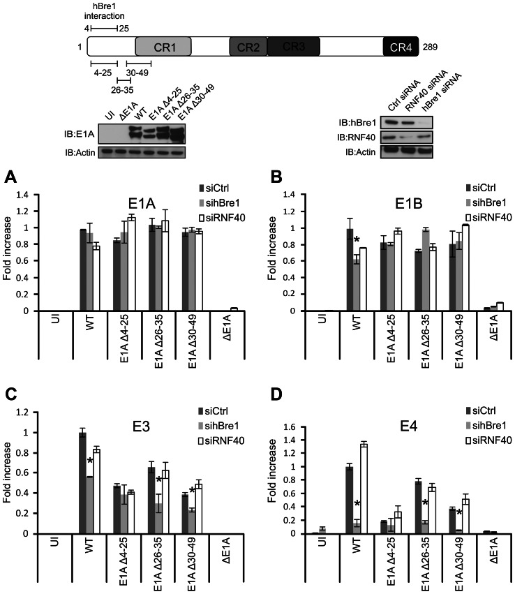
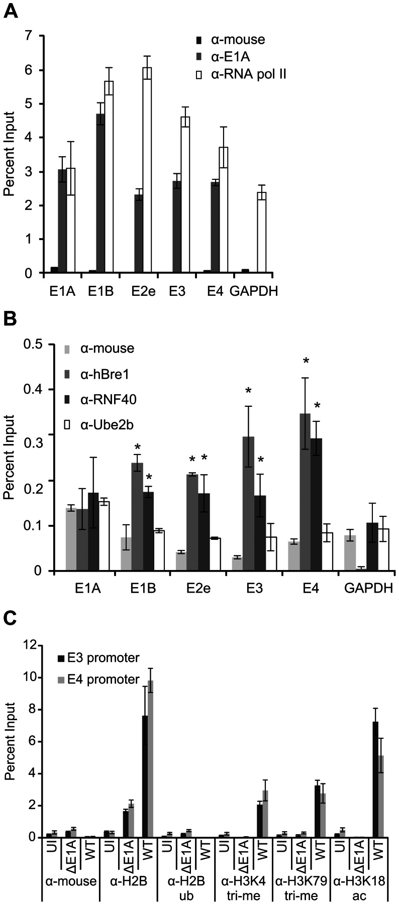
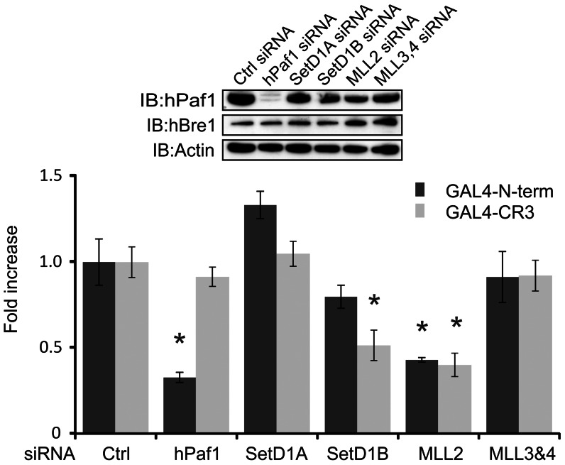
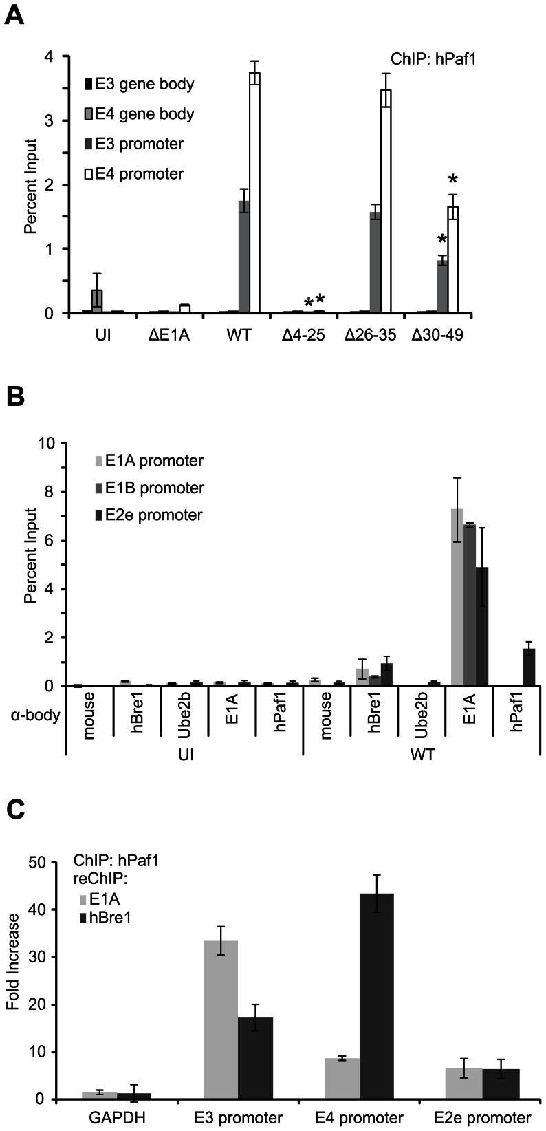
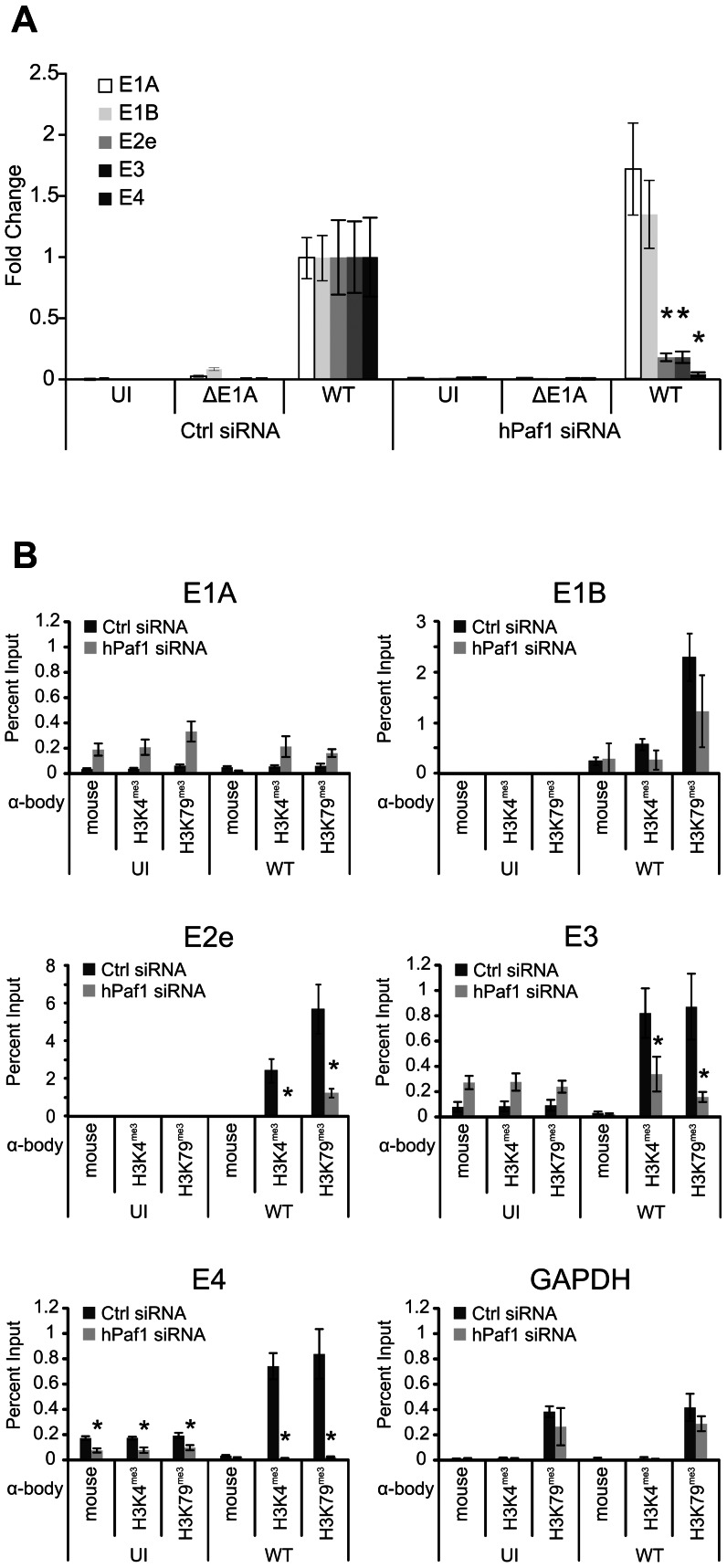
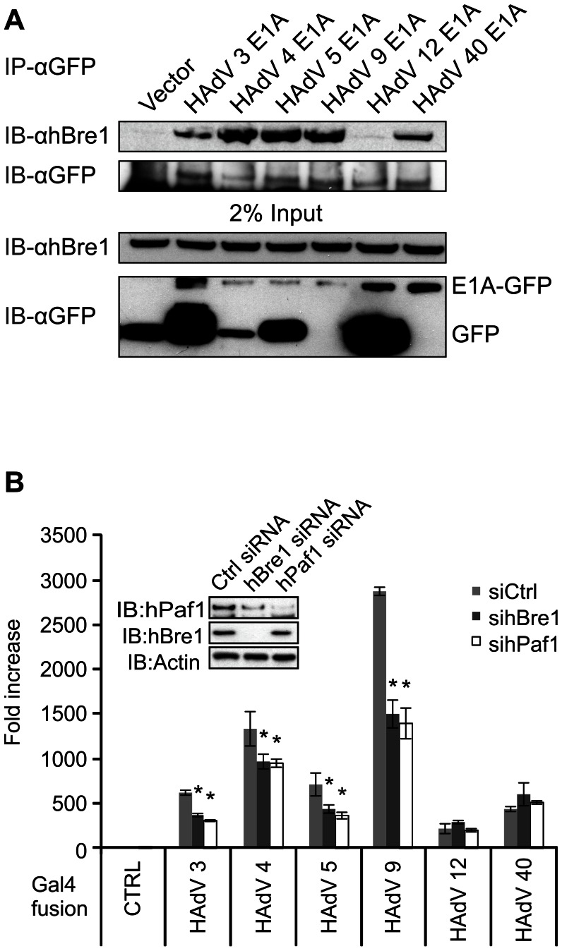
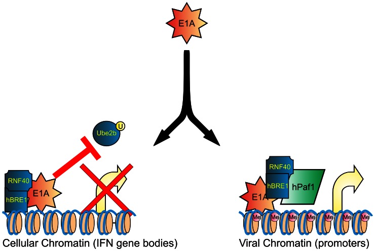
Upon infection, human adenovirus (HAdV) must activate the expression of its early genes to reprogram the cellular environment to support virus replication. This activation is orchestrated in large part by the first HAdV gene expressed during infection, early region 1A (E1A). E1A binds and appropriates components of the cellular transcriptional machinery to modulate cellular gene transcription and activate viral early genes transcription. Previously, we identified hBre1/RNF20 as a target for E1A. The interaction between E1A and hBre1 antagonizes the innate antiviral response by blocking H2B monoubiquitination, a chromatin modification necessary for the interferon (IFN) response. Here, we describe a second distinct role for the interaction of E1A with hBre1 in transcriptional activation of HAdV early genes. Furthermore, we show that E1A changes the function of hBre1 from a ubiquitin ligase involved in substrate selection to a scaffold which recruits hPaf1 as a means to stimulate transcription and transcription-coupled histone modifications. By using hBre1 to recruit hPaf1, E1A is able to optimally activate viral early transcription and begin the cycle of viral replication. The ability of E1A to target hBre1 to simultaneously repress cellular IFN dependent transcription while activating viral transcription, represents an elegant example of the incredible economy of action accomplished by a viral regulatory protein through a single protein interaction.


Conflict of interest statement

The authors have declared that no competing interests exist.

Figures

Figure 1
Figure 1. The N-terminus of E1A specifically requires hBre1 for full activation of transcription.
U-2 OS cells were transfected with a negative control siRNA or 1 of 4 siRNAs specific for hBre1. Cells were then transfected with a constitutive β-galactosidase reporter, a Gal4 responsive luciferase reporter and a vector expressing the Gal4 DNA binding domain (DBD) alone, the Gal4 DBD fused to the N-terminus of E1A, or the Gal4 DBD fused to E1A CR3. Luciferase activity was measured. Results were normalized to β-galactosidase activity and siRNA treated groups were set as a fold change to the Gal4 only transfected counterpart. A statistically significant decrease from control siRNA treatment is indicated (* P<0.01). n = 3.
Figure 2
Figure 2. hBre1 contributes to E1A mediated activation of E3 and E4 expression during infection with HAdV.
A549 cells were treated with Ctrl siRNA, siRNA specific to RNF40 or siRNA specific for hBre1 (Shown in the inset panel) and infected at an MOI of 5 with WT HAdV, or a series of HAdV containing the indicated deletions within the N-terminus of E1A (Shown in the inset panel). RT-qPCR was performed with a panel of HAdV early genes, normalized to GAPDH and fold change to uninfected Ctrl siRNA treated cells was plotted. Results for the HAdV E1A (A), E1B (B), E3 (C), and E4 (D) transcription units are shown. A statistically significant decrease from siCtrl is indicated (* P<0.01). n = 3.
Figure 3
Figure 3. E1A recruits hBre1 to HAdV early gene promoters, but HAdV chromatin is not monoubiquitinated by hBre1.
A549 cells were infected with WT HAdV or ΔE1A HAdV for 20 hours. Chromatin immunoprecipitation (ChIP) was then performed with antibodies specific for the indicated proteins. DNA was probed via qRT-PCR for the presence of HAdV early gene promoters. Data was normalized to ΔE1A HAdV and a non-specific antibody control. Occupancy by RNA pol II and E1A (A), hBre1 complex members (B), and the indicated histone post-translational modifications (C) at HAdV early gene promoters are shown. In B, a statistically significant increase from mouse antibody ChIP is indicated (* P<0.01). n = 3.
Figure 4
Figure 4. The N-terminus of E1A, but not CR3 requires hPaf1 for efficient transcriptional activation.
U-2 OS cells were transfected with a negative Ctrl siRNA or siRNAs specific for known hBre1 interacting proteins. Cells were then transfected with a constitutive β-galactosidase reporter, a Gal4 responsive luciferase reporter and a vector expressing the Gal4 DBD alone, Gal4 DBD fused to the N-terminus of E1A, or Gal4 DBD fused to E1A CR3. Luciferase activity was measured. Results were normalized to β-galactosidase activity and siRNA treated groups were set as a fold to the Gal4 only transfected counterpart. A statistically significant decrease from Ctrl siRNA treatment is indicated (* P<0.01). n = 3.
Figure 5
Figure 5. E1A recruits hPaf1 to HAdV early gene promoters via hBre1.
(A, B) hPaf1 is localized at the E2e, E3 and E4 promoters. A549 cells were infected with WT HAdV, ΔE1A HAdV or HAdV containing deletions within E1A for 20 hours. Chromatin immunoprecipitation (ChIP) was performed with the indicated antibodies and DNA was probed via qRT-PCR for the presence of HAdV early gene promoters or transcribed regions. hPaf1 localization requires residues 4–25 of E1A and s found on the E2e, E3 and E4 but not the E1A or E1B promoter regions. A statistically significant decrease from WT in A is indicated (* P<0.01). (C) hPaf1 colocalizes with E1A and hBre1 on the E3 and E4 promoters. hPaf1 ChIP was followed by re-ChIP with either hBre1 or E1A specific antibodies to determine co-occupancy. Data was normalized to ΔE1A HAdV and a non-specific antibody control. (D and E) hPaf1 recruitment to the E3 and E4 promoter requires hBre1. A549 cells were treated with a non-specific siRNA, or siRNA specific for hBre1 (D), or hPaf1 (E) prior to virus infection. ChIP assays were then performed using hBre1 or hPaf1 specific antibodies. hPaf1 and hBre1 occupancy was then determined at the HAdV E3 and E4 promoters as described above. n = 3.
Figure 6
Figure 6. hPaf1 is required for expression of the HAdV E2e, E3 and E4 genes during infection and chromatin marks associated with active gene expression.
(A) Knockdown of hPaf1 does not affect E1A and E1B expression, but greatly reduces E3 and E4 expression during infection. A549 cells were treated with Ctrl siRNA or hPaf1 specific siRNA and infected with wildtype (WT) or ΔE1A HAdV. qRT-PCR was then performed for E3 and E4 expression. Data was normalized to GAPDH and fold was set to the WT CTRL siRNA of the same gene. (B) hPaf1 is required for H3K4 and H3K79 trimethylation at the E3 and E4 promoters. ChIP assays were performed on A549 cells treated with siRNA as indicated and infected for 20 hours at a MOI of 5. ChIP was then performed using antibodies specific for trimethylated H3K4 or H3K79 and qRT-PCR was performed for the indicated HAdV early gene promoter region. A statistically significant decrease from Ctrl siRNA treatment is indicated (* P<0.01). n = 3.
Figure 7
Figure 7. hBre1 is a conserved target of the E1A proteins of multiple types of HAdV.
(A) Evolutionary conservation of hBre1 binding with the E1A proteins of multiple HAdV types. HT1080 cells were transfected with hBre1 and either empty control vector, or the E1A genes from representative members of the 6 types of HAdV. E1A was immunoprecipitated and Western blots were performed using hBre1 specific antibody or HA to detect E1A proteins. (B) The E1A proteins from several types utilize hBre1 and hPaf1 for transcriptional activation. U-2 OS cells were treated with Ctrl non-specific siRNA, or an hBre1 or hPaf1 specific siRNA (shown in the inset panel). Cells were then transfected with a constitutive β-galactosidase reporter, a Gal4 responsive luciferase reporter and a vector expressing the Gal4 DBD alone, or Gal4 DBD fused to the N-terminus of the indicated HAdV E1A. Luciferase activity was measured. Results were normalized to β-galactosidase activity and siRNA treated groups were set as a fold to the Gal4 only transfected counterpart. A statistically significant decrease from Ctrl siRNA treatment within the Gal4 fusion group is indicated (* P<0.01). n = 3.
Figure 8
Figure 8. The interaction of E1A with hBre1 serves two completely different purposes during HAdV infection.
By disassociating the catalytically active Ube2b component from the hBre1 complex, E1A inhibits H2B monoubiquitination and suppresses transcription of IFN responsive genes (left panel). However, E1A then retasks the catalytically inactive hBre1 complex by using it as a scaffold to recruit hPaf1, leading to localized H3K4 and H3K79 trimethylation and stimulation of viral gene expression.

References

    1. Frisch SM, Mymryk JS (2002) Adenovirus-5 E1A: paradox and paradigm. Nature Reviews Molecular Cell Biology 3: 441–452. - PubMed
    1. Ferrari R, Berk AJ, Kurdistani SK (2009) Viral manipulation of the host epigenome for oncogenic transformation. Nature reviews Genetics 10: 290–294. - PMC - PubMed
    1. Hearing P, Shenk T (1986) The adenovirus type 5 E1A enhancer contains two functionally distinct domains: one is specific for E1A and the other modulates all early units in cis. Cell 45: 229–236. - PubMed
    1. Avvakumov N, Wheeler R, D'Halluin JC, Mymryk JS (2002) Comparative Sequence Analysis of the Largest E1A Proteins of Human and Simian Adenoviruses. Journal of Virology 76: 7968–7975. - PMC - PubMed
    1. Pelka P, Ablack JNG, Fonseca GJ, Yousef AF, Mymryk JS (2008) Intrinsic structural disorder in adenovirus E1A: a viral molecular hub linking multiple diverse processes. Journal of Virology 82: 7252–7263. - PMC - PubMed

Publication types

MeSH terms

LinkOut - more resources