. 2013 Jun 18:4:148.
doi: 10.3389/fimmu.2013.00148. eCollection 2013.

The vitamin d receptor and T cell function

Affiliations

The vitamin d receptor and T cell function

Martin Kongsbak et al. Front Immunol. .

Abstract

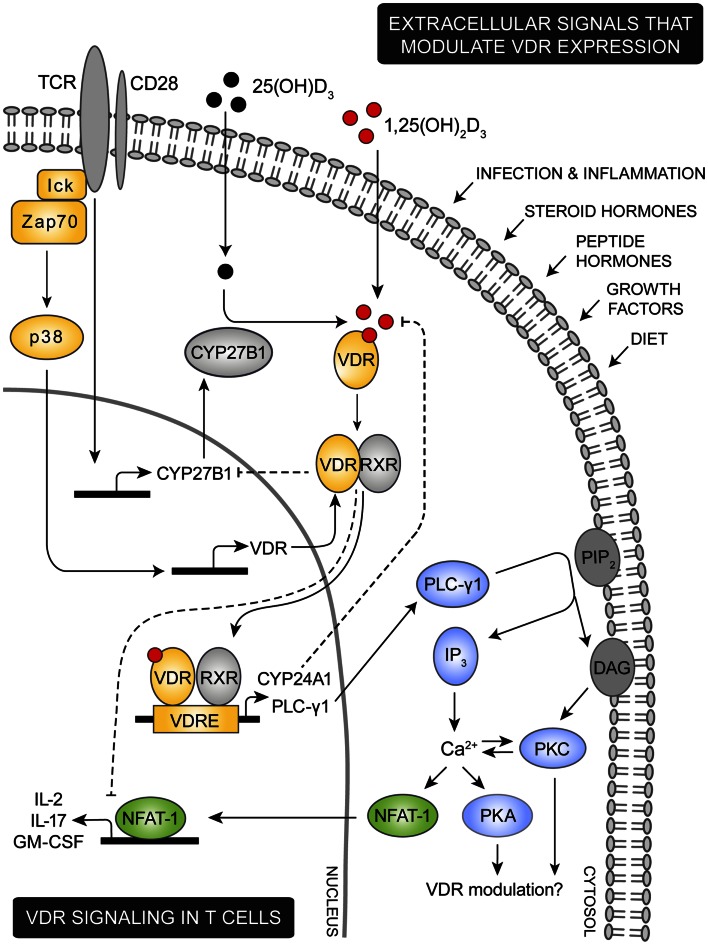
The vitamin D receptor (VDR) is a nuclear, ligand-dependent transcription factor that in complex with hormonally active vitamin D, 1,25(OH)2D3, regulates the expression of more than 900 genes involved in a wide array of physiological functions. The impact of 1,25(OH)2D3-VDR signaling on immune function has been the focus of many recent studies as a link between 1,25(OH)2D3 and susceptibility to various infections and to development of a variety of inflammatory diseases has been suggested. It is also becoming increasingly clear that microbes slow down immune reactivity by dysregulating the VDR ultimately to increase their chance of survival. Immune modulatory therapies that enhance VDR expression and activity are therefore considered in the clinic today to a greater extent. As T cells are of great importance for both protective immunity and development of inflammatory diseases a variety of studies have been engaged investigating the impact of VDR expression in T cells and found that VDR expression and activity plays an important role in both T cell development, differentiation and effector function. In this review we will analyze current knowledge of VDR regulation and function in T cells and discuss its importance for immune activity.

Keywords: T cell function; activity; expression; signaling; vitamin D; vitamin D receptor.


Figures

Figure 1
Figure 1
Proposed model for VDR signaling in T cells. Various extracellular signals including infection, inflammation, steroid and peptide hormones, and diet are involved in regulation of the intracellular VDR level. During an immune response the TCR is triggered by specific antigens, inducing a cascade of intracellular signaling events. Among these, lck and ZAP-70 are activated leading to activation of the p38 kinase which in naïve human T cells induce expression of VDR. TCR triggering also promotes expression of the 1,25(OH)2D3 synthesis enzyme CYP27B1. Through intrinsic synthesis of 1,25(OH)2D3 and uptake of 1,25(OH)2D3 from the extracellular environment, VDR is activated and translocated into the nucleus where it either induce or suppress transcription of a variety of genes. As an example, VDR induce upregulation of PLC-γ1 in naïve human T cells. Once PLC-γ1 is expressed, TCR induced activation of PLC-γ1 leads to activation of PKA and PKC and an increase in the intracellular calcium level. In other cell types PKA and PKC has been shown to modulate expression of VDR, depending on the particular cell type and cellular differentiation state investigated. An increase in intracellular calcium concentration activates NFAT1 a necessary transcription factor for expression of IL-2 and other cytokines. IL-2 is a cytokine required for proliferation of T cells and one mechanism by which VDR adjust T cell activity is to outcompete NFAT1’s binding to the IL-2 promoter and furthermore to down-regulate the actual expression of NFAT1. To control VDR activity a series of negative feedback loops exists; activated VDR both induce expression of the 1,25(OH)2D3 degrading enzyme CYP24A1 and down-regulates expression of the 1,25(OH)2D3 synthesizing enzyme CYP27B1.

References

    1. Achiron A., Grotto I., Balicer R., Magalashvili D., Feldman A., Gurevich M. (2010). Microarray analysis identifies altered regulation of nuclear receptor family members in the pre-disease state of multiple sclerosis. Neurobiol. Dis. 38, 201–209 10.1016/j.nbd.2009.12.029 - DOI - PubMed
    1. Adorini L., Penna G. (2009). Dendritic cell tolerogenicity: a key mechanism in immunomodulation by vitamin D receptor agonists. Hum. Immunol. 70, 345–352 10.1016/j.humimm.2009.01.016 - DOI - PubMed
    1. Adorini L., Penna G., Giarratana N., Uskokovic M. (2003). Tolerogenic dendritic cells induced by vitamin D receptor ligands enhance regulatory T cells inhibiting allograft rejection and autoimmune diseases. J. Cell. Biochem. 88, 227–233 10.1002/jcb.10340 - DOI - PubMed
    1. Alroy I., Towers T. L., Freedman L. P. (1995). Transcriptional repression of the interleukin-2 gene by vitamin D3: direct inhibition of NFATp/AP-1 complex formation by a nuclear hormone receptor. Mol. Cell. Biol. 15, 5789–5799 - PMC - PubMed
    1. Araki M., Kondo T., Gumperz J. E., Brenner M. B., Miyake S., Yamamura T. (2003). Th2 bias of CD4+ NKT cells derived from multiple sclerosis in remission. Int. Immunol. 15, 279–288 10.1093/intimm/dxg029 - DOI - PubMed