. 2013 Jun 13;8(6):e65740.
doi: 10.1371/journal.pone.0065740. Print 2013.

RNA-interference components are dispensable for transcriptional silencing of the drosophila bithorax-complex

Affiliations

RNA-interference components are dispensable for transcriptional silencing of the drosophila bithorax-complex

Filippo M Cernilogar et al. PLoS One. .

Abstract

Background: Beyond their role in post-transcriptional gene silencing, Dicer and Argonaute, two components of the RNA interference (RNAi) machinery, were shown to be involved in epigenetic regulation of centromeric heterochromatin and transcriptional gene silencing. In particular, RNAi mechanisms appear to play a role in repeat induced silencing and some aspects of Polycomb-mediated gene silencing. However, the functional interplay of RNAi mechanisms and Polycomb group (PcG) pathways at endogenous loci remains to be elucidated.

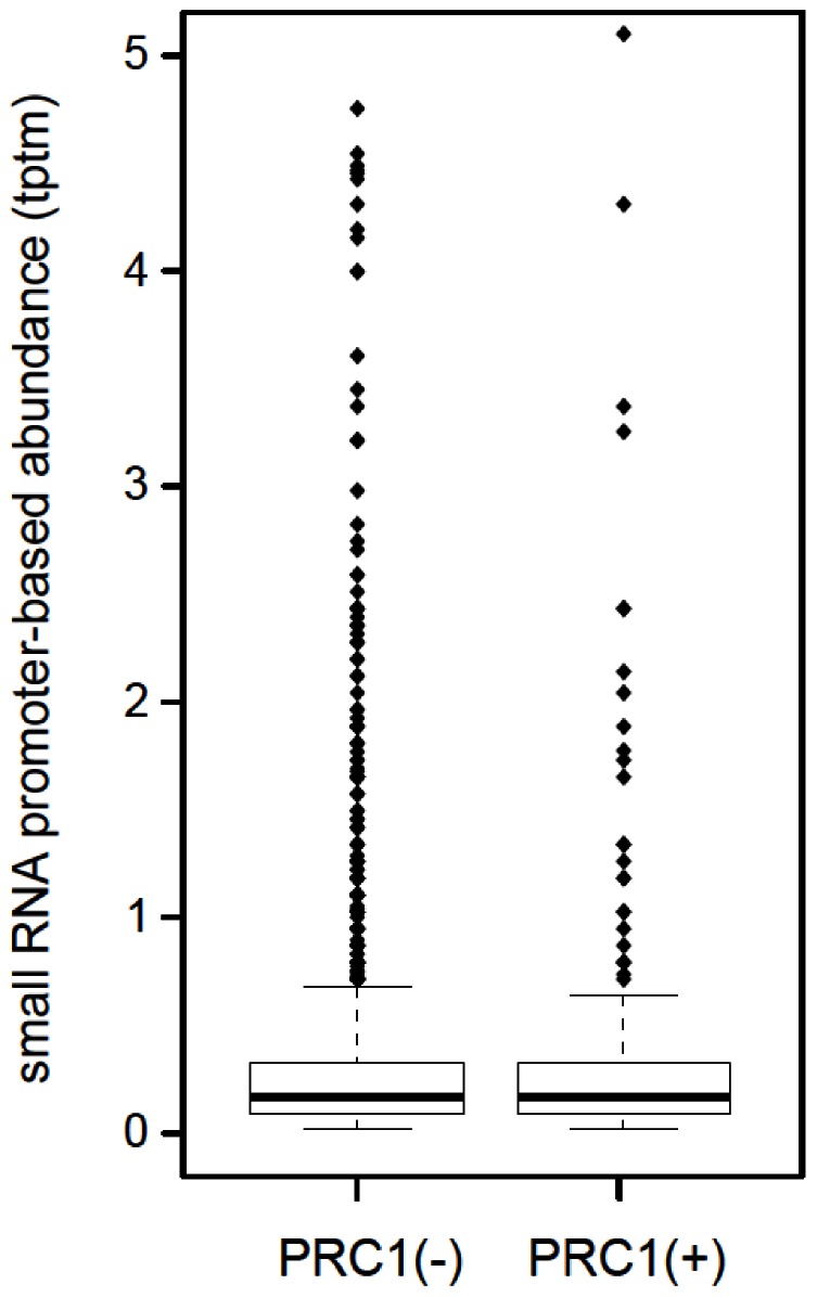
Principal findings: Here we show that the endogenous Dicer-2/Argonaute-2 RNAi pathway is dispensable for the PcG mediated silencing of the homeotic Bithorax Complex (BX-C). Although Dicer-2 depletion triggers mild transcriptional activation at Polycomb Response Elements (PREs), this does not induce transcriptional changes at PcG-repressed genes. Moreover, Dicer-2 is not needed to maintain global levels of methylation of lysine 27 of histone H3 and does not affect PRE-mediated higher order chromatin structures within the BX-C. Finally bioinformatic analysis, comparing published data sets of PcG targets with Argonaute-2-bound small RNAs reveals no enrichment of these small RNAs at promoter regions associated with PcG proteins.

Conclusions: We conclude that the Dicer-2/Argonaute-2 RNAi pathway, despite its role in pairing sensitive gene silencing of transgenes, does not have a role in PcG dependent silencing of major homeotic gene cluster loci in Drosophila.


Conflict of interest statement

Competing Interests: Axel Imhof is a PLOS ONE Editorial Board member. This does not alter the authors' adherence to all the PLOS ONE policies on sharing data and materials.

Figures

Figure 1
Figure 1. Potential sources of dsRNA at PREs.
(A) Schematic representation of the BX-C. Fab-7, Mcp and bxd regions are highlighted and the positions of the primers used in this study are indicated by arrows. All amplicons are about 500 bp long. Core PRE regions are highlighted in red. A: ApaI restriction site (Fab-7) or AccI site (Mcp); E: EcoRI site; P: PstI site; X: XbaI. (B) Analysis of strand specific transcripts at BX-C PREs. RT-PCR was performed on total RNA (DNAse treated) from embryos (0–18 h after egg laying), S2 and S3 cells. Primers complementary to forward (F) or reverse (R) strand transcripts (the homeotic genes of the BX-C are transcribed from the forward strand) were used to initiate cDNA synthesis; subsequently, the other primer was added for PCR amplification. As input control a primer pair specific for transcripts of the Argonaute-1 (Ago1) gene was included (top). (C) To check for genomic contamination total RNA was PCR amplified using primers f9 without the RT step (-RT). As positive control genomic (G) DNA was also amplified.
Figure 2
Figure 2. Dcr2 depletion does not cause transcriptional activation of homeotic genes.
(A, B) The samples analyzed were mock-treated S2 cells (control transfection without dsRNA) or S2 cells treated with Dcr2 dsRNA. (A) Western blot with the indicated antibodies. (B) RT-PCR analysis of glyceraldehyde-3-phosphate dehydrogenase (gapdh) and core PRE transcripts. To check for genomic DNA contamination total RNA from each sample was PCR amplified without the RT step (-RT). (C) RT-PCR analysis in S3 cells and embryos (0–18 h after egg laying). To check for genomic DNA contamination total RNA from each source was PCR amplified without the RT step (-RT). (D) Quantitative RT-PCR of the BX-C homeotic genes. The samples analyzed were mock-treated S2 cells (control without dsRNA) or S2 cells treated with Dcr2 dsRNA. The results shown are from three independent experiments; error bars show the standard deviation.
Figure 3
Figure 3. Consequences of Dcr2 depletion on chromatin marks at PREs.
Crosslinked chromatin from mock-treated S2 cells (control transfection without the dsRNA) or S2 cells treated with Dcr2 dsRNA was immunoprecipitated with the antibodies indicated below the bars (H3K27m3, PC, Pol II). The immunoprecipitated DNA was analyzed by quantitative PCR with primers specific for the Fab-7, Mcp and bxd core PRE and for an intergenic control region (3,2 kb upstream of CG14356 [62]). Protein binding is expressed as the percentage of input minus the background signal. The results shown are from three independent experiments; error bars show the standard deviation.
Figure 4
Figure 4. Contribution of the RNAi pathway to H3K27 methylation.
Histones were acid extracted from mock treated S2 cells (control transfection without the dsRNA) or S2 cells treated with Dcr2 dsRNA, separated by SDS-PAGE, isolated from the gel and analyzed by MALDI-TOF mass spectrometry. The graph shows the amount of methylation within the peptide containing aminoacids 27–40 of H3 from two independent experiments.
Figure 5
Figure 5. BX-C higher order interactions are not impaired after Dcr2 depletion.
(A) The scheme shows the Bithorax Complex (BX-C), including transcription units and genetically characterized regulatory regions (black). (B–C) Chromosome Conformation Capture (3C) on S2 cells. Cells were treated with EGFP-dsRNA (control) and Dcr2-dsRNA. Crosslinking frequencies between PcG targets are shown. All data points were generated from an average of at least three independent experiments. The standard error of the mean is indicated. Two-tailed t-test was applied for statistical analysis. Asterisks indicate statistically relevant differences; α = 0.05. (B) Crosslinking frequencies, normalized on the control, between the fixed fragments spanning homeotic promoters (Abd-Bγ; abd-A) and BX-C PREs. (C) PRE/PRE crosslinking frequencies, normalized on the control. Standard error of the mean is indicated.
Figure 6
Figure 6. PcG-occupied promoters are not enriched in AGO2-bound proximal promoter-derived small RNA sequences.
No significant enrichment in small RNAs observed in PcG-bound promoters relative to promoters without PcG-binding (Kolmogorov–Smirnov test, p-value = 1.0); tag counts are normalized to tags per ten million tags (tptm). Promoter regions were defined as the genome region 50 nucleotides upstream and downstream of the Refseq-defined transcriptional start site, which were linked to PcG occupancy as previously described .

References

    1. Bartel DP (2004) MicroRNAs: genomics, biogenesis, mechanism, and function. Cell 116: 281–297. - PubMed
    1. Baulcombe D (2004) RNA silencing in plants. Nature 431: 356–363. - PubMed
    1. Meister G, Tuschl T (2004) Mechanisms of gene silencing by double-stranded RNA. Nature 431: 343–349. - PubMed
    1. Lee YS, Nakahara K, Pham JW, Kim K, He Z, et al. (2004) Distinct roles for Drosophila Dicer-1 and Dicer-2 in the siRNA/miRNA silencing pathways. Cell 117: 69–81. - PubMed
    1. Okamura K, Ishizuka A, Siomi H, Siomi MC (2004) Distinct roles for Argonaute proteins in small RNA-directed RNA cleavage pathways. Genes Dev 18: 1655–1666. - PMC - PubMed

Publication types

LinkOut - more resources