. 2013 Jun 13;8(6):e66137.
doi: 10.1371/journal.pone.0066137. Print 2013.

The nervous systems of basally branching nemertea (palaeonemertea)

Affiliations

The nervous systems of basally branching nemertea (palaeonemertea)

Patrick Beckers et al. PLoS One. .

Abstract

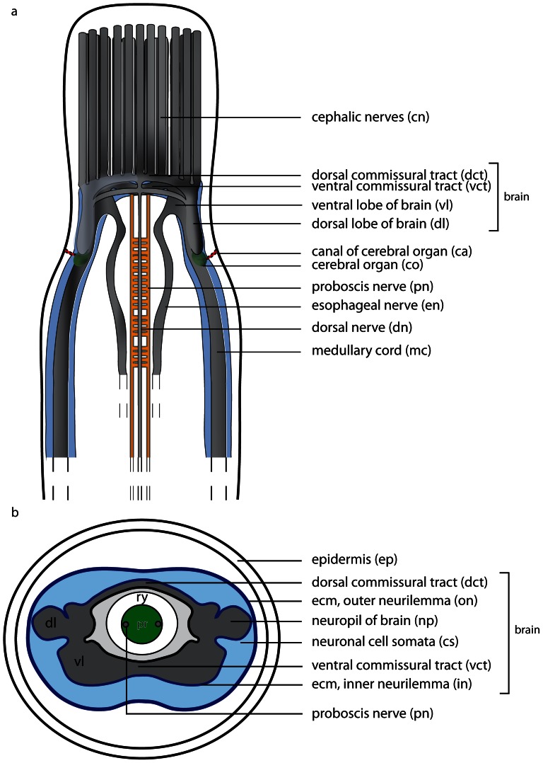
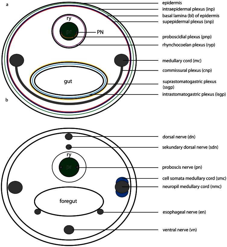
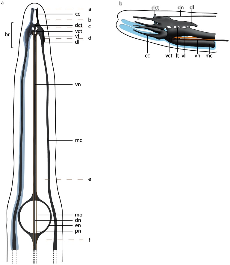
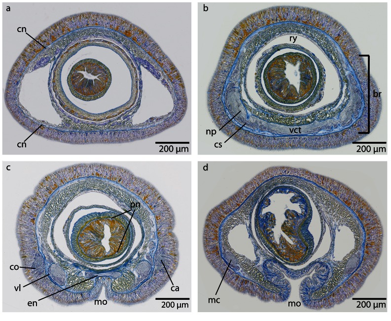
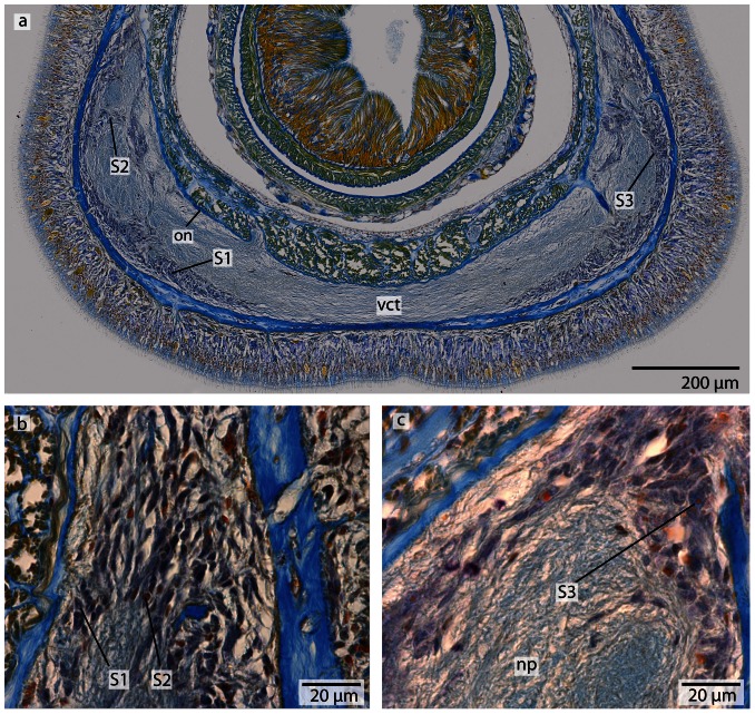
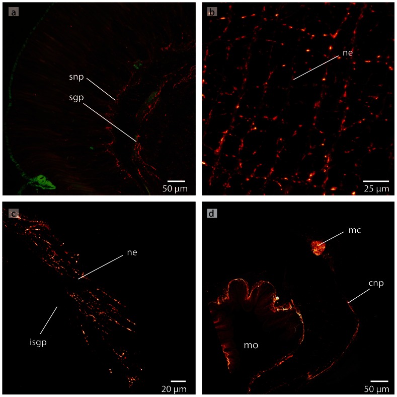
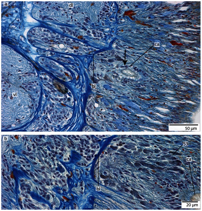
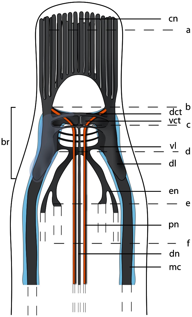
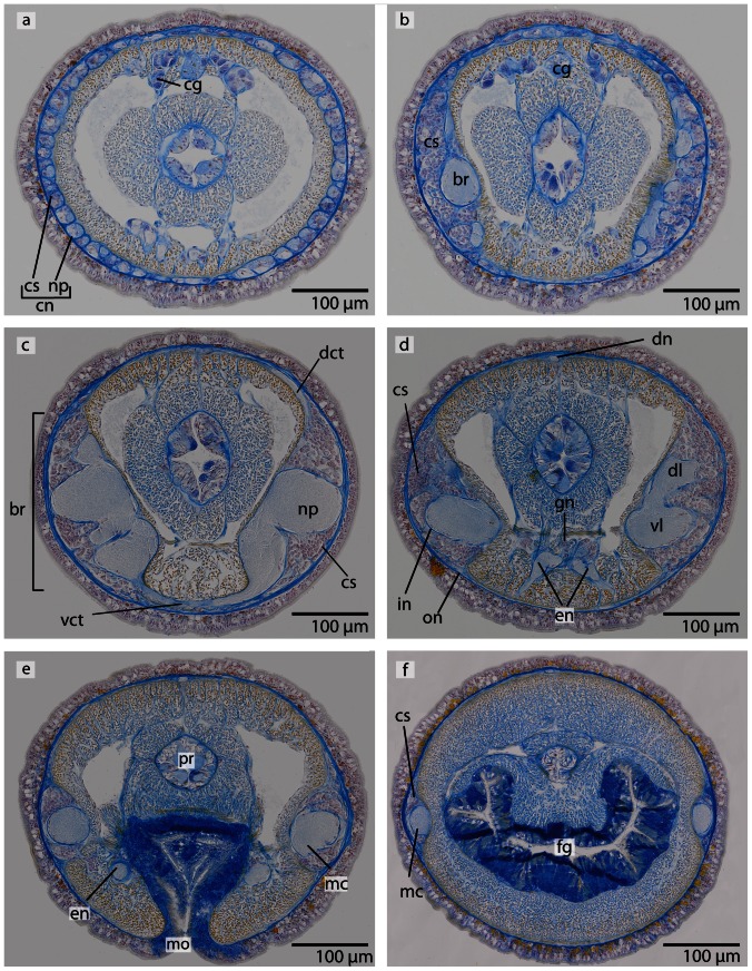
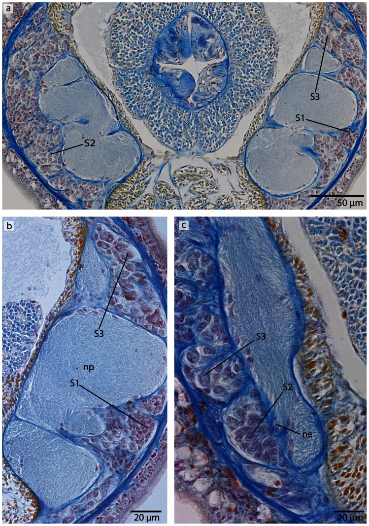
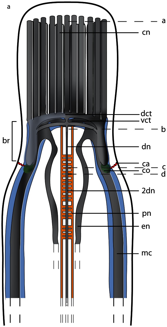
In recent years, a lot of studies have been published dealing with the anatomy of the nervous system in different spiralian species. The only nemertean species investigated in this context probably shows derived characters and thus the conditions found there are not useful in inferring the relationship between nemerteans and other spiralian taxa. Ingroup relationships within Nemertea are still unclear, but there is some agreement that the palaeonemerteans form a basal, paraphyletic grade. Thus, palaeonemertean species are likely the most informative when comparing with other invertebrate groups. We therefore analyzed the nervous system of several palaeonemertean species by combining histology and immunostaining. 3D reconstructions based on the aligned slices were performed to get an overall impression of the central nervous system, and immunohistochemistry was chosen to reveal fine structures and to be able to compare the data with recently published results. The insights presented here permit a first attempt to reconstruct the primary organization of the nemertean nervous system. This comparative analysis allows substantiating homology hypotheses for nerves of the peripheral nervous system. This study also provides evidence that the nemertean brain primarily consists of two lobes connected by a strong ventral commissure and one to several dorsal commissures. During nemertean evolution, the brain underwent continuous compartmentalization into a pair of dorsal and ventral lobes interconnected by commissures and lateral tracts. Given that this conclusion can be corroborated by cladistic analyses, nemerteans should share a common ancestor with spiralians that primarily have a simple brain consisting of paired medullary, frontally commissurized and reinforced cords. Such an organization resembles the situation found in presumably basally branching annelids or mollusks.


Conflict of interest statement

Competing Interests: The authors have declared that no competing interests exist.

Figures

Figure 1
Figure 1. Schematic drawings of the nemertean central nervous system.
a: Dorsal view of the central nervous system, the neuronal cell somata are shown in bright blue. b: Transverse section of the brain. The brain is composed of a central neuropil (gray), which is surrounded by neuronal cell somata (bright blue). The neuronal cell somata may be separated from the neuropil by an inner neurilemma. The whole brain may be enclosed by an outer neurilemma. The two halves of the brain are interconnected by a dorsal (dct) and a ventral commissural tract (vct) which form a ring around the rhynchocoel (ry). dl dorsal lobe of brain, pr proboscis, vl ventral lobe of brain.
Figure 2
Figure 2. Schematic drawings of the peripheral nervous system in nemerteans.
a: Scheme of the different nerve plexus in nemerteans. b: Scheme of the different major nerves present in nemerteans. Note the location of the neuronal cell somata (blue) of the medullary cords. The neuronal cell somata overlie the neuropil of the medullary cords dorsally and ventrally. pr proboscis, ry rhynchocoel.
Figure 3
Figure 3. Living specimens of a: Procephalothrix filiformis.
b: Cephalothrix linearis. c: Tubulanus superbus. d: Tubulanus polymorphus. e: Callinera grandis. f: Carinina ochracea. br brain.
Figure 4
Figure 4. Procephalothrix filiformis, schematic drawings of the central nervous system based on 3D-reconstruction of 501 aligned 0.5 µm sections, dorsal (a) and lateral (b) view.
a: The nervous system is composed of neuropil (np, gray) which may be surrounded by cell somata (cs, blue). Cephalic cords (cc) are circular arranged around the head of the animals. The paired proboscidial nerves (pn, yellow) originate from the ventral commissural tract (vct). A dorsal nerve (dn) originates from the dorsal commissural tract (dct), a ventral nerve (vn) from the ventral commissural tract. The branching esophageal nerves (en) originate from the ventral nerve and surround the mouth opening (mo). The lateral medullary cords (mc) originate in the ventral lobes of the brain (br). Letters on the right (af) refer to the histological sections in figure 5 . The nerve plexus originating in the dorsal nerve (see fig. 5e ) is omitted. b: Central nervous system lateral view. The dorsal lobe (dl) of the brain and the ventral lobe (vl) of the brain are connected by several lateral tracts (lt).
Figure 5
Figure 5. Procephalothrix filiformis, light micrographs of Azan stained transverse sections of the brain.
a: Frontal part showing the huge layer of neuronal cell somata (cs). b: Four cephalic cords (cc) extend towards the tip of the head. c: A dorsal commissural tract (dct) connects the two halves of the brain (br). The neuronal cell somata are separated from the neuropil of the brain by an inner neurilemma (in). The brain is embedded in a layer of longitudinal muscles (lm), which lie underneath a layer of circular muscles (cm). d: The brain (br) is divided into dorsal (dl) and ventral (vl) lobes in its posterior part and composed of a central neuropil (np) which is surrounded by cell somata (cs). The somata are separated from the neuropil by an inner neurilemma (in), and two proboscidial nerves (pn) are present. e: A dorsal nerve (dn) arises from the dorsal commissural tract inside the musculature and migrates dorsally, a ventral nerve (vn) from the ventral commissural tract. Neurites (ne) of the dorsal nerve extends as a roof-like structure around the rhynchocoel. mc: medullary cord. f: The lateral medullary cords (mc) are embedded into the longitudinal muscle layer, and the dorsal nerve (dn) is located subepidermally. bv blood vessel, fg foregut, vn ventral nerve.
Figure 6
Figure 6. Procephalothrix filiformis, light micrographs of Azan stained neurons.
a: Transverse section, overview of the brain, position of the three different types of neurons (S1–S3). b: Higher magnification of somata of type 1 and 2 neurons (S1, S2). Nuclei of S1 dye orange and the cell bodies are beak shaped. Cell bodies of S2 are circular and enlarged. Nuclei dye orange. c: The nuclei of cell somata type 3 (S3) also stain orange. The cell bodies are circular but more prominent than in S1S2. in inner neurilemma, np neuropil.
Figure 7
Figure 7. Cephalothrix linearis, light micrographs of Azan stained transverse sections of the brain.
a: The 4 cephalic cords (cc) are frontally covered by an enormous layer of cell somata (cs). b: The brain (br) is composed of a central neuropil (np) surrounded by cell somata (cs). The somata are separated from the neuropil by an inner neurilemma (in). A ventral commissural tract (vct) connects the two parts of the brain. c: The posterior part of the brain is divided into dorsal (dl) and ventral (vl) lobes. d: the ventral lobes are confluent with the lateral medullary cords (mc), two proboscidial nerves (pn) and esophageal nerves (en) are present. A dorsal nerve (dn) extends to the posterior underneath the basal lamina of the epidermis. fg foregut.
Figure 8
Figure 8. Cephalothrix linearis, light micrographs of Azan stained neurons.
a: Transverse section, overview of the brain, position of the three different types of neurons (S1S3). b: Frontally, only type 1 and type 2 neurons are present. c: Nuclei of type 1 neurons stain red. Nuclei of type 2 neurons stain bright purple; the cell body is slightly enlarged. The cell body of type 3 neurons is the most prominent. Their nuclei stain red. in inner neurilemma, np neuropil.
Figure 9
Figure 9. Procephalothrix filiformis, confocal laserscanning (cLSM) micrographs of differently immunostained whole mounts.
a: Anti-FMRF. The cephalic cords (cc) originate in the lateral aspects of the brain (br). The lateral medullary cords (mc) extend the full length of the animal. There are two proboscidial nerves (pn) that run opposed to each other, along both sides of the proboscis. The esophageal nerves (en) originate at the ventral nerve (vn) and branch shortly before the mouth opening (mo). b: Anti-FMRF. Only few neuronal cell somata (cs) of the brain (br) are immunoreactive against FMRF. c: Anti-FMRF. Four minor nerves (arrow) unite in the tip of the animals' head (arrowhead). The nerves are interconnected by a circular nerve (asterisk) d: Anti-FMRF. The dorsal nerve (dn) is connected to the medullary cords (mc) in the very posterior part of the animal; bottle shaped sensory cells (sc) are distributed all over the body. e: Anti serotonin. The medullary cords (mc) are connected by serial arranged circular nerves (scn). Note that the proboscis nerves show no immunoreactivity against serotonin. pr proboscis.
Figure 10
Figure 10. Procephalothrix filiformis, confocal laserscanning (cLSM) micrographs of differently immunostained whole mounts of.
a: Anti-FMRF, z-coded stack. The major lateral medullary cords (mc), some minor lateral nerves (mln) as well as a ventral nerve (vn) are visible. pn: proboscis nerves. b: Anti-FMRF. The neurites (ne) of the intraepidermal plexus are arranged in a ladder-like way. c: Anti-FMRF. On the very tip of the head is a cluster of cells showing immunoreactivity against FMRF (arrow).
Figure 11
Figure 11. Carinoma mutabilis, schematic drawings of the central nervous system based on 3D-reconstruction of 137 aligned 0.5 µm sections, dorsal (a) and lateral (b) view.
a: The nervous system is composed of neuropil (np, gray), which may be surrounded by cell somata (cs, blue). Cephalic nerves (cn) are circular arranged around the animal's head, and the paired proboscidial nerves (pn, yellow) originate from the ventral commissural tract (vct). The posterior part of the brain is divided into a dorsal (dl) and ventral lobe (vl). A dorso-lateral nerve (dln) connects the posterior part of the dorsal lobe with the dorsal commissural tract. A dorsal nerve strand (dn) originates from the dorsal commissural tract (dct). The branching esophageal nerves (en) originate from the ventral commissural tract. The lateral medullary cords (mc) originate in the ventral lobes of the brain. The letters on the right (ad) refer to the histological sections in figure 12 . b: The cephalic nerves (cn) originate in the lateral parts of the brain. Only the posterior region of the brain is divided into a ventral (vl) and dorsal lobe (dl).
Figure 12
Figure 12. Carinoma mutabilis, light micrographs of Azan stained transverse sections of the brain.
a: The cephalic nerves (cn) are circularly arranged around the inner margins of the head. b: The brain (br) is composed of a central neuropil (np) surrounded by cell somata (cs). The whole brain is encircled by an outer neurilemma (on). The two proboscidial nerves (pn) originate from the ventral commissural tract (vct). c: A dorsal nerve (dn) arises from the dorsal commissural tract, the esophageal nerves (en) originate from the ventral commissural tract, the brain is in its posterior part divided into a dorsal (dl) and ventral (vl) lobe. Dorso-lateral nerves (dln) arise from the dorsal commissural tract and merge with the dorsal lobes. d: The ventral lobes are confluent with the lateral medullary cords (mc), two proboscidial nerves are present (pn). Note the different positions of the dorsal nerve (dn1–dn3). e: Higher magnification of the anterior head region, showing the location of the cephalic nerves (cn). dn dorsal nerve, mo mouth opening.
Figure 13
Figure 13. Carinoma mutabilis, light micrographs of Azan stained neurons.
a: Transverse section, overview of the brain, position of the two different types of neurons (S1S2). Another type of cells is located in the dorsal tip of the brain (arrow). b: Higher magnification of somata of type 2 neurons (S2). The nuclei of S2 dye purple. The neurites (ne) of the brain neuropil branch into the somata layer. c: In the dorsal and ventral tip of the brain, cells with red-stained nuclei and very prominent cell bodies (arrows) are found. Note the close association of these cells with the blood vessels (bv). These cells resemble glandular cells. pn proboscis nerve.
Figure 14
Figure 14. Tubulanus superbus, light micrographs of Azan stained transverse sections of the brain.
a: The cephalic nerves (cn) are circularly arranged around the margins of the head. b: The brain is composed of a central neuropil (np) and surrounding cell somata (cs). An outer neurilemma (on) surrounds the whole brain. The two proboscidial nerves (pn) originate in the ventral commissural tract. c: Posteriorly the brain is divided into a dorsal and ventral lobe (vl). The canal (ca) of the cerebral organ does not pierce the basal lamina of the epidermis. d: The canal ends in a layer of neuronal cell somata (arrow) which have contact to the brain. Two esophageal nerves (en) are present. The dorsal nerve (dn) originates in a dorsal commissural tract. The ventral lobes of the brain are confluent with the lateral medullary cords (mc). mo: mouth opening.
Figure 15
Figure 15. Tubulanus superbus, light micrographs of Azan stained neurons.
a: Transverse section, overview of the brain, position of the different neuronal cell somata (S1S3). b: The nuclei of type 1 neurons stain purple. The perikarya are not enlarged. c: Somata of type 2 neurons exhibit orange-stained nuclei. The perikarya are enlarged. d: Cell bodies of type 3 neurons are very prominent; the nuclei stain orange. on outer neurilemma.
Figure 16
Figure 16. Tubulanus polymorphus, drawing of a dorsal view on the central nervous system, based on 3D-reconstruction of 173 aligned 0.5 µm sections.
The nervous system is composed of neuropil (np, gray) which may be surrounded by cell somata (cs, blue). Cephalic nerves (cn) are circularly arranged around the animal's head, and the paired proboscidial (pn, yellow) nerves originate from the ventral commissural tract (vct). A dorsal nerve (dn) originates from the dorsal commissural tract (dct). The cerebral organs (co, green) contact to the environment via small canals (ca) which open laterally. The cerebral organ is connected to the dorsal lobe of the brain. The branching esophageal nerves (en) originate from the ventral commissural tract. The lateral medullary cords (mc) originate in the ventral lobes of the brain. The letters on the right (ad) refer to the histological sections in figure 17 .
Figure 17
Figure 17. Tubulanus polymorphus, light micrographs of Azan stained transverse sections of the brain.
a: The cephalic nerves (cn) are circularly arranged around the tip of the head. b: The brain (br) is composed of a central neuropil (np) which is surrounded by cell somata (cs), the ventral commissural tract (vct) connects the two halves of the brain below the rhynchocoel (ry). c: The canals (ca) of the cerebral organ (co) open ventro- laterally to the environment. The esophageal nerves (en) originate in the ventral commissural tract and are connected again shortly in front of the mouth opening (mo). Two proboscidial nerves (pn) are present. d: The ventral lobes of the brain are confluent with the lateral medullary cords. mo mouth opening, vl ventral lobes of brain.
Figure 18
Figure 18. Tubulanus polymorphus, light micrographs of Azan stained neurons.
a: Transverse section, overview of the brain; position of the three different types of neurons (S1S3). b: Higher magnification of the somata of neuron types 1 and 2. Nuclei of type 1 neurons (S1) stain purple; the cell body is not enlarged. Nuclei of S2 stain red. Type 2 neurons are solitarily distributed all over the brain. c: Nuclei of type 3 neurons stain red and the cell body is more prominent compared to S1 and S2. np neuropil, on outer neurilemma, vct ventral commissural tract.
Figure 19
Figure 19. Tubulanus polymorphus, confocal laserscanning (cLSM) micrographs of differently immunostained vibratome sections.
a: Transverse section, anti-FMRF (red), anti-α-tubulin (green) showing the subepidermal (snp) and the stomatogastric (sgp) nerve plexus. b: Horizontal section, anti-FMRF showing the regular arranged neurites (ne) of the subepidermal nerve plexus. c: Horizontal section, anti-FMRF. The neurites (ne) of the intrastomatogastric nerve plexus (isgp) are arranged in a regular ladder-like fashion. d: Transverse section, anti-serotonin. The lateral medullary cords (mc) are interconnected by a commissural plexus (cnp). The commissural plexus surrounds the mouth opening (mo).
Figure 20
Figure 20. Tubulanus superbus, light micrographs of Azan stained transverse sections of the brain and cerebral organ.
a: The sensory cells (arrow) surrounding the canal (ca) of the cerebral organ are connected to the dorsal lobe (dl) of the brain. The canal ends anterior to the basal lamina (bl) of the epidermis. b: The cells adjacent to the cerebral organ are type 1 brain cells. vl: ventral lobe.
Figure 21
Figure 21. Callinera grandis, schematic drawing of a dorsal view on the central nervous system, based on 3D-reconstruction of 140 aligned 0.5 µm sections.
The central nervous system is composed of neuropil (np, gray), which is surrounded by cell somata (cs, blue). Cephalic nerves (cn) are circularly arranged; the paired proboscidial nerves (pn, yellow) originate from the ventral commissural tract (vct). A dorsal nerve strand (dn) originates from the dorsal commissural tract. The branching esophageal nerves (en) originate from the ventral commissural tract. The lateral medullary cords (mc) originate ventro-caudally in the brain. The letters on the right (af) refer to the histological sections in figure 22 .
Figure 22
Figure 22. Callinera grandis, light micrographs of Azan stained transverse sections of the brain.
a: The cephalic nerves (cn) are composed of neuropil (np) and neuronal cell somata (cs), a cephalic gland (cg) is located dorsally. b: The anterior region of the brain (br) is covered by a enormous layer of cell somata (cs). c: The brain is composed of a central neuropil (np) and a surrounding layer of cell somata (cs). The two halves of the brain are connected by ventral (vct) and dorsal (dct) commissural tracts. d: Posteriorly the brain is divided into a ventral (vl) and dorsal (dl) section. A dorsal nerve (dn) arises from the dorsal commissural tract, and the paired esophageal nerves (en) arise from the ventral commissural tract. Shortly anterior to the foregut a concentration of neurons (gn) occurs which are associated with the esophageal nerves. The somata are separated from the neuropil by an inner neurilemma (in), and the whole brain is enclosed by an outer neurilemma (on). e: The ventral lobes of the brain are confluent with the lateral medullary cords (mc). mo: mouth opening, pr: proboscis. f: The lateral medullary cords (mc) run to the posterior of the animal. The cell somata (cs) cover the neuropil in a C-shaped manner. fg foregut.
Figure 23
Figure 23. Callinera grandis, light micrographs of Azan stained neurons.
a: Transverse section, overview of the brain; position of the three different types of neurons (S1S3); all nuclei stain purple. b: Higher magnification of somata of type 1 and 3 neurons (S1, S3). Cell bodies are circular; those of S1 are arranged in clusters, those of S3 are most prominent. c: Higher magnifcation of somata of type 2 and 3 neurons. S2 cell bodies are pear shaped and slightly enlarged. The neurites (ne) of the brain branch into the cell somata layer. np neuropil.
Figure 24
Figure 24. Carinina ochracea, schematic drawing of a dorsal view on the central nervous system, based on 3D-reconstruction of 160 aligned 0.5 µm sections.
The nervous system is composed of neuropil (np, gray) which may be surrounded by cell somata (cs, blue). Cephalic nerves (cn) form basiepidermal basket, the paired proboscidial nerves (pn, yellow) originate from the ventral commissural tract (vct). A dorsal nerve strand (dn) originates from the dorsal commissural tract (dct). A second dorsal nerve (2dn) runs underneath the dorsal nerve. The cerebral organs (co, green) gain contact to the environment via small canals (ca, red), which open laterally. The cerebral organ is connected to the dorsal section of the brain. The branching esophageal nerves (en) originate from the ventral commissural tract (vct). The lateral medullary cords (mc) originate in the ventral part of the brain. The letters on the right (ad) refer to the histological sections in figure 25 .
Figure 25
Figure 25. Carinina ochracea, light micrographs of Azan stained transverse sections of the brain.
a: Frontal region showing the circularly arranged cephalic nerves (cn). bv: blood vessel. b: The brain (br) is composed of a central neuropil (np) which is surrounded by cell somata (cs). A ventral commissural tract (vct) connects the two halves of the brain, and an outer neurilemma (on) encloses the whole brain. (pn): proboscidial nerves. c: The canals (ca) of the cerebral organs open laterally, where the brain divides into a dorsal (dl) and ventral (vl) sections. Two proboscidial nerves (pn) are present. d: The canals of the cerebral organs (co) end dorsally. A dorsal nerve strand (dn) arises from the dorsal commissural tract, the ventral part of the brain is confluent with the lateral medullary cords (mc). Note the ecm (arrow) branching into the neuropil of the brain. mo mouth opening.
Figure 26
Figure 26. Carinina ochracea, light micrographs of Azan stained neurons.
a: Cell bodies of type 1 neurons (S1) are not enlarged while those of type 2 neurons (S2) are; nuclei stain purple. b: Neuronal cell somata of the medullary cord (mc) are of the second type described for the brain. c: The canal of the cerebral organ ends in a layer of special neuronal cell somata (sco), which are not present in the brain.
Figure 27
Figure 27. Carinina ochracea, confocal laserscanning (cLSM) micrographs of differently immunostained vibratome sections.
a: Anti-serotonin, the brain is composed of neuropil (np) and cell somata (cs). Note the few neuronal cell somata showing immunoreactivity against serotonin. b: Anti-α-tubulin, the neurites of the neuropil (np) of the brain are horizontally arranged in the ventral commissural tract (vct). vl: ventral lobe. c: Anti-serotonin; only few neurites show immunoreactivity against serotonin. d: Anti-FMRF. The cell somata (cs) may represent glia cells. a: anterior np: neuropil, p: posterior.
Figure 28
Figure 28. Carinina ochracea, confocal laserscanning (cLSM) micrographs of differently immunostained vibratome sections.
a: Anti-FMRF, horizontal section. A sensory cell (sc) originates in the subepidermal plexus (snp) and proceeds into the epidermis (ep). b: Anti-FMRF, horizontal section. The neurites (ne) of the intraepidermal nerve plexus are arranged in a ladder-like manner. c: Anti-α-tubulin, sagital section. The lateral medullary cords (mc) are interconnected to the subepidermal plexus (snp) by branches of neurites (ne). d: Higher magnification of c. Note the concentration of neurites in the subepidermal nerve plexus (arrows) e: Anti-FMRF, horizontal section. The neurites (ne) of the intrastomatogastric nerve plexus are irregular arranged. f: Anti FMRF, horizontal section. Multipolar neurons (cs) are distributed in the intraepidermal nerve plexus.
Figure 29
Figure 29. Carinina ochracea, confocal laserscanning (cLSM) micrographs of anti α-tubulin immunostained vibratome sections.
a: Sagital section. The neurites (ne) of the cells lining the canal of the cerebral organ are connected to the neuropil of the dorsal lobe (dl) of the brain (br). Note the unstained structures in the neuropil of the brain (arrowheads). b: sagital section. The canal terminates in a pit with a great number of cilia (arrow). c: horizontal section. The neurites (ne) of the sensory cells built a dense meshwork which is connected to the brain (br). ca canal of the cerebral organ, mc medullary cord.

Similar articles

Cited by

References

    1. Nordhausen W (1988) Impact of the nemertean Lineus viridis on its polychaete prey on an intertidal sandflat. Hydrobiologia 156: 39–46.
    1. Thiel M (1998) Nemertines as predators on tidal flats - High noon at low tide. Hydrobiologia 365: 241–250.
    1. Thiel M, Kruse I (2001) Status of the Nemertea as predators in marine ecosystems. Hydrobiologia 456: 21–32.
    1. Bürger O (1895) Die Nemertinen des Golfes von Neapel und der angrenzenden Meeres-Abschnitte. In Fauna und Flora des Golfes von Neapel und der angrenzenden Meeres-Abschnitte. Berlin: R Friedländer und Sohn. 743 p.
    1. Gibson R (1972) Nemerteans. London: Hutchinson University Library. 224 p.

Publication types

MeSH terms

LinkOut - more resources