

Accuracy of the first step of the dermatoscopic 2-step algorithm for pigmented skin lesions
- PMID: 23785610
- PMCID: PMC3663352
- DOI: 10.5826/dpc.0203a08
Accuracy of the first step of the dermatoscopic 2-step algorithm for pigmented skin lesions
Abstract
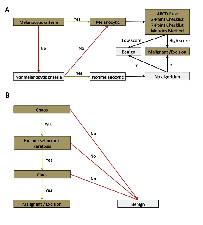
Objectives: To evaluate the frequency of misclassifications of equivocal pigmented lesions according to the first step of the dermatoscopic 2-step algorithm.
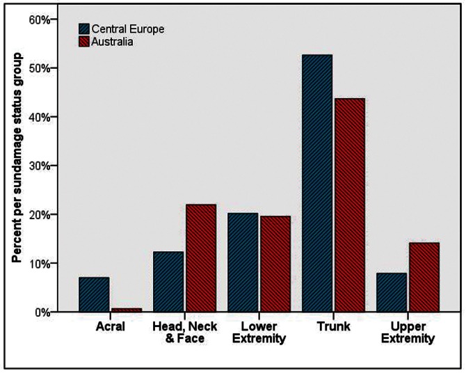
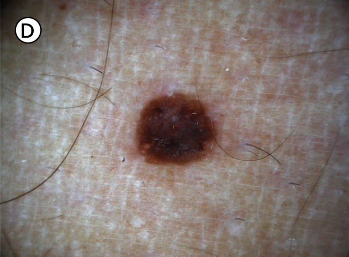
Patients and methods: 707 consecutive cases from 553 patients of central Europe and Australia were included in the study. Dermatoscopic images were evaluated in a blinded fashion for the presence of features described in the 2-step algorithm to determine their melanocytic or non-melanocytic origin. Mucosal, genital and non-pigmented lesions were excluded.
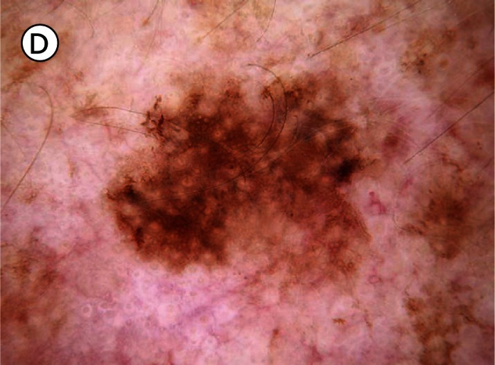
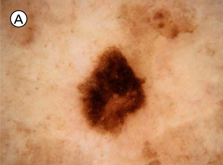
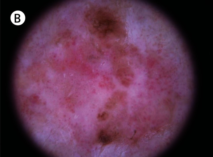
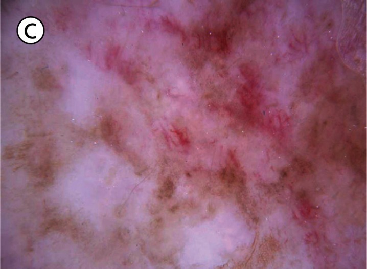
Results: The sensitivity of the first step was 97.1% for patients from Australia and 96.8% for patients from central Europe. The specificity was 33.6% for Australian patients and 67.9% for European patients. The most common reasons for misclassification were the presence of a pigmented network in a non-melanocytic lesion (n=68, 25.2%) and the absence of dermatoscopic features of melanocytic and non-melanocytic lesions in 69 (25.6%) non-melanocytic lesions.
Conclusion: The first step of the dermatoscopic 2-step algorithm, if applied consistently, has high sensitivity but low specificity. Many non-melanocytic lesions, especially solar lentigines and seborrheic keratoses, are wrongly classified as melanocytic. The worse performance of the first step algorithm in Australian patients is probably due to a higher rate of solar lentigines in patients with severely sun-damaged skin.
Figures
References
-
- Kittler H, Pehamberger H, Wolff K, Binder M. Diagnostic accuracy of dermoscopy. Lancet Oncol. 2002;3(3):159–65. - PubMed
-
- Vestergaard ME, Macaskill P, Holt PE, Menzies SW. Dermoscopy compared with naked eye examination for the diagnosis of primary melanoma: a meta-analysis of studies performed in a clinical setting. Br J Dermatol. 2008;159(3):669–76. - PubMed
-
- Rosendahl C, Tschandl P, Cameron A, Kittler H. Diagnostic accuracy of dermatoscopy for melanocytic and nonmelanocytic pigmented lesions. J Am Acad Dermatol. 2011;64(6):1068–73. - PubMed
-
- Argenziano G, Fabbrocini G, Carli P, De Giorgi V, Sammarco E, Delfino M. Epiluminescence microscopy for the diagnosis of doubtful melanocytic skin lesions. Comparison of the ABCD rule of dermatoscopy and a new 7-point checklist based on pattern analysis. Arch Dermatol. 1998;134(12):1563–70. - PubMed
-
- Henning JS, Dusza SW, Wang SQ, et al. The CASH (color, architecture, symmetry, and homogeneity) algorithm for dermoscopy. J Am Acad Dermatol. 2007;56(1):45–52. - PubMed
LinkOut - more resources
Full Text Sources