

doi: 10.5826/dpc.0204a08.
Print 2012 Oct.
Embryology of a melanoma? A case report with speculation based on dermatoscopic and histologic evidence
Affiliations
- PMID: 23785622
- PMCID: PMC3663368
- DOI: 10.5826/dpc.0204a08
Item in Clipboard
Embryology of a melanoma? A case report with speculation based on dermatoscopic and histologic evidence
Dermatol Pract Concept.
.
No abstract available
Keywords: dermatoscopy; histologic criteria; melanoma; nevus; small melanoma.
Figures
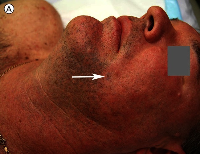
Clinical and close-up image of a (reportedly new) 2 mm pigmented skin lesion on the face of a 51-year-old man. [Copyright: ©2012 Rosendahl et al.]
Clinical and close-up image of a (reportedly new) 2 mm pigmented skin lesion on the face of a 51-year-old man. [Copyright: ©2012 Rosendahl et al.]

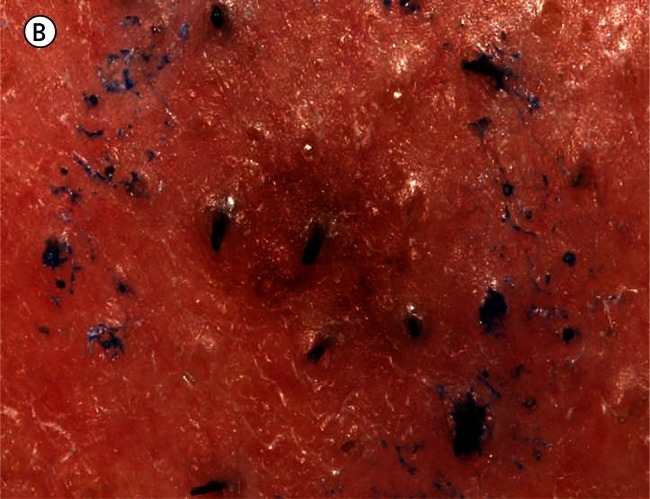
A dermatoscopic image reveals the presence of gray circles (three of which are indicated by arrows). [Copyright: ©2012 Rosendahl et al.]
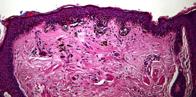
Low power view of the pigmented skin lesion illustrated above showing a melanocytic proliferation with some lentiginous array of single melanocytes as well as nesting. On the left a lentiginous array of single melanocytes extending down a hair follicle is clearly seen. [Copyright: ©2012 Rosendahl et al.]
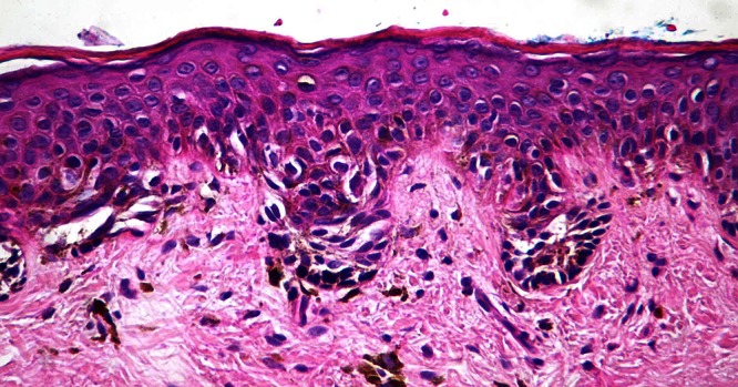
Higher power view showing nesting of (small) melanocytes with pleomorphic hyperchromatic nuclei and apparent partial thickness Pagetoid spread. [Copyright: ©2012 Rosendahl et al.]
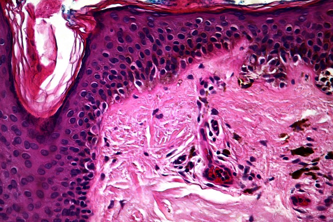
Higher power view of focally confluent lentiginous single melanocytes extending into the superficial part of a follicle. [Copyright: ©2012 Rosendahl et al.]
Similar articles
-
Integrating clinical/dermatoscopic findings and fluorescence in situ hybridization in diagnosing melanocytic neoplasms with less than definitive histopathologic features.J Am Acad Dermatol. 2012 Jun;66(6):917-22. doi: 10.1016/j.jaad.2011.05.051. Epub 2011 Oct 1. J Am Acad Dermatol. 2012. PMID: 21962759
-
Comparison of dermatoscopic diagnostic algorithms based on calculation: The ABCD rule of dermatoscopy, the seven-point checklist, the three-point checklist and the CASH algorithm in dermatoscopic evaluation of melanocytic lesions.J Dermatol. 2014 Jul;41(7):598-603. doi: 10.1111/1346-8138.12491. Epub 2014 May 8. J Dermatol. 2014. PMID: 24807635
-
Dermatoscopy turns histopathologist's attention to the suspicious area in melanocytic lesions.Arch Dermatol. 2001 Oct;137(10):1338-40. doi: 10.1001/archderm.137.10.1338. Arch Dermatol. 2001. PMID: 11594859
-
Dysplastic nevus: why this term should be abandoned in dermatoscopy.Dermatol Clin. 2013 Oct;31(4):579-88, viii. doi: 10.1016/j.det.2013.06.009. Epub 2013 Jul 23. Dermatol Clin. 2013. PMID: 24075546 Review.
-
Dermatoscopy for melanoma and pigmented lesions.Dermatol Clin. 2012 Jul;30(3):413-34. doi: 10.1016/j.det.2012.04.005. Dermatol Clin. 2012. PMID: 22800549 Review.
Cited by
-
Evolution of the Clinical, Dermoscopic and Pathologic Diagnosis of Melanoma.Dermatol Pract Concept. 2021 Jul 1;11(Suppl 1):e2021163S. doi: 10.5826/dpc.11S1a163S. eCollection 2021 Jul. Dermatol Pract Concept. 2021. PMID: 34447612 Free PMC article. Review.
References
-
- Massi G, Leboit PE. Histological Diagnosis of Nevi and Melanoma. Darmstadt, Germany: Steinkopff Verlag; 2004.
-
- Weedon D. Weedon’s Skin Pathology. 3rd ed. London, England: Churchill Livingstone Elsevier; 2010.
-
- Beer J, Xu L, Tschandl P, Kittler H. Growth rate of melanoma in vivo and correlation with dermatoscopic and dermatopathologic findings. Dermatol Pract Conc. 2011;1(1):13. http://dx.doi.org/10.5826/dpc.0101a13. - DOI - PMC - PubMed
-
- Abbasi NR, Shaw HM, Rigel DS, et al. Early diagnosis of cutaneous melanoma: revisiting the ABCD criteria. JAMA. 2004;292(22):2771–6. - PubMed
-
- Teng PP, Hofmann-Wellenhof R, Campbell TM, Soyer HP. Dermoscopic presentation of a 2-mm melanoma in situ. Australas J Dermatol. 2010;51(2):152–3. - PubMed
LinkOut - more resources
Full Text Sources