. 2013 Oct;94(5):305-11.
doi: 10.1111/iep.12033. Epub 2013 Jun 21.

Characterization of joint disease in mucopolysaccharidosis type I mice

Affiliations

Characterization of joint disease in mucopolysaccharidosis type I mice

Patricia G de Oliveira et al. Int J Exp Pathol. 2013 Oct.

Abstract

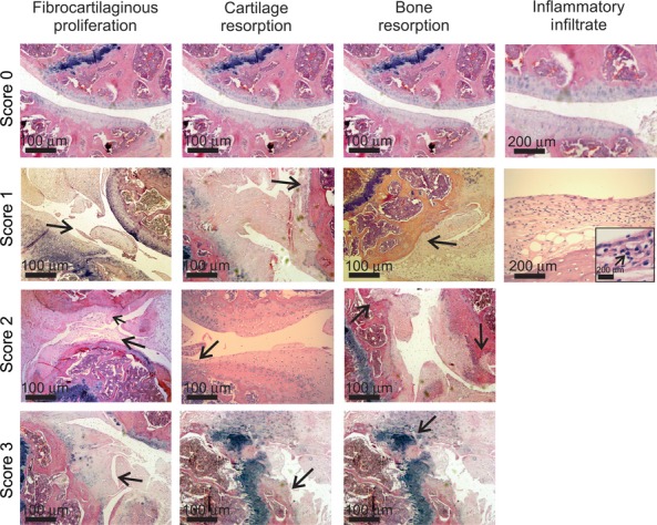
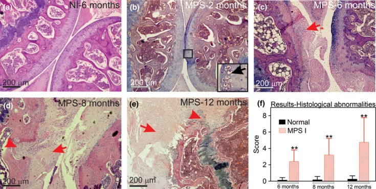
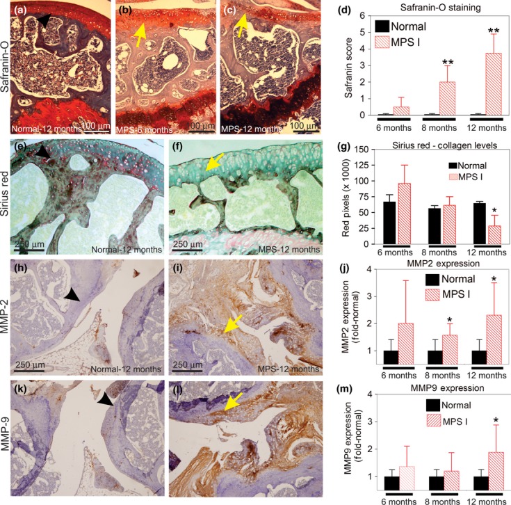
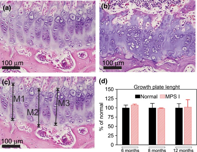
Mucopolysaccharidoses (MPS) are lysosomal storage disorders characterized by mutations in enzymes that degrade glycosaminoglycans (GAGs). Joint disease is present in most forms of MPS, including MPS I. This work aimed to describe the joint disease progression in the murine model of MPS I. Normal (wild-type) and MPS I mice were sacrificed at different time points (from 2 to 12 months). The knee joints were collected, and haematoxylin-eosin staining was used to evaluate the articular architecture. Safranin-O and Sirius Red staining was used to analyse the proteoglycan and collagen content. Additionally, we analysed the expression of the matrix-degrading metalloproteinases (MMPs), MMP-2 and MMP-9, using immunohistochemistry. We observed progressive joint alterations from 6 months, including the presence of synovial inflammatory infiltrate, the destruction and thickening of the cartilage extracellular matrix, as well as proteoglycan and collagen depletion. Furthermore, we observed an increase in the expression of MMP-2 and MMP-9, which could conceivably explain the degenerative changes. Our results suggest that the joint disease in MPS I mice may be caused by a degenerative process due to increase in proteases expression, leading to loss of collagen and proteoglycans. These results may guide the development of ancillary therapies for joint disease in MPS I.

Keywords: alpha-L-iduronidase; joint disease; matrix metalloproteinases; mucopolysaccharidoses type I.


Figures

Figure 1
Figure 1
Example of abnormalities observed in the score used for evaluation of joint disease in MPS I mice (H–E stain). Arrows indicate the specific abnormality shown in each picture. In the inflammatory infiltrate picture grade 1, an insert of higher magnification was used to better exemplify the mononuclear cells observed in the synovium area (arrow). In some cases, the same picture was used more than once, when it contained more than one abnormality or in the case of normal architecture.
Figure 2
Figure 2
Aspect of knee joints using H–E stain. (a) Joint sections representative from a wt mouse at 6 months; (b) MPS I mice at 2 months, insert indicates GAG storage; (c) MPS I mouse at 6 months, with the arrow indicating damage to the articular surface; (d) MPS I mouse at 8 months, with the arrowhead representing bone resorption and the arrow indicating fibrocartilaginous proliferation; (e) MPS I mouse at 12 months, with the arrowhead showing intense bone resorption and the arrow indicating intense fibrocartilaginous proliferation; and (f) results from histological scores obtained in wt and MPS I mice joints at 6, 8 and 12 months (N = 6–11). Score was based on abnormalities described in Figure 1 and detailed in the methods section, where the higher the score, the worse the joint. The maximum score is 10. **P < 0.01, using Student's t-test. N = 6–11 mice/group.
Figure 3
Figure 3
Mechanisms of joint degeneration in MPS I. (a-d) Safranin-O staining. (a) Representative section of a wt joint at 12 months. (b) MPS I mouse at 6 months. (c) MPS I mouse at 12 months, with loss of red signal (yellow arrow) showing loss of proteoglycans. (d) Results from the histological scores obtained with the Safranin-O stain at 6, 8 and 12 months. **P = 0.01, using Student's t-test. N = 3–4 mice/group. (e–g) Collagen levels evaluated by Sirius Red stain. (e) Wt joint at 12 months. (f) MPS I mouse at 12 months, showing loss of red stain (yellow arrow). (g) Quantification of the red signal in wt and MPS I mice. A significant reduction in the collagen levels was observed in the MPS I mice at 12 months (**P < 0.01, Student's t-test). (h–m) Immunohistochemistry for the matrix metalloproteinases (MMP). (h) MMP-2 expression in 12 months wt mouse; (i) MMP-2 expression in 12 months' MPS I mouse, yellow arrow denotes positive staining (brown areas). (j) Quantification of MMP-2 positive signal.*P < 0.05, t-test. N = 6–9 mice/group. (k) MMP-9 expression in Wt mouse at 12 months. (l) MMP-nine expression in MPS I joint at 12 months. (m) Quantification of MMP-2 positive signal. *P < 0.05, t-test. Results are shown as fold-normal, with the normal values being considered 1. Black arrows represent the normal histology, while yellow arrows represent the abnormal findings. N = 6–9 mice/group.
Figure 4
Figure 4
Measurements of the length of the growth plate. On the upper panels, an example of a growth plate from a normal (a) and a MPS I (b) mouse, with disorganization of the chondrocyte axis being shown in MPS I mice. On the lower panel, an example on how the growth plate length was measured (c) and the results for growth plate length (d) are visualized. No differences in length were found between normal and MPS I mice at any time points analysed. N = 6 mice/group. M, measure.

Similar articles

Cited by

References

    1. Baldo G, Wu S, Howe RA, et al. Pathogenesis of aortic dilatation in mucopolysaccharidosis VII mice may involve complement activation. Mol. Genet. Metab. 2011a;104:608–619. - PMC - PubMed
    1. Baldo G, Kretzmann NA, Tieppo J, et al. Bone marrow cells reduce collagen deposition in the rat model of common bile duct ligation. Cell. Sci. Ther. 2011b;2:1–6.
    1. Baldo G, Mayer FQ, Martinelli B, et al. Evidence of a progressive motor dysfunction in Mucopolysaccharidosis type I mice. Behav. Brain Res. 2012;233:169–175. - PMC - PubMed
    1. Clarke LA, Russell CS, Pownall S, et al. Murine mucopolysaccharidosis type I: targeted disruption of the murine alpha-L-iduronidase gene. Hum. Mol. Genet. 1997;6:503–511. - PubMed
    1. Giugliani R, Federhen A, Muñoz Rojas MV, et al. Enzyme replacement therapy for mucopolysaccharidoses I, II and VI: recommendations from a group of Brazilian F experts. Rev. Assoc. Med. Bras. 2010;56:271–277. [Article in Portuguese] - PubMed

Publication types

MeSH terms

LinkOut - more resources