

Defective CD8+CD28+ regulatory T cell suppressor function in rheumatoid arthritis is restored by tumour necrosis factor inhibitor therapy
- PMID: 23786396
- PMCID: PMC3784209
- DOI: 10.1111/cei.12161
Defective CD8+CD28+ regulatory T cell suppressor function in rheumatoid arthritis is restored by tumour necrosis factor inhibitor therapy
Abstract
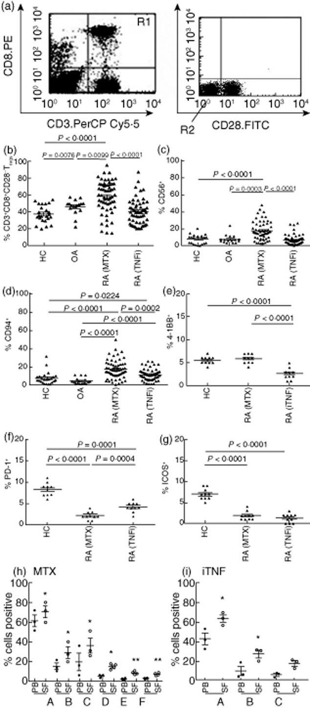
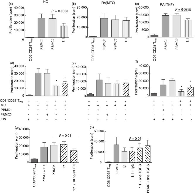
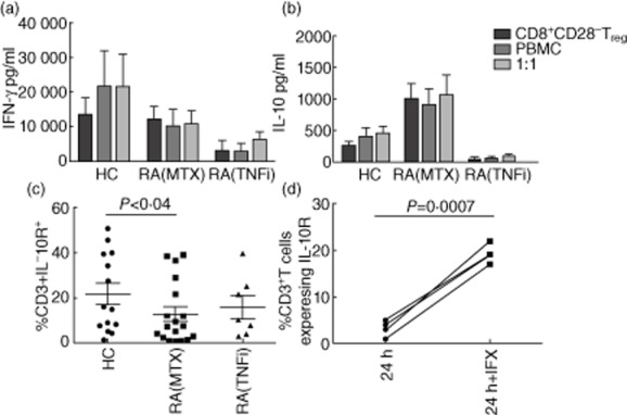
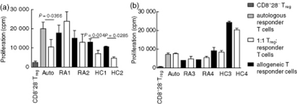
Balanced immunoregulatory networks are essential for maintenance of systemic tolerance. Disturbances in the homeostatic equilibrium between inflammatory mediators, immune regulators and immune effector cells are implicated directly in the pathogenesis of autoimmune diseases, including rheumatoid arthritis (RA). In this study we characterize the peripheral blood CD8(+) CD28(-) regulatory T cells (Treg) contribution to the immunoregulatory network in health and in RA. In health, CD8(+) CD28(-) Treg are suppressive but, unlike CD4(+) Treg , they function predominantly through the action of soluble mediators such as interleukin (IL)-10 and transforming growth factor (TGF)-β. Neutralization of TGF-β consistently reduced CD8(+) CD28(-) Treg suppressor function in vitro. RA, CD8(+) CD28(-) Treg are increased numerically, but have reduced expression of inducible co-stimulator (ICOS) and programmed death 1 (PD-1) compared to healthy or disease controls. They produce more IL-10 but autologous T cells express less IL-10R. This expression was found to be restored following in-vitro addition of a tumour necrosis factor inhibitor (TNFi). Deficiencies in both the CD8(+) CD28(-) Treg population and reduced sensitivity of the T responder cells impact upon their regulatory function in RA. TNFi therapy partially restores CD8(+) CD28(-) Treg ability in vivo and in vitro, despite the defects in expression of functionally relevant molecules by RA CD8(+) CD28(-) Treg compared to healthy controls. This study places CD8(+) CD28(-) Treg cells in the scheme of immune regulation alongside CD4(+) Treg cells, and highlights the importance of understanding impaired responsiveness to regulation that is common to these suppressor subsets and their restored function in response to TNFi therapy.
Keywords: CD8+ T cells; anti-TNF therapy; immunotherapy; regulatory T cells; rheumatoid arthritis.
© 2013 British Society for Immunology.
Figures
Similar articles
-
CD45RA-Foxp3(high) activated/effector regulatory T cells in the CCR7 + CD45RA-CD27 + CD28+central memory subset are decreased in peripheral blood from patients with rheumatoid arthritis.Biochem Biophys Res Commun. 2013 Sep 6;438(4):778-83. doi: 10.1016/j.bbrc.2013.05.120. Epub 2013 Jun 6. Biochem Biophys Res Commun. 2013. PMID: 23747721
-
Interferon-γ and tumor necrosis factor-α promote the ability of human placenta-derived mesenchymal stromal cells to express programmed death ligand-2 and induce the differentiation of CD4(+)interleukin-10(+) and CD8(+)interleukin-10(+)Treg subsets.Cytotherapy. 2015 Nov;17(11):1560-71. doi: 10.1016/j.jcyt.2015.07.018. Cytotherapy. 2015. PMID: 26432559
-
Proinflammatory mediator-induced reversal of CD4+,CD25+ regulatory T cell-mediated suppression in rheumatoid arthritis.Arthritis Rheum. 2007 Mar;56(3):732-42. doi: 10.1002/art.22414. Arthritis Rheum. 2007. PMID: 17328044
-
Harnessing CD8+CD28- Regulatory T Cells as a Tool to Treat Autoimmune Disease.Cells. 2021 Nov 1;10(11):2973. doi: 10.3390/cells10112973. Cells. 2021. PMID: 34831195 Free PMC article. Review.
-
The effect of autoimmune arthritis treatment strategies on regulatory T-cell dynamics.Curr Opin Rheumatol. 2013 Mar;25(2):260-7. doi: 10.1097/BOR.0b013e32835d0ee4. Curr Opin Rheumatol. 2013. PMID: 23274520 Review.
Cited by
-
A high frequency of CD8+CD28- T-suppressor cells contributes to maintaining stable graft function and reducing immunosuppressant dosage after liver transplantation.Int J Med Sci. 2018 Jun 4;15(9):892-899. doi: 10.7150/ijms.24042. eCollection 2018. Int J Med Sci. 2018. PMID: 30008601 Free PMC article.
-
CD4+ /CD8+ T-cell ratio correlates with the graft fate in pig-to-non-human primate islet xenotransplantation.Xenotransplantation. 2020 Mar;27(2):e12562. doi: 10.1111/xen.12562. Epub 2019 Oct 23. Xenotransplantation. 2020. PMID: 31642566 Free PMC article.
-
Expression patterns of CD28 and CTLA-4 in early, chronic, and untreated rheumatoid arthritis.J Clin Lab Anal. 2020 May;34(5):e23188. doi: 10.1002/jcla.23188. Epub 2020 Jan 6. J Clin Lab Anal. 2020. PMID: 31907973 Free PMC article.
-
Non-tumor necrosis factor-based biologic therapies for rheumatoid arthritis: present, future, and insights into pathogenesis.Biologics. 2014;8:1-12. doi: 10.2147/BTT.S35475. Epub 2013 Dec 9. Biologics. 2014. PMID: 24353404 Free PMC article. Review.
-
[Biomarkers for prognosis of response to anti-TNF therapy of rheumatoid arthritis: Where do we stand?].Z Rheumatol. 2015 Nov;74(9):812-8. doi: 10.1007/s00393-014-1543-4. Z Rheumatol. 2015. PMID: 26347122 Review. German. No abstract available.
References
-
- Feldmann M, Brennan FM, Maini RN. Rheumatoid arthritis. Cell. 1996;85:307–310. - PubMed
-
- Ponchel F, Morgan AW, Bingham SJ, et al. Dysregulated lymphocyte proliferation and differentiation in patients with rheumatoid arthritis. Blood. 2002;100:4550–4556. - PubMed
-
- Davila E, Kang YM, Park YW, et al. Cell-based immunotherapy with suppressor CD8+ T cells in rheumatoid arthritis. J Immunol. 2005;174:7292–7301. - PubMed
-
- Filaci G, Fenoglio D, Fravega M, et al. CD8+ CD28- T regulatory lymphocytes inhibiting T cell proliferative and cytotoxic functions infiltrate human cancers. J Immunol. 2007;179:4323–4334. - PubMed
Publication types
MeSH terms
Substances
Grants and funding
LinkOut - more resources
Full Text Sources
Other Literature Sources
Medical
Research Materials
