. 2013 Sep;126(5):591-603.
doi: 10.1111/jnc.12345. Epub 2013 Jul 22.

Determinants of buildup of the toxic dopamine metabolite DOPAL in Parkinson's disease

Affiliations

Determinants of buildup of the toxic dopamine metabolite DOPAL in Parkinson's disease

David S Goldstein et al. J Neurochem. 2013 Sep.

Abstract

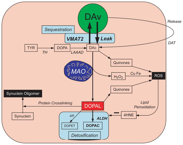
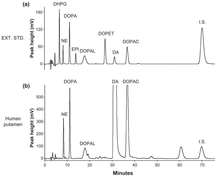
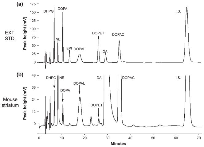
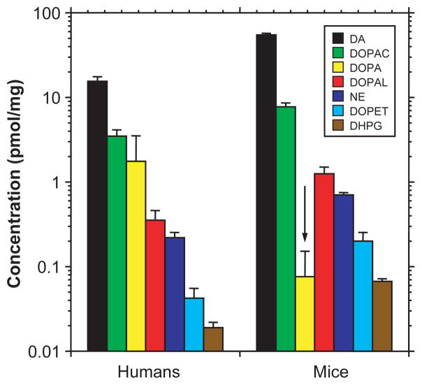
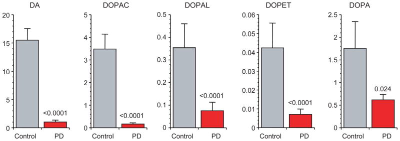
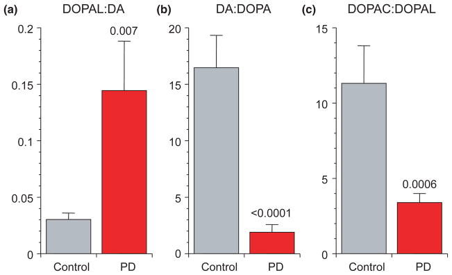
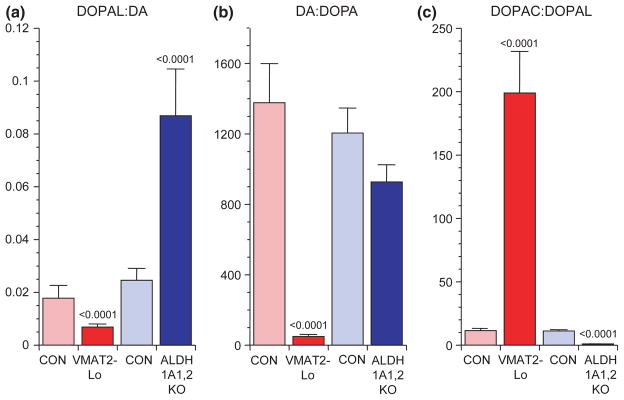
Intra-neuronal metabolism of dopamine (DA) begins with production of 3,4-dihydroxyphenylacetaldehyde (DOPAL),which is toxic. According to the 'catecholaldehyde hypothesis', DOPAL destroys nigrostriatal DA terminals and contributes to the profound putamen DA deficiency that characterizes Parkinson’s disease (PD). We tested the feasibility of using post-mortem patterns of putamen tissue catechols to examine contributions of altered activities of the type 2 vesicular monoamine transporter (VMAT2) and aldehyde dehydrogenase(ALDH) to the increased DOPAL levels found in PD. Theoretically, the DA : DOPA concentration ratio indicates vesicular uptake, and the 3,4-dihydroxyphenylacetic acid: DOPAL ratio indicates ALDH activity. We validated these indices in transgenic mice with very low vesicular uptake VMAT2-Lo) or with knockouts of the genes encoding ALDH1A1 and ALDH2 (ALDH1A1,2 KO), applied these indices in PD putamen, and estimated the percent decreases in vesicular uptake and ALDH activity in PD. VMAT2-Lo mice had markedly decreased DA:DOPA (50 vs. 1377, p < 0.0001),and ALDH1A1,2 KO mice had decreased 3,4-dihydroxyphenylacetic acid:DOPAL (1.0 vs. 11.2, p < 0.0001). In PD putamen, vesicular uptake was estimated to be decreased by 89% and ALDH activity by 70%. Elevated DOPAL levels in PD putamen reflect a combination of decreased vesicular uptake of cytosolic DA and decreased DOPAL detoxification by ALDH.

Keywords: DOPAC; DOPAL; DOPET; Parkinson's disease; dopamine; monoamine oxidase.


Conflict of interest statement

The Authors have no conflicts of interest to disclose.

Figures

Fig. 1
Fig. 1
Concept diagram about sources and metabolic fates of 3,4-dihydroxyphenylacetaldehyde (DOPAL) in dopaminergic neurons. Under resting conditions, most of the irreversible loss of dopamine (DA) from the neurons is because of passive leakage from vesicles (DAv) into the cytosol (DAc), followed by enzymatic deamination catalyzed by monoamine oxidase (MAO). Cytosolic DA is taken up into the vesicles via the type 2 vesicular monoamine transporter (VMAT2). Under resting conditions, release by exocytosis from the vesicles, with escape of reuptake via the cell membrane DA transporter (DAT), constitutes only a minor determinant of DA turnover. The loss of DA is balanced by ongoing catecholamine biosynthesis from the action of L-aromatic-amino-acid decarboxylase on DOPA produced from tyrosine (TYR) by tyrosine hydroxylase (TH). The action of MAO on cytosolic DA produces the catecholaldehyde, DOPAL. DOPAL is detoxified mainly by aldehyde dehydrogenase (ALDH), to form the acid, 3,4-dihydroxyphenylacetic acid (DOPAC), with 3,4-dihydroxyph-enylethanol (DOPET) a minor metabolite formed via aldehyde/aldose reductase. Both DAc and DOPAL (and, at least theoretically, DOPAC and DOPET) spontaneously auto-oxidize to quinones, which augment generation of reactive oxygen species, resulting in lipid peroxidation. 4-Hydroxynonenal (4HNE), a major lipid peroxidation product, inhibits ALDH. DOPAL cross-links with proteins, augmenting oligomerization of alpha-synuclein. Tissue DOPAL : DA indicates DOPAL content adjusted for DA stores. As explained in the Appendix, DA : DOPA provides an index of vesicular uptake and DOPAC : DOPAL an index of ALDH activity.
Fig. 2
Fig. 2
Chromatographs of extracted catechol standards and catechols in putamen tissue from a control subject. (a) Chromatograph after injection of alumina eluate from 1000 pg of 3,4-dihydroxyphenylglycol, 250 pg norepinephrine, 1000 pg DOPA, 250 pg epinephrine (EPI), 1000 pg 3,4-dihydroxyphenylethanol, 250 pg dopamine, 1000 pg 3,4-dihydroxyphenylacetic acid, and internal standard (I.S.); (b) Chromatograph after injection of alumina eluate from a sample of putamen tissue homogenate of a control subject.
Fig. 3
Fig. 3
Chromatographs of extracted catechol standards and catechols in striatum tissue from a control mouse. (a) Chromatograph after injection of alumina eluate from 1000 pg of 3,4-dihydroxyphenylglycol, 250 pg norepinephrine, 1000 pg DOPA, 250 pg epinephrine (EPI), 1000 pg 3,4-dihydroxyphenylethanol, 250 pg dopamine, 1000 pg 3,4-dihydroxyphenylacetic acid, and internal standard (I.S.); (b) Chromatograph after injection of alumina eluate from a sample of striatum tissue homogenate of a control mouse.
Fig. 4
Fig. 4
Comparison of human putamen with mouse striatal tissue concentrations of catechols. Control mouse data were for the ALDH1A1,2 knockouts (n = 54). Note that mice have higher striatal mean concentrations of catecholamines and deaminated metabolites but a lower mean concentration of DOPA than found in human putamen.
Fig. 5
Fig. 5
Putamen mean (± SEM) concentrations (pmol/mg wet weight) of catechols in post-mortem putamen from patients with Parkinson’s disease (PD, red) and controls (gray). Abbreviations: DA = dopamine; DOPAC = 3,4-dihydroxyphenylacetic acid; DOPAL = 3,4-dihydroxyphenylacetaldehyde; DOPET = 3,4-dihydroxyphenylethanol; DOPA = 3,4-dihydroxyphenylalanine. Note severely decreased DA and DOPAC, less severely decreased DOPAL and DOPET, and even less severely decreased DOPA in PD relative to controls.
Fig. 6
Fig. 6
Putamen mean (± SEM) ratios of (a) 3,4-dihydroxyphenylacetaldehyde (DOPAL): dopamine (DA), (b) DA : DOPA, and (c) 3,4-dihydroxyphenylacetic acid (DOPAC): DOPAL in post-mortem putamen from patients with Parkinson’s disease (PD, red) and controls (gray). PD features neurochemical evidence for decreased vesicular sequestration and decreased aldehyde dehydrogenase activity.
Fig. 7
Fig. 7
Striatal mean (± SEM) ratios of (a) 3,4-dihydroxyphenylacetaldehyde (DOPAL): dopamine (DA), (b) DA : DOPA, and (c) 3,4-dihydroxyphenylacetic acid (DOPAC) : DOPAL in mice with very low activity of the type 2 vesicular monoamine transporter (VMAT2-Lo, red) and mice with double knockout of the ALDH1A1 and ALDH2 genes (ALDH1A1,2 KO, blue). Light colors represent the corresponding control groups. The results show that low VMAT2 activity decreases DOPAL : DA and DA : DOPA and increases DOPAC : DOPAL, whereas low aldehyde dehydrogenase activity increases DOPAL : DA, does not affect DA : DOPA, and decreases DOPAC : DOPAL.
Fig. 8
Fig. 8
Putamen tissue DOPA content expressed as a function of the maximum levodopa dose prior to death in Parkinson’s disease patients. Tissue DOPA content was unrelated to the maximum levodopa dose prior to death, with or without exclusion of two outliers who had relatively high tissue DOPA levels.

References

    1. Anderson DG, Mariappan SV, Buettner GR, Doorn JA. Oxidation of 3,4-dihydroxyphenylacetaldehyde, a toxic dopaminergic metabolite, to a semiquinone radical and an orthoquinone. J Biol Chem. 2011;286:26978–26986. - PMC - PubMed
    1. Bohnen NI, Albin RL, Koeppe RA, Wernette KA, Kilbourn MR, Minoshima S, Frey KA. Positron emission tomography of monoaminergic vesicular binding in aging and Parkinson disease. J Cereb Blood Flow Metab. 2006;26:1198–1212. - PubMed
    1. Bonifacio MJ, Archer M, Rodrigues ML, Matias PM, Learmonth DA, Carrondo MA, Soares-Da-Silva P. Kinetics and crystal structure of catechol-o-methyltransferase complex with co-substrate and a novel inhibitor with potential therapeutic application. Mol Pharmacol. 2002;62:795–805. - PubMed
    1. Burke WJ, Li SW, Williams EA, Nonneman R, Zahm DS. 3,4-Dihydroxyphenylacetaldehyde is the toxic dopamine metabolite in vivo: implications for Parkinson’s disease pathogenesis. Brain Res. 2003;989:205–213. - PubMed
    1. Burke WJ, Li SW, Chung HD, et al. Neurotoxicity of MAO metabolites of catecholamine neurotransmitters: role in neurodegenerative diseases. Neurotoxicology. 2004;25:101–115. - PubMed

Publication types

MeSH terms