. 2013 Jun 20:13:142.
doi: 10.1186/1471-2180-13-142.

Genomic and phenotypic characterization of in vitro-generated Chlamydia trachomatis recombinants

Affiliations

Genomic and phenotypic characterization of in vitro-generated Chlamydia trachomatis recombinants

Brendan M Jeffrey et al. BMC Microbiol. .

Abstract

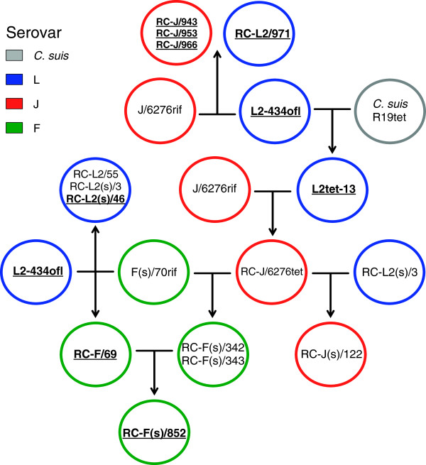
Background: Pre-genomic and post-genomic studies demonstrate that chlamydiae actively recombine in vitro and in vivo, although the molecular and cellular biology of this process is not well understood. In this study, we determined the genome sequence of twelve Chlamydia trachomatis recombinants that were generated in vitro under antibiotic selection. These strains were used to explore the process of recombination in Chlamydia spp., including analysis of candidate recombination hotspots, and to correlate known C. trachomatis in vitro phenotypes with parental phenotypes and genotypes.

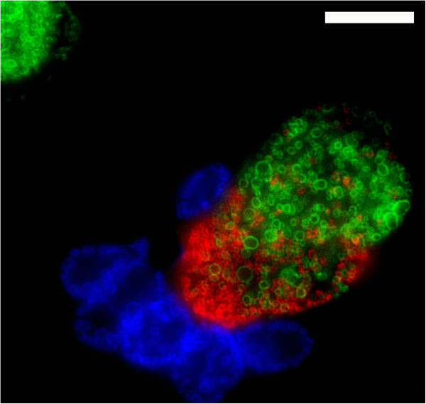
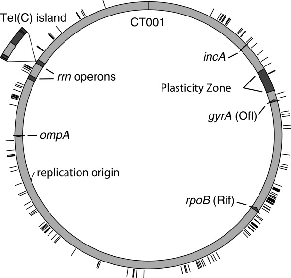
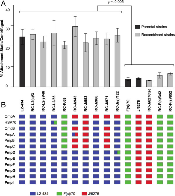
Results: Each of the 190 examined recombination events was the product of homologous recombination, and no candidate targeting motifs were identified at recombination sites. There was a single deletion event in one recombinant progeny that resulted in the removal of 17.1 kilobases between two rRNA operons. There was no evidence for preference for any specific region of the chromosome for recombination, and analyses of a total of over 200 individual recombination events do not provide any support for recombination hotspots in vitro. Two measurable phenotypes were analyzed in these studies. First, the efficiency of attachment to host cells in the absence of centrifugation was examined, and this property segregated to regions of the chromosome that carry the polymorphic membrane protein (Pmp) genes. Second, the formation of secondary inclusions within cells varied among recombinant progeny, but this did not cleanly segregate to specific regions of the chromosome.

Conclusions: These experiments examined the process of recombination in C. trachomatis and identified tools that can be used to associate phenotype with genotype in recombinant progeny. There were no data supporting the hypothesis that particular nucleotide sequences are preferentially used for recombination in vitro. Selected phenotypes can be segregated by analysis of recombination, and this technology may be useful in preliminary analysis of the relationship of genetic variation to phenotypic variation in the chlamydiae.


Figures

Figure 1
Figure 1
The genealogy of recombinant strains generated and explored in this study. The figure shows the parental strains used to generate recombinant strains. In each case, the parents in a cross are connected by a horizontal line, and the progeny are at the end of the arrow. In some cases, the progeny of one cross was used as a parent in a subsequent cross. Primary parental strain names includes drug resistance, and all recombinant strains (indicated by prefix RC- ) are both rifampicin and ofloxacin resistant. The colors used indicate the OmpA phenotype of each strain, as determined by fluorescence microscopy and genome sequence analysis. Strains containing the plasmid are shown in bold face and underlined. Crosses involving three parents are not shown because no triply drug resistant strains could be recovered.
Figure 2
Figure 2
Fluorescent microscopy showing host cells infected with three C. trachomatis strains. Strains were labeled with primary antibodies against OmpA. Cells are infected with L2-434 (green), J/6276 (red), and the inclusion fusion negative strain F(s)/70 (blue). Scale bar, 5 μm.
Figure 3
Figure 3
Genome maps of recombinant strains, derived from complete nucleotide sequence analysis. The colors used in recombinant maps indicate the parental genotype, as is indicated at the top of the figure. The Tet(C) island is originally from C. suis R19. The approximate location of the genetic markers used in the construction of the recombinant genomes is shown above the RC-J/6276tet genome map. Below each strain name is the antibiotic resistance markers that the recombinant strain carries. The bracket and number below each genome map indicate the largest size of contiguous integrated DNA. The small brackets above each genome map indicate crossover regions that were confirmed by PCR amplification and Sanger sequencing.
Figure 4
Figure 4
Schematic diagram of the CT740 to CT749 regions in selected recombinant sequences. The colors used indicate the genotype of a given region. The ribosomal operons are shown in yellow, and crossover sites are shown in black. The deletion of the C. trachomatis homologous region of CT740 to CT749 in the RC-J(s)/122 sequence is indicated by the delta symbol.
Figure 5
Figure 5
The genomic location of crossover regions in each of the twelve sequenced recombinant progeny strains. The sequenced strain D/UW3Cx gene designations were used as the reference, with the location of gene CT001 indicated at the top of representative genome. The black tick marks indicates the location of a crossover region. The location of the plasticity zone is shown in gray.
Figure 6
Figure 6
Attachment efficiency and subsequent genomic analysis of parental and progeny recombinant strains. Panel A: Measurement of the attachment efficiency for parental and recombinant strains. The specific strains analyzed are represented on the x-axis (center of figure), and the percent attachment efficiency is represented on the y-axis. Dark gray bars represent parental strains, and light gray bars represent recombinant strains. Panel B: The genotype of each strain for the 9 pmp genes and 3 other genes previously discussed as being associated with attachment are shown below each strain in graph. The colored boxes indicate the parental genotype of each gene, as indicated at the bottom of the figure. The pmp genes that are associated with attachment efficiency are indicated in bold. Boxes containing two colors indicate that a crossover event occurred within the gene in this strain.
Figure 7
Figure 7
Fluorescent microscopic analysis of the secondary inclusion formation phenotype of recombinant strain RC-J/953. McCoy cells were infected at an MOI of ~0.5, and images were taken 48 h post-infection. All cells were labeled with anti-IncA (green), and anti-OmpA (red), and DNA is labeled with DAPI (blue). A representative secondary inclusion is indicated by the white arrow in the bottom panel. The strain being analyzed is shown at the right of each image. Scale bar, 10 μm.

References

    1. Mabey D. Trachoma: recent developments. Adv Exp Med Biol. 2008;609:98–107. doi: 10.1007/978-0-387-73960-1_8. - DOI - PubMed
    1. Kari L, Goheen MM, Randall LB, Taylor LD, Carlson JH, Whitmire WM, Virok D, Rajaram K, Endresz V, McClarty G. et al.Generation of targeted Chlamydia trachomatis null mutants. Proc Natl Acad Sci USA. 2011;108(17):7189–7193. doi: 10.1073/pnas.1102229108. - DOI - PMC - PubMed
    1. Wang Y, Kahane S, Cutcliffe LT, Skilton RJ, Lambden PR, Clarke IN. Development of a transformation system for Chlamydia trachomatis: restoration of glycogen biosynthesis by acquisition of a plasmid shuttle vector. PLoS Pathog. 2011;7(9):e1002258. doi: 10.1371/journal.ppat.1002258. - DOI - PMC - PubMed
    1. Demars R, Weinfurter J, Guex E, Lin J, Potucek Y. Lateral gene transfer in vitro in the intracellular pathogen Chlamydia trachomatis. J Bacteriol. 2007;189(3):991–1003. doi: 10.1128/JB.00845-06. - DOI - PMC - PubMed
    1. Suchland RJ, Sandoz KM, Jeffrey BM, Stamm WE, Rockey DD. Horizontal transfer of tetracycline resistance among Chlamydia spp. in vitro. Antimicrob Agents Chemother. 2009;53(11):4604–4611. doi: 10.1128/AAC.00477-09. - DOI - PMC - PubMed

Publication types

LinkOut - more resources