. 2013 Oct 1;100(1):151-9.
doi: 10.1093/cvr/cvt157. Epub 2013 Jun 19.

Role of caveolae in shear stress-mediated endothelium-dependent dilation in coronary arteries

Affiliations

Role of caveolae in shear stress-mediated endothelium-dependent dilation in coronary arteries

Qiang Chai et al. Cardiovasc Res. .

Abstract

Aims: Caveolae are membrane microdomains where important signalling pathways are assembled and molecular effects transduced. In this study, we hypothesized that shear stress-mediated vasodilation (SSD) of mouse small coronary arteries (MCA) is caveolae-dependent.

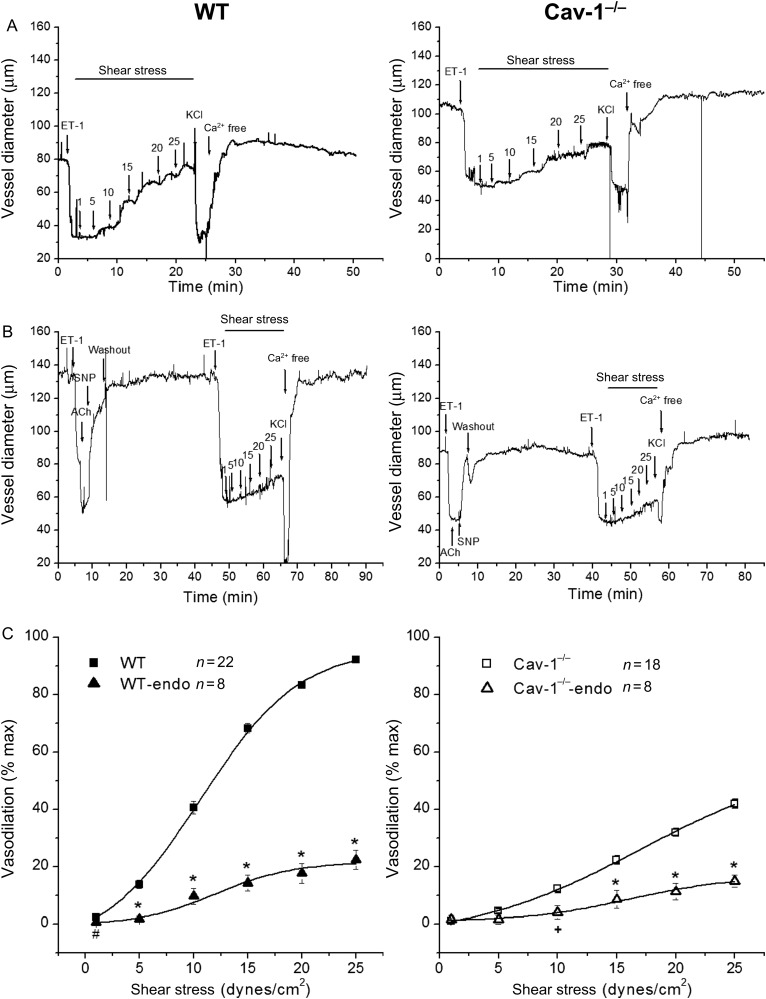
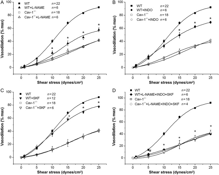
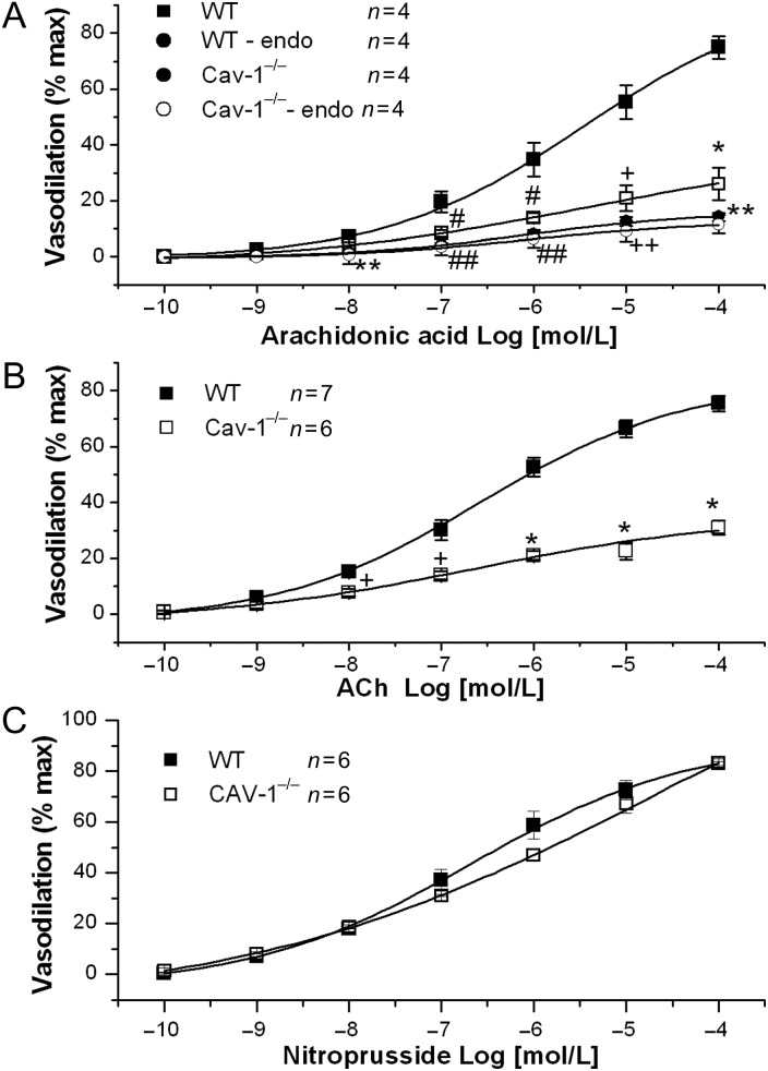
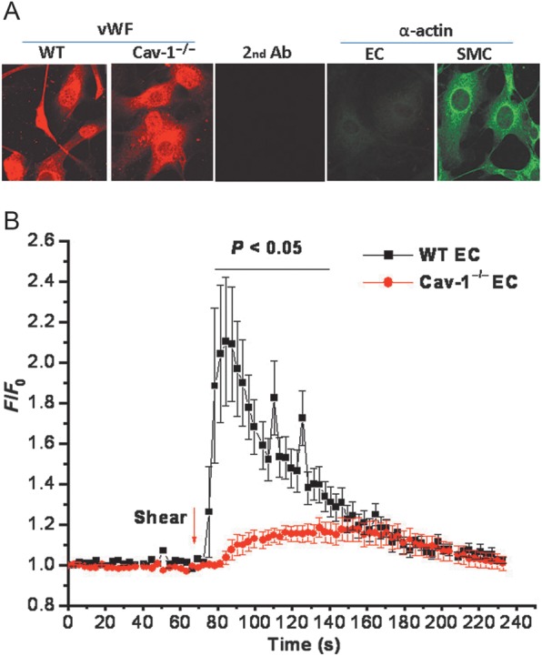
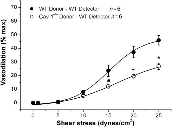
Methods and results: MCA (80-150 μm) isolated from wild-type (WT) and caveolin-1 null (Cav-1(-/-)) mice were subjected to physiological levels of shear stress (1-25 dynes/cm(2)) with and without pre-incubation of inhibitors of nitric oxide synthase (L-NAME), cyclooxygenase (indomethacin, INDO), or cytochrome P450 epoxygenase (SKF 525A). SSD was endothelium-dependent in WT and Cav-1(-/-) coronaries but that in Cav-1(-/-) was significantly diminished compared with WT. Pre-incubation with L-NAME, INDO, or SKF 525A significantly reduced SSD in WT but not in Cav-1(-/-) mice. Vessels from the soluble epoxide hydrolase null (Ephx2(-/-)) mice showed enhanced SSD, which was further augmented by the presence of arachidonic acid. In donor-detector-coupled vessel experiments, Cav-1(-/-) donor vessels produced diminished dilation in WT endothelium-denuded detector vessels compared with WT donor vessels. Shear stress elicited a robust intracellular Ca(2+) increase in vascular endothelial cells isolated from WT but not those from Cav-1(-/-) mice.

Conclusion: Integrity of caveolae is critical for endothelium-dependent SSD in MCA. Cav-1(-/-) endothelium is deficient in shear stress-mediated generation of vasodilators including NO, prostaglandins, and epoxyeicosatrienoic acids. Caveolae plays a critical role in endothelial signal transduction from shear stress to vasodilator production and release.

Keywords: Calcium; Caveolae; Coronary artery; Shear stress; Soluble epoxide hydrolase.


Figures

Figure 1
Figure 1
Role of caveolae and effects of endothelium denudation on SSD. (A) Representative continuous recordings of vessel diameters of isolated coronary arteries from WT (left panel) and Cav-1−/− (right panel) mouse vessels were pre-contracted with ET-1 followed by shear stress-induced dilation. At the end of the experiments, vessels were exposed to 100 mmol/L KCl and then to zero Ca2+ Kreb’ solution. (B) Endothelium-denuded WT (left panel) and Cav-1−/− (right panel) mouse coronary arteries failed to dilate in response to acetylcholine (ACh 1 μmol/L) while the effects of sodium nitroprusside (SNP 100 μmol/L) remained intact. After endothelium removal, shear stress-induced dilation was significantly attenuated in both WT and Cav-1−/− vessels. (C) Group data showing SSD in WT (n = 22) and Cav-1−/− mice (n = 18) and the effects of endothelium denudation. WT-endo and Cav-1−/−-endo are vessels without endothelium, n = 8 for both, #P < 0.05, +P < 0.01, and *P < 0.001 vs. vessels with intact endothelium.
Figure 2
Figure 2
Effects of eNOS, COX, and CYP inhibition on SSD. Vessels were pre-treated with (A) L-NAME (100 μmol/L) for 30 min, with (B) Indomethacin (10 μmol/L) for 30 min, with (C) SKF 525A (10 μmol/L) for 60 min, and with (D) L-NAME + INDO + SKF 525A. #P < 0.05, +P < 0.01, and *P < 0.001 vs. no treatment.
Figure 3
Figure 3
Role of soluble epoxide hydrolase on SSD. (A) In the presence of L-NAME (100 μmol/L) and INDO (10 μmol/L), the same level of shear stress produced greater vasodilation in Ephx2−/− compared with WT vessels (WT n = 8 and Ephx2−/− n = 6, *P < 0.001 vs. WT). After endothelium denudation, SSD was significantly reduced in both Ephx2−/− and WT mice (n = 6 for both Ephx2−/−-Endo and WT–Endo; +P < 0.01 and *P < 0.001 vs. vessels with intact endothelium). In the presence of arachidonic acid (AA 10 μmol/L) in vessels pre-incubated with with L-NAME and INDO, SSD is increased both in WT (n = 10) (B) and in Ephx2−/− vessels (n = 4) (C) #P < 0.05 and *P < 0.001 vs. no AA).
Figure 4
Figure 4
Impaired endothelial function in Cav-1−/− coronary arteries. (A) AA-induced (10−10 to 10−4 mol/L) vasodilation in WT and Cav-1−/− coronary arteries with and without intact endothelium. #P < 0.05, +P < 0.01, and *P < 0.001 for Cav-1−/− vs. WT vessels. ##P < 0.05, ++P < 0.01, and **P < 0.001 for WT vessels denuded of endothelium (WT-endo) vs. WT vessels. (B) ACh-induced (10−10 to 10−4 mol/L) vasodilation in WT and Cav-1−/− coronary arteries. +P < 0.01 and *P < 0.001 for Cav-1−/− vs. WT vessels. (C) Sodium nitroprusside-induced (10−10 to 10−4 mol/L) vasodilation in WT and Cav-1−/− coronary arteries.
Figure 6
Figure 6
Impaired shear stress-induced Ca2+ response in vascular endothelial cells from Cav-1 −/− mice. (A) Characterization of endothelial cells explanted from WT and Cav-1−/− aortas. Endothelial cells were stained with antibodies against the endothelial cell marker, von Willebrand factor (vWF, red), and the smooth muscle cell marker, α-actin (green). (B) Increase in intracellular Ca2+ in response to shear stress (11 dynes/cm2) in fura-2 loaded endothelial cells from WT (n = 7) and Cav-1−/− (n = 8) vessels. Statistical difference between WT and Cav-1−/− cells was indicated by the bar above the tracings with P < 0.05.
Figure 5
Figure 5
Effect of shear stress-induced coronary vasodilation in donor-detector-coupled vessels with WT and Cav-1−/− as donor vessels and endothelium-denuded WT vessels as detectors. SSD in detector vessels with WT donor vessels (closed circle) and Cav-1−/− donor vessels (open circle). #P < 0.05, +P < 0.01, and *P < 0.001 vs. WT donor vessels.

References

    1. Razani B, Woodman SE, Lisanti MP. Caveolae: from cell biology to animal physiology. Pharmacol Rev. 2002;54:431–467. - PubMed
    1. Gratton JP, Bernatchez P, Sessa WC. Caveolae and caveolins in the cardiovascular system. Circulation Research. 2004;94:1408–1417. - PubMed
    1. Patel HH, Murray F, Insel PA. Caveolae as organizers of pharmacologically relevant signal transduction molecules. Annu Rev Pharmacol Toxicol. 2008;48:359–391. - PMC - PubMed
    1. Adebiyi A, Zhao G, Cheranov SY, Ahmed A, Jaggar JH. Caveolin-1 abolishment attenuates the myogenic response in murine cerebral arteries. Am J Physiol. 2007;292:H1584–H1592. - PMC - PubMed
    1. Shaw L, Sweeney MA, O'Neill SC, Jones CJ, Austin C, Taggart MJ. Caveolae and sarcoplasmic reticular coupling in smooth muscle cells of pressurised arteries: the relevance for Ca2+ oscillations and tone. Cardiovasc Res. 2006;69:825–835. - PubMed

Publication types