. 2013 Sep;34(28):6862-70.
doi: 10.1016/j.biomaterials.2013.05.053. Epub 2013 Jun 17.

The effect of lipid monolayer in-plane rigidity on in vivo microbubble circulation persistence

Affiliations

The effect of lipid monolayer in-plane rigidity on in vivo microbubble circulation persistence

Sumit Garg et al. Biomaterials. 2013 Sep.

Abstract

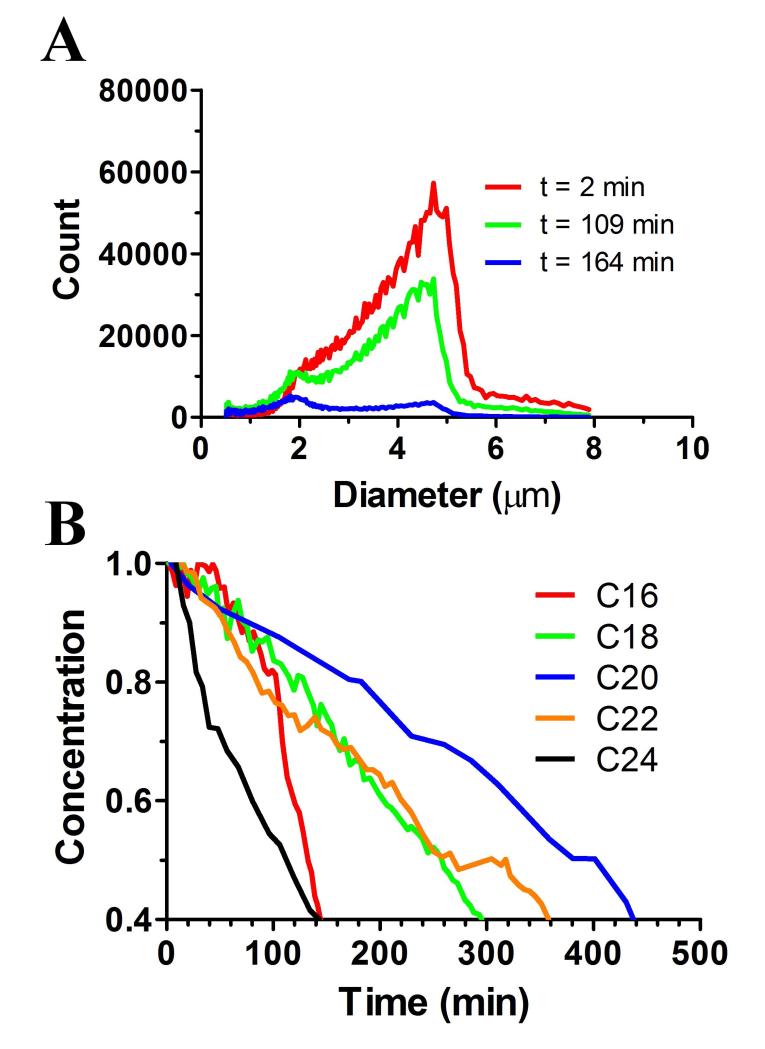
The goal of this study was to increase in vivo microbubble circulation persistence for applications in medical imaging and targeted drug delivery. Our approach was to investigate the effect of lipid monolayer in-plane rigidity to reduce the rate of microbubble dissolution, while holding constant the microbubble size, concentration and surface architecture. We first estimated the impact of acyl chain length of the main diacyl phosphatidylcholine (PC) lipid and inter-lipid distance on the cohesive surface energy and, based on these results, we hypothesized that microbubble stability and in vivo ultrasound contrast persistence would increase monotonically with increasing acyl chain length. We therefore measured microbubble in vitro stability to dilution with and without ultrasound exposure, as well as in vivo ultrasound contrast persistence. All measurements showed a sharp rise in stability between DPPC (C16:0) and DSPC (C18:0), which correlates to the wrinkling transition, signaling the onset of significant surface shear and gas permeation resistance, observed in prior single-bubble dissolution studies. Further evidence for the effect of the wrinkling transition came from an in vitro and in vivo stability comparison of microbubbles coated with pure DPPC with those of lung surfactant extract. Microbubble stability against dilution without ultrasound and in vivo ultrasound contrast persistence showed a monotonic increase with acyl chain length from DSPC to DBPC (C22:0). However, we also observed that stability dropped precipitously for all measurements on further increasing lipid acyl chain length from DBPC to DLiPC (C24:0). This result suggests that hydrophobic mismatch between the main PC lipid and the lipopolymer emulsifier, DSPE-PEG5000, may drive a less stable surface microstructure. Overall, these results support our general hypothesis of the role of in-plane rigidity for increasing the lifetime of microbubble circulation.


Figures

Figure 1
Figure 1
Schematic representation of the two key mechanisms for removal of lipid-coated microbubbles from in vivo circulation. (Top) shows phagocytic removal by macrophages, which is mediated by complement C3b binding to the microbubble surface. (Bottom) shows dissolution into the bloodstream. Rigid lipid monolayers show wrinkling during dissolution owing to enhanced cohesiveness between the lipid constituents.
Figure 2
Figure 2
Inter-lipid interactions. A) Schematic showing van der Waals and hydrophobic forces balancing hydration and steric repulsion forces. B) Attractive van der Waals forces between the lipids were calculated using the London equation, Equation 1. Also shown is the expected area per molecule for lipid-coated microbubbles (A0). The attractive van der Waals force increases with acyl chain length. This effect is more pronounced for monolayers compared to bilayers.
Figure 3
Figure 3
Effect of lipid acyl chain length on microbubble stability to dilution. (A) Representative size distributions of microbubbles coated with DPPC. At time=0, microbubbles were diluted to the concentration of 106 mL−1 in PBS, and the size distribution was tracked as function of time using an Accusizer. (B) Representative microbubble concentration versus time curves for each lipid acyl chain length. Only microbubbles between 0 and 8 μm were counted. As hypothesized, the rate of microbubble dissolution decreased with increasing acyl chain length, but only for DPPC (C16:0) through DBPC (C22:0). Surprisingly, the dissolution rate increased significantly from DBPC to DiLiPC (C24:0).
Figure 4
Figure 4
Effect of lipid acyl chain length on microbubble stability to ultrasound. (A) Schematic of the agarose phantom with a flow channel used to measure the in vitro ultrasound contrast persistence of microbubbles. Ultrasound images are shown of the agarose phantom before and after the injection of microbubbles at concentration of 106 mL−1. (B) Typical average video intensity as function of time for each microbubble encapsulation (n=4). (C) In vitro half-life t1/2 for ultrasound contrast. Half-life was determined by fitting the pharmacokinetic model (Equation 1) to the time-intensity curves. As hypothesized, the rate of microbubble dissolution decreased with increasing acyl chain length, but only for DPPC (C16:0) through DBPC (C22:0). Microbubbles coated with DPPC and DiLiPC were significantly less stable to ultrasound exposure than those coated with the other lipids.
Figure 5
Figure 5
Effect of lipid acyl chain length on in vivo ultrasound contrast persistence. (A) Ultrasound images of the rat kidney in B-mode and CPS mode before and after injection of microbubbles. The red line shows the region of interest (ROI) drawn around kidney (identified from B-mode imaging). (B) Time-intensity curves for ROI plotted as a function of time for each microbubble encapsulation. (C) In vivo half-life t1/2 for ultrasound contrast. Half-life was determined by fitting the pharmacokinetic model (Equation 1) to the time-intensity curves. As hypothesized, the rate of microbubble dissolution decreased with increasing acyl chain length, but only for DPPC (C16:0) through DBPC (C22:0). Microbubbles coated with DPPC and DiLiPC were significantly less stable to in vivo than those coated with the other lipids.
Figure 6
Figure 6
Effect of surfactant proteins on microbubble dissolution. Shown are sequential bright-field microscope images for dissolution of (A) DPPC and (B) Survanta microbubbles. DPPC microbubbles remained spherical, while Survanta microbubbles attained non-spherical, wrinkled shapes during dissolution. Plots show diameter versus time for SF6 microbubbles coated with C) DPPC and D) Survanta after exposure to an air-saturated environment. DPPC shows continuous dissolution, while Survanta shows discontinuous “wrinkling” dissolution. The plots also show the rate of change in area as bars. For DPPC, the rate of change in area increased monotonically as the microbubble shrank. For Survanta, the rate of change in area was intermittent and independent of the microbubble diameter.
Figure 7
Figure 7
Effect of surfactant proteins on ultrasound contrast persistence of microbubbles. Shown are time-intensity curves for (A) in vitro and (B) in vivo CPS imaging. The data were fit to the pharmacokinetic model in Equation 1. Using the fit, the half-life was determined (C) in vitro and (B) in vivo. In both cases, the Survanta microbubbles, which contain surfactant proteins and exhibit wrinkling during dissolution, were significantly more stable than pure DPPC microbubbles.

Similar articles

Cited by

References

    1. Lindner JR. Microbubbles in medical imaging: current applications and future directions. Nat Rev Drug Discov. 2004;3:527–32. - PubMed
    1. Klibanov AL, Hughes MS, Wojdyla JK, Wible JH, Brandenburger GH. Destruction of contrast agent microbubbles in the ultrasound field: the fate of the microbubble shell and the importance of the bubble gas content. Acad Radiol. 2002;9:S41–S5. - PubMed
    1. Unger EC, Porter T, Culp W, Labell R, Matsunaga T, Zutshi R. Therapeutic applications of lipid-coated microbubbles. Adv Drug Deliv Rev. 2004;56:1291–314. - PubMed
    1. Kwan JJ, Borden MA. Lipid monolayer dilatational mechanics during microbubble gas exchange. Soft Matter. 2012;8:4756–66.
    1. Stride E. Physical principles of microbubbles for ultrasound imaging and therapy. Cerebrovasc Dis. 2009;27(Suppl 2):1–13. - PubMed

Publication types