

Review
doi: 10.1038/ijos.2013.36.
Epub 2013 Jun 21.
Biology of tooth replacement in amniotes
Affiliations
- PMID: 23788284
- PMCID: PMC3707075
- DOI: 10.1038/ijos.2013.36
Item in Clipboard
Review
Biology of tooth replacement in amniotes
Int J Oral Sci.
2013 Jun.
Abstract
Tooth replacement is a common trait to most vertebrates, including mammals. Mammals, however, have lost the capacity for continuous tooth renewal seen in most other vertebrates, and typically have only 1-2 generations of teeth. Here, we review the mechanisms of tooth replacement in reptiles and mammals, and discuss in detail the current and historical theories on control of timing and pattern of tooth replacement and development.
Figures
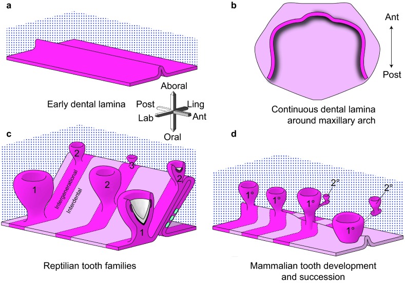
Development of generational teeth in a diphyodont mammal and a polyphyodont reptile. (a, b) The early dental lamina in both reptiles and at least some mammals, including humans, is a continuous invagination of oral epithelium (pink tones) into the dental mesenchyme (blue stippling) that will give rise to the all teeth of the primary dentition. b is redrawn from ref. 6. (c) In reptiles, the dental lamina remains continuous in both the intergenerational (dark pink) and interdental (lighter pink) regions. Tooth families contain several generations of teeth (1, 2, 3) at progressive stages of development. The lingual side or non-tooth forming side of the dental lamina contains the label-retaining putative stem cells (green circles). The successional lamina continues as an extension of the dental lamina off the newest forming tooth. (d) In diphyodont mammals, the dental lamina is continuous and connects adjacent teeth of the primary dentition until the dental lamina degrades in the bell stage, disconnecting the enamel organ from the oral epithelium. The permanent tooth bud is also disconnected from the primary tooth once the primary tooth begins root formation. Three-axis compass rose orients a, c and d; single axis rose orients b.
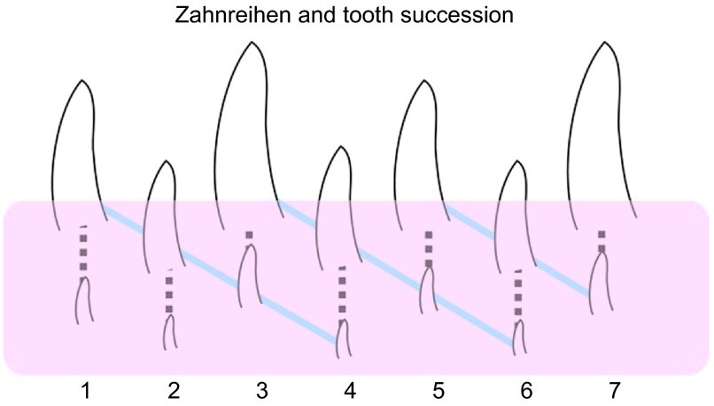
Diagrammatic representation of the first seven tooth families in a hypothetical reptile jaw. Diagonal lines connecting teeth in decreasing stages of development, or Zahnreihen, can be drawn across tooth families (shown in blue), illustrating the apparent ‘waves' of tooth replacement. The dashed line shows succession within a single tooth family. The pink layer indicates the teeth that are not visible in the oral cavity and are still forming. Consequently, Zahnreihen can only be seen on radiographs or in histology.
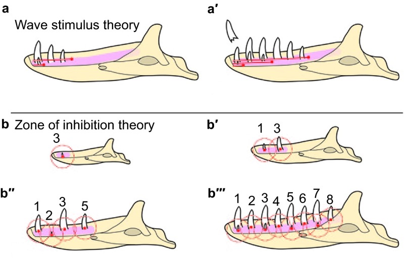
Schematic of the two main theories controlling the pattern of tooth replacement observed in reptiles. (a–a′) Edmund's ‘wave stimulus', an extrinsic chemical signal, originates from near the first tooth position and disperses distally along the odontogenic anlage, inducing the initiation of tooth development as it proceeds. In a, the first wave stimulus has just passed tooth position 3, resulting in the development of the first three teeth. The second wave stimulus has just passed tooth position 1, initiating a second wave of tooth development. In a′, the first two wave stimuli have continued posteriorly, and a third stimulus has just passed the first tooth position, where an erupted tooth has just been shed. (b–b′′′) Osborn's ‘zone of inhibition' theory. In b, the first tooth to develop (future tooth position 3) generates an as-yet-unknown signal inhibiting tooth development (red dot) diffusing out in all directions. As the odontogenic anlage is still small, the secreted molecule inhibits tooth development throughout the entire structure. b′–b′′′ illustrate how growth influences the physical spacing of teeth and thereby the distance between ZOI. ZOI, zone of inhibition.
References
-
- Jernvall J, Thesleff I. Tooth shape formation and tooth renewal: evolving with the same signals. Development. 2012;139 19:3487–3497. - PubMed
-
- Handrigan GR, Richman JM. A network of Wnt, hedgehog and BMP signalling pathways regulates tooth replacement in snakes. Dev Biol. 2010;348 1:130–141. - PubMed
-
- Handrigan GR, Richman JM. Autocrine and paracrine Shh signaling are necessary for tooth morphogenesis, but not tooth replacement in snakes and lizards (Squamata) Dev Biol. 2010;337 1:171–186. - PubMed
-
- Richman JM, Handrigan GR. Reptilian tooth development. Genesis. 2011;49 4:247–260. - PubMed
-
- Järvinen E, Tummers M, Thesleff I. The role of the dental lamina in mammalian tooth replacement. J Exp Zool B Mol Dev Evol. 2009;312 4:281–291. - PubMed
Publication types
MeSH terms
LinkOut - more resources
Full Text Sources
Other Literature Sources
