. 2013 Sep;84(3):393-406.
doi: 10.1124/mol.113.087346. Epub 2013 Jun 20.

α4α6β2* nicotinic acetylcholine receptor activation on ventral tegmental area dopamine neurons is sufficient to stimulate a depolarizing conductance and enhance surface AMPA receptor function

Affiliations

α4α6β2* nicotinic acetylcholine receptor activation on ventral tegmental area dopamine neurons is sufficient to stimulate a depolarizing conductance and enhance surface AMPA receptor function

Staci E Engle et al. Mol Pharmacol. 2013 Sep.

Abstract

Tobacco addiction is a serious threat to public health in the United States and abroad, and development of new therapeutic approaches is a major priority. Nicotine activates and/or desensitizes nicotinic acetylcholine receptors (nAChRs) throughout the brain. nAChRs in ventral tegmental area (VTA) dopamine (DA) neurons are crucial for the rewarding and reinforcing properties of nicotine in rodents, suggesting that they may be key mediators of nicotine's action in humans. However, it is unknown which nAChR subtypes are sufficient to activate these neurons. To test the hypothesis that nAChRs containing α6 subunits are sufficient to activate VTA DA neurons, we studied mice expressing hypersensitive, gain-of-function α6 nAChRs (α6L9'S mice). In voltage-clamp recordings in brain slices from adult mice, 100 nM nicotine was sufficient to elicit inward currents in VTA DA neurons via α6β2* nAChRs. In addition, we found that low concentrations of nicotine could act selectively through α6β2* nAChRs to enhance the function of 2-amino-3-(3-hydroxy-5-methyl-isoxazol-4-yl)propanoic acid (AMPA) receptors on the surface of these cells. In contrast, α6β2* activation did not enhance N-methyl-D-aspartic acid receptor function. Finally, AMPA receptor (AMPAR) function was not similarly enhanced in brain slices from α6L9'S mice lacking α4 nAChR subunits, suggesting that α4α6β2* nAChRs are important for enhancing AMPAR function in VTA DA neurons. Together, these data suggest that activation of α4α6β2* nAChRs in VTA DA neurons is sufficient to support the initiation of cellular changes that play a role in addiction to nicotine. α4α6β2* nAChRs may be a promising target for future smoking cessation pharmacotherapy.


Figures

Fig. 1.
Fig. 1.
Electrophysiological identification of VTA DA neurons. (A) Whole-cell current-clamp recordings of VTA DA neurons show spontaneous (I = 0 pA), pacemaker firing (1–5 Hz), and “sag” responses in the membrane potential in response to hyperpolarizing (I = −120 pA) current injections. (B) VTA DA neurons have wide action potentials. The neuron in (A) indicated with an arrow is shown on an expanded time scale to better view the action potential width (typically 2–5 milliseconds) seen in the neurons under study. (C) Ih currents in VTA DA neurons. VTA cells were held at −60 mV in voltage-clamp mode and membrane current was recorded at baseline and during a voltage step to −120 mV. (D) Single-cell RT-PCR. VTA neurons recorded in whole-cell mode were aspirated into the recording pipette, followed by RT of RNA and subsequent PCR reactions to detect TH and GAPDH (positive control) expression. Expected band sizes are as follows: TH = 207 bp and GAPDH = 138 bp (the asterisk indicates a spurious PCR reaction, possibly generated from external primer pairs). As a negative control, a pipette was lowered into the slice and mild negative pressure was applied. The pipette was removed from the slice and assayed with RT-PCR as for a recorded cell.
Fig. 2.
Fig. 2.
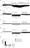
A low concentration of nicotine is sufficient to increase inward currents in VTA DA neurons. (A) α6L9′S or non-Tg control neurons were voltage clamped in whole-cell mode. Inhibitor cocktail [10 μM CNQX, 75 μM picrotoxin, 0.5 μM tetrodotoxin (TTX)] was superfused, followed by nicotine (100 nM) and then αCtxMII (100 nM). A representative experiment from a α6L9′S neuron is shown. Expanded recordings from time points (i), (ii), and (iii) are shown in B–D. (B–D) Voltage-clamp recording segments from α6L9′S (B; 100 nM nicotine), non-Tg (C; 100 nM nicotine), and non-Tg (D; 300 nM nicotine) VTA DA neurons at (i) baseline with inhibitor cocktail present, (ii) inhibitor cocktail plus nicotine, and (iii) inhibitor cocktail/nicotine plus αCtxMII. (E) Summary showing mean holding current (−pA) change from baseline in response to nicotine, and nicotine plus αCtxMII in the indicated mouse strain (α6L9′S and non-Tg littermate). *P < 0.05.
Fig. 3.
Fig. 3.
AMPA-evoked current methodology. (A) A drug-filled pipette is positioned above/next to the cell being recorded. A piezoelectric translator brings the pipette close (20–40 µm) to the cell, a TTL pulse triggers a pressure ejection that dispenses drug (AMPA) onto the cell, and the piezoelectric translator withdraws the pipette away from the cell. (B) Representative recording showing the timing of the TTL pulse, piezo drive movement, and resulting inward current elicited by application of 100 µM AMPA to a VTA DA neuron.
Fig. 4.
Fig. 4.
Activation of α6* nAChRs is sufficient to enhance AMPAR function on the surface of VTA DA neurons. (A) Slice treatment procedure. Brain slices from adult α6L9′S and non-Tg littermate mice were cut, recovered for 60 minutes, and incubated for 60 minutes in control recording solution or recording solution plus nicotine (100 nM). Nicotine was washed out for ≥60 minutes, and whole-cell recordings were established in VTA DA neurons. (B) AMPA currents were evoked by puff-application of AMPA (100 μM) at holding potentials of −60, 0, and +40 mV. Representative recordings from incubation of slices in control and nicotine solutions are shown for α6L9′S and non-Tg littermate mice. (C and D) Summary showing mean AMPA-evoked currents ([AMPA] = 100 μM) in non-Tg littermate (C) and α6L9′S (D) VTA DA neurons in response to control incubation or nicotine incubation at the indicated concentration. The numbers of observations were as follows: non-Tg control (−60 mV, n = 10; 0 mV, n = 7; +40 mV, n = 7); non-Tg 100 nM nicotine (−60 mV, n = 4; 0 mV, n = 4; +40 mV, n = 4), non-Tg 500 nM nicotine (−60 mV, n = 16; 0 mV, n = 12; +40 mV, n = 12), α6L9′S control (−60 mV, n = 14; 0 mV, n = 13; +40 mV, n = 13), and α6L9′S 100 nM nicotine (−60 mV, n = 11; 0 mV, n = 11; +40 mV, n = 11). (E) AMPA concentration-response curve in VTA DA neurons. AMPA-evoked currents were measured in non-Tg and α6L9′S neurons. AMPA concentrations and number of observations at each data point are as follows: non-Tg (1 μM, n = 2; 10 μM, n = 6; 50 μM, n = 5; 100 μM, n = 10; 250 μM, n = 5; 500 μM, n = 14; 1000 μM, n = 11), and α6L9′S (1 μM, n = 2; 10 μM, n = 4; 50 μM, n = 4; 100 μM, n = 14; 250 μM, n = 5; 500 μM, n = 4; 1000 μM, n = 5; 3000 μM, n = 2). Data (mean ± S.E.M.) were fitted to the Hill equation, and the EC50 (±95% confidence interval) for each curve is plotted. (F) AMPA concentration-response curve in α6L9′S VTA DA neurons. AMPA-evoked currents were measured in α6L9′S control slices or slices incubated in 100 nM nicotine for 60 minutes followed by >60 minutes washout prior to recording. Control treated α6L9′S data from (E) are replotted here for reference. AMPA concentrations and number of observations at each data point for α6L9′S slices treated with nicotine are as follows: α6L9′S (1 μM, n = 2; 10 μM, n = 3; 50 μM, n = 2; 100 μM, n = 11; 300 μM, n = 3; 1000 μM, n = 3). Data (mean ± S.E.M.) were fitted to the Hill equation and the EC50 (± 95% confidence interval) for each curve is plotted. *P < 0.05; **P < 0.01.
Fig. 5.
Fig. 5.
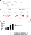
Time dependence for enhancement of AMPA-evoked currents in α6L9′S VTA DA neurons. (A) Slice treatment procedure. Brain slices from α6L9′S mice were cut and recovered for 60 minutes. Slices were then incubated in nicotine (100 nM) for either 10 or 60 minutes, followed in either case by a washout period of ≥60 minutes. Some slices treated with nicotine for 60 minutes were allowed >240 minutes of washout prior to recording. (B) Representative AMPA-evoked currents ([AMPA] = 100 μM) at +40 and −60 mV in VTA DA neurons in response to treatment detailed in (A). (C) Summary showing mean ± S.E.M. AMPA-evoked ([AMPA] = 100 μM) current in α6L9′S VTA DA neurons in response to the conditions described in (A). *P < 0.05.
Fig. 6.
Fig. 6.
Pharmacology of AMPA-evoked current induction in α6L9′S VTA DA neurons. (A) Slice treatment procedure. α6L9′S brain slices were cut and recovered for 60 minutes. Slices were pretreated for 10 minutes with one of the drugs indicated in B, followed by cotreatment with the drug plus nicotine (100 nM) for 60 minutes. Slices were washed out for >60 minutes prior to recording. (B) Representative AMPA-evoked currents ([AMPA] = 100 μM) at +40 and −60 mV in VTA DA neurons from α6L9′S brain slices pre-exposed for 10 minutes to either control recording solution or the following drugs followed by incubation in 100 nM nicotine for 60 minutes: αCtxMII (MII), SCH23390 (SCH), AP-5, and MLA. (C) Summary showing mean ± S.E.M. AMPA-evoked currents ([AMPA] = 100 μM) in α6L9′S VTA DA neurons in response to the conditions described in (A). *P < 0.05; **P < 0.01.
Fig. 7.
Fig. 7.
Enhanced AMPA-evoked currents in α6L9′S VTA DA neurons are mediated, in part, by α4 nAChR subunits. (A) Slice treatment procedure. Brain slices from adult α6L9′S and α6L9′Sα4KO littermate mice were cut, recovered for 60 minutes, and incubated for 60 minutes in control recording solution or recording solution plus nicotine (100 nM). Nicotine was washed out for ≥60 minutes, and whole-cell recordings were established in VTA DA neurons. (B) Representative AMPA-evoked currents ([AMPA] = 100 μM) at +40 and −60 mV in VTA DA neurons from α6L9′S and α6L9′Sα4KO brain slices after incubation in 100 nM nicotine for 60 minutes. (C) Summary showing mean ± S.E.M. AMPA-evoked currents ([AMPA] = 100 μM) in α6L9′S and α6L9′Sα4KO VTA DA neurons in response to the conditions described in A. ***P < 0.001.
Fig. 8.
Fig. 8.
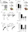
α6* nAChR function is reduced in α4KO mice. (A) Schematic of α6GFP transgenic mice and α6GFP nAChRs. (B) The resulting α6* nAChR that remains after crossing α6GFP mice to α4KO mice is shown. (C) α6* nAChRs were quantified in α6GFP and α6GFPα4KO VTA DA neurons using anti-GFP immunohistochemistry and confocal microscopy. Mean per-cell pixel intensity for each genotype is shown. (D) The resulting α6* nAChR that remains after crossing α6L9′S mice to α4KO mice is shown. (E) Representative ACh-evoked currents in α6L9′S and α6L9′Sα4KO VTA DA neurons. VTA DA neurons from both genotypes were patch clamped and ACh was puff-applied (250 milliseconds) at the indicated concentration. (F) Summary showing mean ± S.E.M. ACh-evoked current in α6L9′S and α6L9′Sα4KO VTA DA neurons in response to the indicated concentration of ACh. (G) Representative nicotine-evoked currents in α6L9′S and α6L9′Sα4KO VTA DA neurons. VTA DA neurons from both genotypes were patch clamped and nicotine was puff-applied at the indicated concentration. (H) Summary showing mean ± S.E.M. nicotine-evoked current in α6L9′S and α6L9′Sα4KO VTA DA neurons in response to the indicated concentration of nicotine. **P < 0.01.
Fig. 9.
Fig. 9.
NMDA-evoked currents are not changed by nicotine in α6L9′S VTA DA neurons. (A) Slice treatment procedure. Brain slices from adult α6L9′S and non-Tg littermate mice were cut, recovered for 60 minutes, and incubated for 60 minutes in control recording solution or recording solution plus nicotine (100 nM). Nicotine was washed out for ≥60 minutes, and whole-cell recordings were established in VTA DA neurons. (B) Representative NMDA-evoked currents ([NMDA] = 100 μM) at +40 mV in VTA DA neurons from α6L9′S and non-Tg littermate brain slices in response to control incubation or incubation in 100 nM nicotine for 60 minutes. (C) Summary showing mean ± S.E.M. NMDA-evoked currents ([NMDA] = 100 μM) in α6L9′S and non-Tg littermate VTA DA neurons in response to the conditions described in A.

References

    1. Azam L, Maskos U, Changeux JP, Dowell CD, Christensen S, De Biasi M, McIntosh JM. (2010) α-Conotoxin BuIA[T5A;P6O]: a novel ligand that discriminates between α6ß4 and α6ß2 nicotinic acetylcholine receptors and blocks nicotine-stimulated norepinephrine release. FASEB J 24:5113–5123 - PMC - PubMed
    1. Baker LK, Mao D, Chi H, Govind AP, Vallejo YF, Iacoviello M, Herrera S, Cortright JJ, Green WN, McGehee DS, et al. (2013) Intermittent nicotine exposure upregulates nAChRs in VTA dopamine neurons and sensitises locomotor responding to the drug. Eur J Neurosci 37:1004–1011 - PMC - PubMed
    1. Bellone C, Lüscher C. (2006) Cocaine triggered AMPA receptor redistribution is reversed in vivo by mGluR-dependent long-term depression. Nat Neurosci 9:636–641 - PubMed
    1. Brown MT, Bellone C, Mameli M, Labouèbe G, Bocklisch C, Balland B, Dahan L, Luján R, Deisseroth K, Lüscher C. (2010) Drug-driven AMPA receptor redistribution mimicked by selective dopamine neuron stimulation. PLoS ONE 5:e15870. - PMC - PubMed
    1. Calabresi P, Lacey MG, North RA. (1989) Nicotinic excitation of rat ventral tegmental neurones in vitro studied by intracellular recording. Br J Pharmacol 98:135–140 - PMC - PubMed

Publication types