. 2013 Aug;19(8):1147-58.
doi: 10.1261/rna.036871.112. Epub 2013 Jun 20.

A novel insight into the mechanism of mammalian selenoprotein synthesis

Affiliations

A novel insight into the mechanism of mammalian selenoprotein synthesis

Olga Kossinova et al. RNA. 2013 Aug.

Abstract

The amino acid selenocysteine is encoded by UGA, usually a stop codon, thus requiring a specialized machinery to enable its incorporation into selenoproteins. The machinery comprises the tRNA(Sec), a 3'-UTR mRNA stem-loop termed SElenoCysteine Insertion Sequence (SECIS), which is mandatory for recoding UGA as a Sec codon, the SECIS Binding Protein 2 (SBP2), and other proteins. Little is known about the molecular mechanism and, in particular, when, where, and how the SECIS and SBP2 contact the ribosome. Previous work by others used the isolated SECIS RNA to address this question. Here, we developed a novel approach using instead engineered minimal selenoprotein mRNAs containing SECIS elements derivatized with photoreactive groups. By cross-linking experiments in rabbit reticulocyte lysate, new information could be gained about the SBP2 and SECIS contacts with components of the translation machinery at various translation steps. In particular, we found that SBP2 was bound only to the SECIS in 48S pre-initiation and 80S pretranslocation complexes. In the complex where the Sec-tRNA(Sec) was accommodated to the A site but transpeptidation was blocked, SBP2 bound the ribosome and possibly the SECIS element as well, and the SECIS had flexible contacts with the 60S ribosomal subunit involving several ribosomal proteins. Altogether, our findings led to broadening our understanding about the unique mechanism of selenocysteine incorporation in mammals.

Keywords: SECIS-binding protein 2; cross-linking approach; mammalian ribosome; selenocysteine incorporation; selenocysteine insertion sequence.


Figures

FIGURE 1.
FIGURE 1.
Schematic representation of the minimal mRNAs and their photoreactive derivatives used in this work. (A) Minimal mRNAs with WT GPx1 SECIS: flSec mRNA, mRNA carrying UGA Sec codon, and flPhe mRNA, mRNA carrying a Phe codon instead of UGA. SECIS-element of Mut1 mRNA contains nucleotide residues shown in the box instead of residues shown in brackets. (B) Minimal selenoprotein mRNA containing photoreactive groups: 4-thiouridine residues (s4U) or aminoallyl-containing uridine residues bearing perfluorophenylazido group (N3R-aaU, where R is perfluorophenyl residue). (C) Schematic description of the synthesis of N3R-aaUTP used as a substrate to obtain N3R-aaU-containing SECIS.
FIGURE 2.
FIGURE 2.
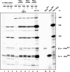
Ribosome binding abilities of the minimal selenoprotein mRNA. (A) Schematic representation of the complexes obtained from RRL with various translation inhibitors (concentration used is indicated). E, P, and A, tRNA binding sites. Initiation factors are displayed on the 48S initiation complex. Charged tRNAs in 48S and 80S-I complexes as well as deacylated tRNA and dipeptidyl tRNA in 80S-II complex are shown. (B) flSec mRNA (closed circles), flPhe mRNA (closed squares), s4U-containing flSec mRNA (s4U-flSec mRNA, open circles), and s4U-containing flPhe mRNA (s4U-flPhe mRNA, open squares) binding to ribosomes in RRL. Note that the concentration of the ribosomes in the RRL-based mixtures (50% of RRL v/v) was ∼0.05 μM. The data are the average of at least three independent experiments. The relative error was ∼10%. (C) Toe-printing assay of 48S and 80S complexes formed in RRL with flPhe mRNA (lanes 14) and flSec mRNA (lanes 58) in the presence of SBP2. Lanes 1 and 5: 48S complex; lanes 2 and 6: 80S-I complex; lanes 3 and 7: 80S-II complex; translation inhibitors were omitted in lanes 4 and 8. (A,C,G,T) Sequencing lanes, (ctrl) primer extension with no ddNTP added. Arrows indicate the bands corresponding to toe-print signals. The two weak bands below the toe-prints in lanes 2 and 6 are assigned to signals corresponding to the post-translocational complexes that could be formed in insignificant amounts because of incomplete immobilization of ribosomes by anisomycin (by analogy with Kozak [1998] where lower concentrations of anisomycin were used). The strong signal in the upper part of the gel in the flPhe RNA toe-printing lanes may reflect partial degradation of the flPhe mRNA since it is present in all lanes with flPhe mRNA.
FIGURE 3.
FIGURE 3.
A-site occupancy by the tRNASec. Analysis was carried out on Cu2+-treated and 3′ end [32P]-labeled RNAs isolated from 48S and 80S complexes assembled in RRL on the endogenous (lanes 13) or flSec mRNAs in the presence (lanes 4, 5, 8, 9) or absence (lane 6) of SBP2, or from the 80S-I complex formed on the Mut1 mRNA (lane 7). Lanes 1 and 4: 48S complex; lanes 2, 57: 80S-I complex; lanes 3, 8, 9 (the sample was preliminarily treated at pH 9.0): 80S-II complex (the weak bands observed in lane 9 but not in the other lanes could result from slight RNA hydrolysis at pH 9.0); lane 10: T7 tRNASec transcript (93 nt; it contains three additional Gs at the 5′ end compared to the authentic tRNASec); lane 11: tRNAMet (76 nt); lane 12: total RNA isolated from 60S subunits (5.8S rRNA, 160 nt; 5S rRNA, 120 nt). Arrows indicate positions of the tRNAMet and tRNASec. The band just above the tRNASec observed in lanes 2, 3 and 59 might correspond to partial hydrolysis of the 28S rRNA. The figure displays the autoradiogram corresponding to the part of the gel where RNA fragments with a length lower than 250 nt were resolved.
FIGURE 4.
FIGURE 4.
SBP2 content in ribosomal complexes formed in RRL. (A) Minimal mRNAs used in the experiments. (B) The presence of SBP2 in complexes was shown by Western blotting with anti-SBP2 antibodies. Lanes 2, 3, 4, and 6, complexes with flSec mRNA: lane 2, 48S pre-initiation complex; lane 3, pretranspeptidation complex 80S-I; lanes 4 and 6, pretranslocation complex 80S-II; lane 7, 80S-II complex formed with the 5′ Phe mRNA; lane 8, 80S ribosomes isolated from RRL in the presence of emetine; lane 10, 80S•SBP2·complex formed in the presence of 1 mM emetine; lane 11, 80S•SBP2·complex formed without emetine; lanes 1, 5, and 9, controls containing purified recombinant SBP2. The amount of SBP2 detected in the complexes is indicated below the panel; the amount of SBP2 detected in the 80S-I complex was taken as 100%. (C) flSec mRNA WT and Mut1 mRNA binding to ribosomes in the presence of 5 mM anisomycin (80S-I complex). The relative error in determining the binding extent of these mRNAs with ribosomes was ∼10%. The data are the average of at least three independent experiments. The presence of SBP2 in 80S-I complexes was assayed by Western blotting with anti-SBP2 antibodies (shown in the lower panel).
FIGURE 5.
FIGURE 5.
Cross-linking of minimal s4U-containing mRNAs in RRL. (A) The presence of SBP2 in complexes formed with the flSec mRNA shown by Western blotting. Lane 1: purified recombinant SBP2 as the control; lane 2: 48S complex; lane 3: 80S-I complex; lane 4: 80S-II complex. (B) Autoradiograms of the SDS-PAGE of the proteins cross-linked with the uniformly 32P-labeled s4U-containing SECIS of flSec mRNA (lanes 18 and 1518) or flPhe mRNA (lanes 1114 and 1922), in the presence (+) or absence (−) of SBP2 and UV light. Exposure time: 12 h for the 48S and 80S-II complexes, 48 h for the 80S-I complex. (+ RNase A or + Prot K) Complexes treated with RNase A or proteinase K, respectively. Lanes SBP2, L30 (ribosomal protein L30), TP80, TP60, and TP40 (total proteins from 80S ribosomes, 60S, and 40S subunits, respectively) were Coomassie-stained. (C) Validation of SBP2 cross-links obtained in 48S and 80S-II complexes by immunoprecipitation using anti-SBP2 antibodies. Bars 1: immunoprecipitation of the total proteins from the irradiated ribosomal complexes separated from unbound components; bars 2: same as in bars 1 but with beads lacking antibodies; bars 3: immunoprecipitation of the total proteins from the irradiated ribosome complexes obtained with flPhe mRNA but without SBP2. The data represent the mean and standard deviation of three independent experiments.
FIGURE 6.
FIGURE 6.
Sucrose gradient sedimentation profile of the 60S and 40S ribosomal subunits isolated from the irradiated 80S complex formed in RRL in the presence of 5 mM anisomycin with flSec mRNA bearing SECIS derivatized with either (A) statistically distributed s4Us or (B) randomly inserted N3R-aaUs. Solid line, A260; dashed line, radioactivity corresponding to irradiated (open circles) and nonirradiated (closed circles) complexes. The relative error in determining the amount of radioactivity in the fractions was ∼10%; the data are the average of the three independent experiments.

References

    1. Allmang C, Carbon P, Krol A 2002. The SBP2 and 15.5 kD/Snu13p proteins share the same RNA binding domain: Identification of SBP2 amino acids important to SECIS RNA binding. RNA 8: 1308–1318 - PMC - PubMed
    1. Allmang C, Wurth L, Krol A 2009. The selenium to selenoproteins pathway in eukaryotes: More molecular pathways than anticipated. Biochim Biophys Acta 1790: 1415–1423 - PubMed
    1. Anthony DD, Merrick WC 1992. Analysis of 40S and 80S complexes with mRNA as measured by sucrose density gradients and primer extension inhibition. J Biol Chem 267: 1554–1562 - PubMed
    1. Babaylova E, Graifer D, Malygin A, Stahl J, Shatsky I, Karpova G 2009. Positioning of subdomain IIId and apical loop of domain II of the hepatitis C IRES on the human 40S ribosome. Nucleic Acids Res 37: 2126–2141 - PMC - PubMed
    1. Böck A, Rother M, Leibundgut M, Ban N 2006. Selenium metabolism in prokaryotes. In Selenium: Its molecular biology and role in human health, 2nd ed. (ed. Hatfield DL, et al.), pp. 9–28 Springer Verlag, New York

Publication types

MeSH terms

LinkOut - more resources