. 2013 Aug;33(8):1911-9.
doi: 10.1161/ATVBAHA.113.301591. Epub 2013 Jun 20.

Mitochondrial oxidative stress corrupts coronary collateral growth by activating adenosine monophosphate activated kinase-α signaling

Affiliations

Mitochondrial oxidative stress corrupts coronary collateral growth by activating adenosine monophosphate activated kinase-α signaling

Yuh Fen Pung et al. Arterioscler Thromb Vasc Biol. 2013 Aug.

Abstract

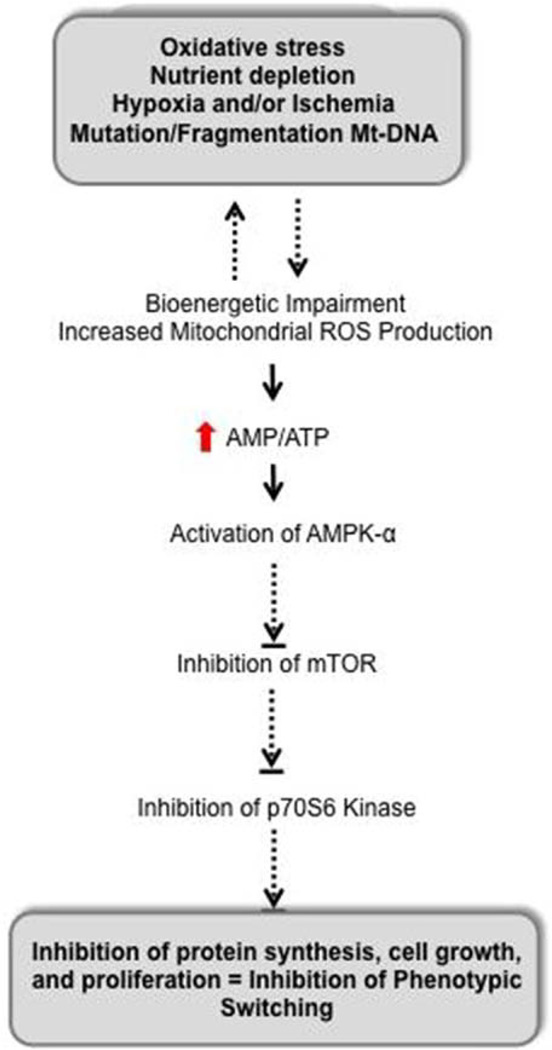
Objective: Our goal was to determine the mechanism by which mitochondrial oxidative stress impairs collateral growth in the heart.

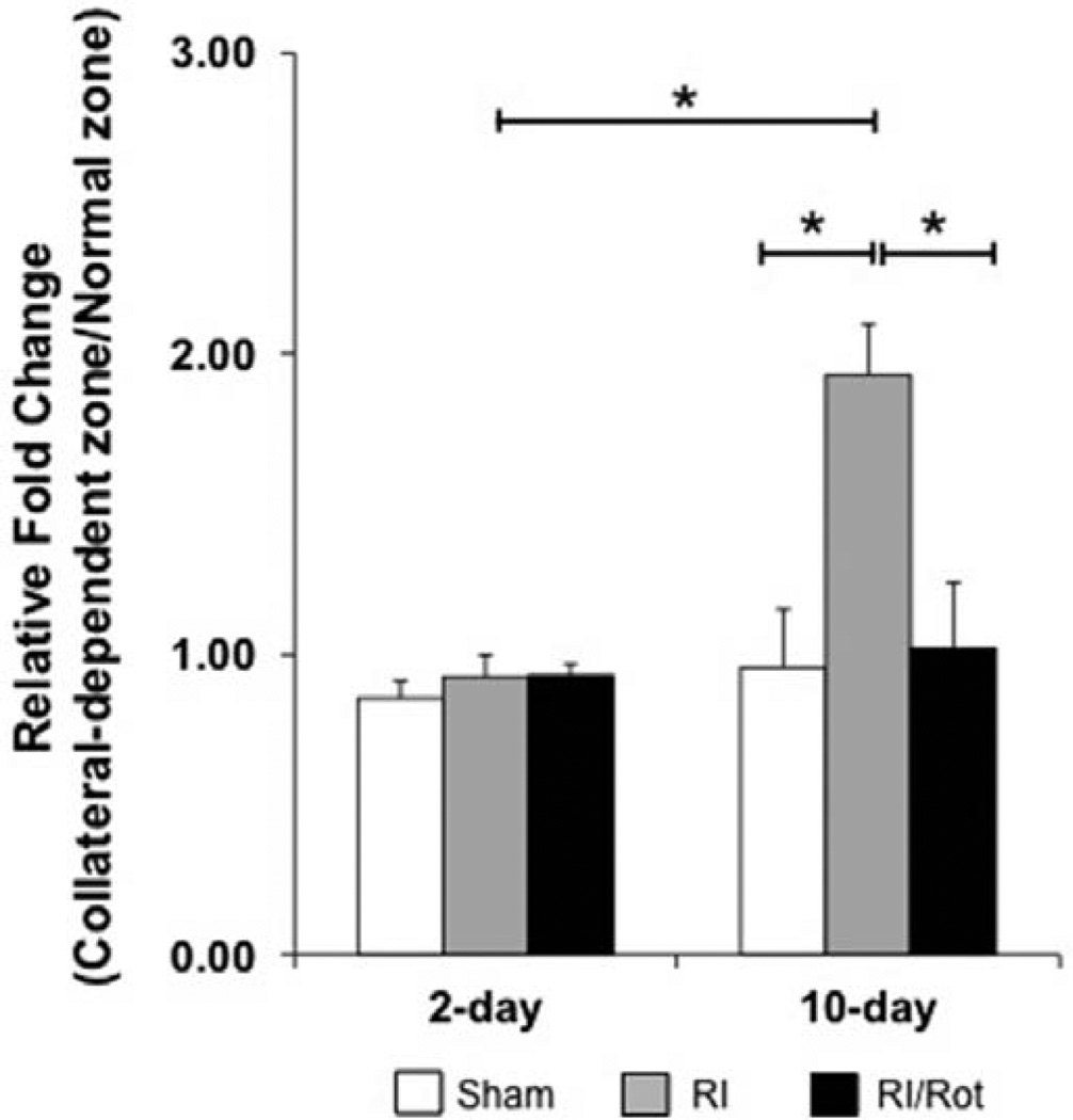
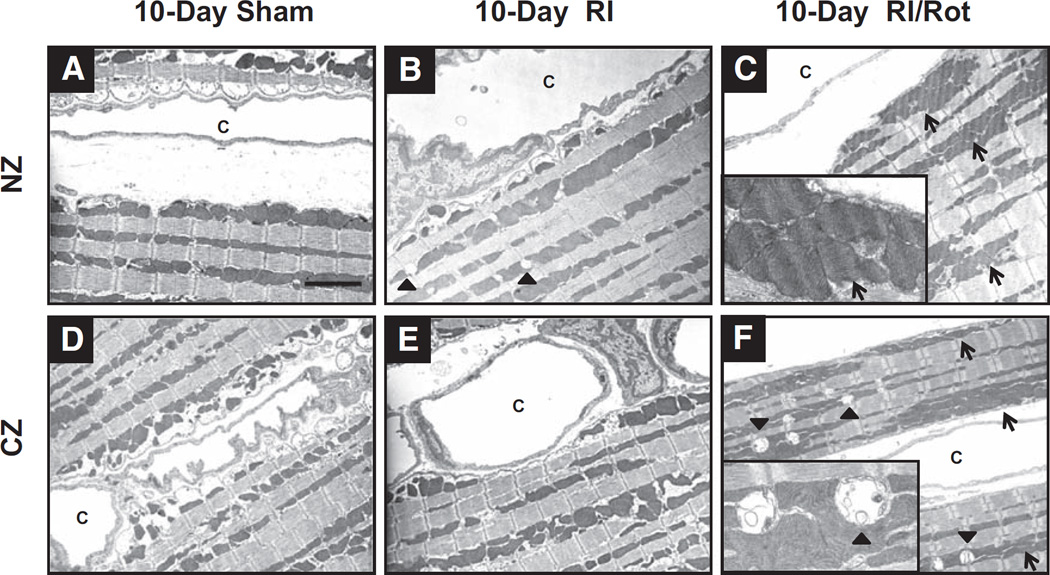
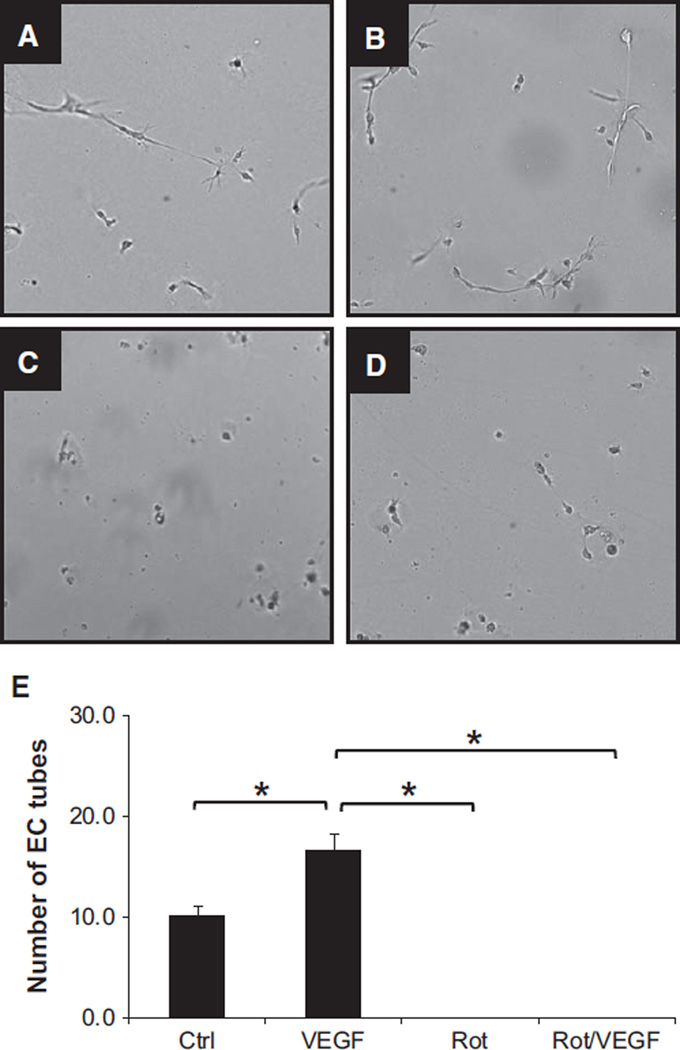
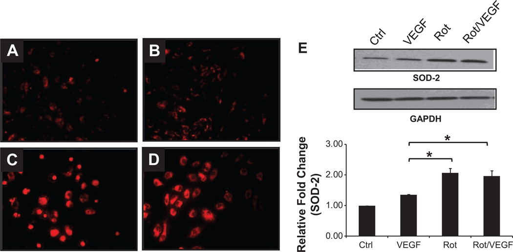
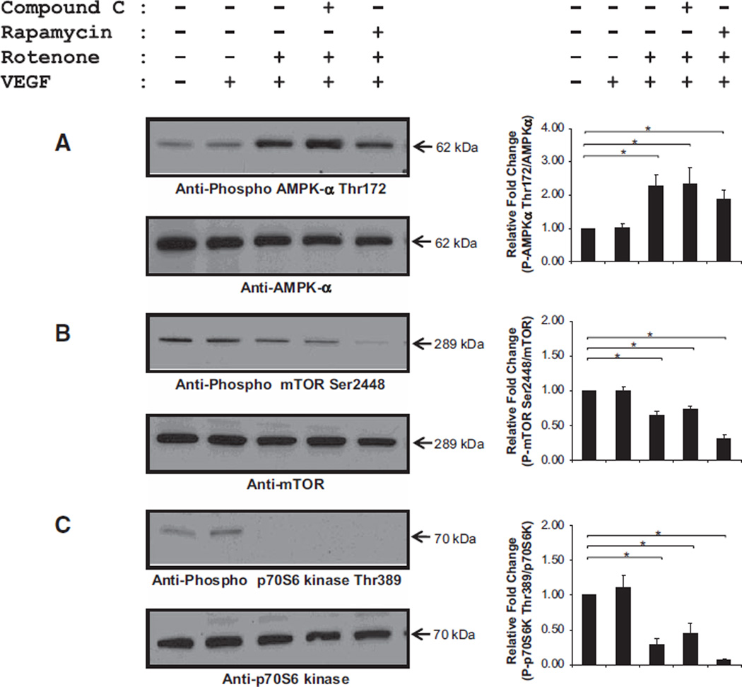
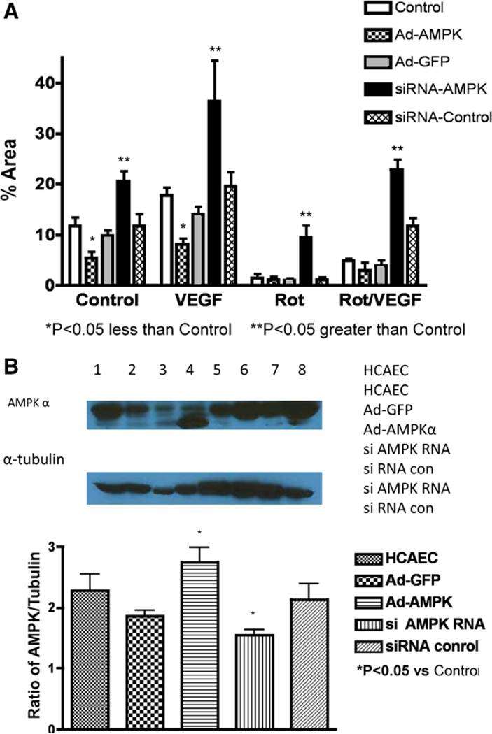
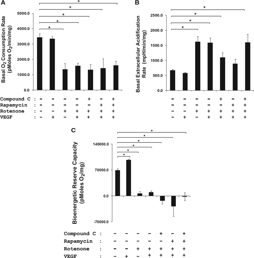
Approach and results: Rats were treated with rotenone (mitochondrial complex I inhibitor that increases reactive oxygen species production) or sham-treated with vehicle and subjected to repetitive ischemia protocol for 10 days to induce coronary collateral growth. In control rats, repetitive ischemia increased flow to the collateral-dependent zone; however, rotenone treatment prevented this increase suggesting that mitochondrial oxidative stress compromises coronary collateral growth. In addition, rotenone also attenuated mitochondrial complex I activity and led to excessive mitochondrial aggregation. To further understand the mechanistic pathway(s) involved, human coronary artery endothelial cells were treated with 50 ng/mL vascular endothelial growth factor, 1 µmol/L rotenone, and rotenone/vascular endothelial growth factor for 48 hours. Vascular endothelial growth factor induced robust tube formation; however, rotenone completely inhibited this effect (P<0.05 rotenone versus vascular endothelial growth factor treatment). Inhibition of tube formation by rotenone was also associated with significant increase in mitochondrial superoxide generation. Immunoblot analyses of human coronary artery endothelial cells with rotenone treatment showed significant activation of adenosine monophosphate activated kinase (AMPK)-α and inhibition of mammalian target of rapamycin and p70 ribosomal S6 kinase. Activation of AMPK-α suggested impairments in energy production, which was reflected by decrease in O2 consumption and bioenergetic reserve capacity of cultured cells. Knockdown of AMPK-α (siRNA) also preserved tube formation during rotenone, suggesting the negative effects were mediated by the activation of AMPK-α. Conversely, expression of a constitutively active AMPK-α blocked tube formation.

Conclusions: We conclude that activation of AMPK-α during mitochondrial oxidative stress inhibits mammalian target of rapamycin signaling, which impairs phenotypic switching necessary for the growth of blood vessels.

Keywords: collateral circulation; coronary circulation; mitochondria; reactive oxygen species.


Figures

Figure 1
Figure 1
Mitochondrial oxidative stress and dysfunction compromise coronary collateral growth in lean rats in response to repetitive ischemia. Wistar Kyoto rats were euthanized at days 2 and 10 and further divided into 3 groups at each time point: sham, repetitive ischemia (RI), and RI/rotenone (Rot). At day 2, the fold change in the collateral-dependent zone (CZ)/normal zone (NZ) blood flow ratio (vs day 0) is unchanged from the basal time point before start of the RI protocol, implying no obvious growth of collaterals (n=5, 4, and 2 rats, respectively, for sham, RI, and RI/Rot groups). After 10 days of RI protocol, the fold increase in the ratio of blood flow compared with day 0 in the CZ/NZ was nearly doubled in the RI group as compared with the sham-operated rats (≈92% increase 10-day RI vs 10-day sham; *P<0.05; n=8 and 6 rats, respectively). Rot prevented this increase in the flow ratio in response to repetitive ischemia.
Figure 2
Figure 2
Mitochondrial oxidative stress and dysfunction induce mitochondrial aggregation and dyad swelling in the myocardium of lean rats in response to repetitive ischemia. Representative electron micrographs from 10-day sham (A, normal zone [NZ]; D, collateral-dependent zone [CZ]); 10-day RI (B, NZ; E, CZ); and 10-day repetitive ischemia (RI)/rotenone (Rot; C, NZ; F, CZ) rats, respectively (n=3 rats per group). Sham-operated rats showed well-preserved intracellular architectures (A and D). Rot induced mitochondrial aggregation (open arrow), indicating mitochondrial network fusion (C and F). This fusion process might be a mechanism for mitochondria to complement impair in function/bioenergetics for survival. It is also important to note that severe dyad swelling (solid arrow) was observed in Rot-treated rats, suggesting alteration in Ca2+ homeostasis in the myocardium (magnification, ×3000; insets, ×10 000). c indicates capillaries.
Figure 3
Figure 3
Mitochondrial oxidative stress and dysfunction inhibit tube formation on 2D Matrigel in human coronary artery endothelial cells (HCAECs). HCAECs were treated with vehicle (A), vascular endothelial growth factor (VEGF; 50 ng/mL; B), rotenone (Rot; 1 µmol/L; C), and Rot/VEGF (D). Bar graph summarizing the number of tubes formed under each condition is shown in E. Although VEGF induced robust formation of tubes on 2D Matrigel, Rot inhibited this process in the presence or absence of VEGF (*P<0.05; n=4; 8–12 wells/condition), implying that mitochondrial bioenergetics is the upstream regulator of growth factor–mediated signaling cascade.
Figure 4
Figure 4
Mitochondrial oxidative stress in human coronary artery endothelial cells (HCAECs). HCAECs were treated with vehicle (A), vascular endothelial growth factor (VEGF; 50 ng/mL; B), rotenone (Rot; 1 µmol/L; C), and Rot/VEGF (D). Rot increases mitochondrial superoxide production as shown by the higher fluorescence intensity of MitoSox Red (A and B vs C and D; n=3). Superoxide dismutase (SOD)-2 expression, normalized against GAPDH, was significantly increased in Rot-treated cells (E; n=3; *P<0.05), suggesting upregulation of this antioxidant as compensation to the elevated mitochondrial oxidative stress.
Figure 5
Figure 5
Impaired growth factor–mediated signaling cascade induced by mitochondrial oxidative stress and dysfunction. Human coronary artery endothelial cells were treated with vehicle, vascular endothelial growth factor (VEGF; 50 ng/mL), rotenone (Rot; 1 µmol/L), and Rot/VEGF, respectively. Rot significantly increased activation of adenosine monophosphate-activated kinase (AMPK)-α refecting energetic limitations (A). Activation of AMPK-α negatively regulates its downstream effectors, mammalian target of rapamycin (mTOR; B), and p70 ribosomal S6 (p70S6; C), which are essential for protein synthesis (n=5–9; *P<0.05).
Figure 6
Figure 6
Effects of AMPK overexpression and knockdown on endothelial tube formation. A, Tube formation by endothelial cells in Matrigel under control conditions, administration of vascular endothelial growth factor (VEGF), rotenone (Rot), and VEGF+Rot. Under these conditions cells were studied without treatment, after transduction of an adenovirus expressing constitutively active AMPK, the adenovirus-expressing green fluorescent protein (GFP), an siRNA to knockdown AMPK, and a scrambled control siRNA. Knockdown of AMPK increased tube formation during Rot, VEGF, and VEGF+Rot. In contrast, overexpression of the active AMPK decreased tube formation during VEGF. The scrambled siRNA and the adenovirus-expressing GFP did not have significant effects. B, AMPK and mutant AMPK protein levels. Knockdown by the siRNA decreased AMPK protein levels but the scrambled siRNA did not have an effect. Transduction with the active mutant resulted in protein levels slightly elevated compared with control levels. The adenovirus-expressing GFP did not have significant effects on AMPK protein levels. HCAECs indicates human coronary artery endothelial cells.
Figure 7
Figure 7
A, Mitochondrial dysfunction in human coronary artery endothelial cells (HCAECs). Mitochondria are the major oxygen consumer, as they consume ≈90% of O2 in the cells. Therefore, oxygen consumption rate (OCR) and extracellular acidification rate (ECAR) are direct representations of mitochondrial function. Basal OCR in pmol/min per milligram protein (A) and ECAR in millipH/min per milligram protein (B) were measured for 30 minutes (n=4; 2–4 wells/condition). Rotenone (Rot; 1 µmol/L) significantly blunted OCR and increased ECAR, as compared with control HCAECs (≈75%), indicating severe mitochondrial dysfunction (*P<0.05). C, Mitochondrial dysfunction and its effect on bioenergetic reserve capacity in HCAECs. Mitochondrial electron transport chain inhibitors, namely oligomycin (1 µg/mL), carbonylcyanide-p-trifuoromethoxyphenylhydrazone (3 µmol/L), and antimycin A (10 µmol/L), were injected sequentially. The reserve capacity was calculated by subtracting the maximal rate of oxygen consumption by the preoligomycin rate. Rot completely attenuated the bioenergetic reserve capacity (n=4; 2–4 wells/condition; *P<0.05). Neither compound C, rapamycin, nor vascular endothelial growth factor (VEGF) reversed the inhibitory effects of Rot.
Figure 8
Figure 8
Transduction of mitochondrial oxidative stress and dysfunction into impaired vascular growth. AMPK-α is a prototypical sensor of energy homeostasis, which is activated when cellular levels of AMP increase and ATP decreases. Activated AMPK then negatively regulates mammalian target of rapamycin (mTOR), which controls energy-demanding cellular processes, for example, protein synthesis, required for cell growth and proliferation. Thus, inhibition of mTOR signaling inhibits tube formation in human coronary artery endothelial cells, and we would also project that mitochondrial dysfunction in vivo would inhibit coronary collateral growth through similar mechanisms. Dephosphorylation/inactivation of p70S6 kinase, even in the presence of growth factor (ie, vascular endothelial growth factor), indicates that impaired mitochondrial bioenergetics would impair new protein synthesis. Thus, proper bioenergetics is critical to control interactions between phenotypic switching that requires new protein synthesis leading to vascular growth and cellular metabolism.

References

    1. Bugger H, Abel ED. Molecular mechanisms for myocardial mitochondrial dysfunction in the metabolic syndrome. Clin Sci (Lond) 2008;114:195–210. - PubMed
    1. Magliano DJ, Shaw JE, Zimmet PZ. How to best define the metabolic syndrome. Ann Med. 2006;38:34–41. - PubMed
    1. Zimmet P, Magliano D, Matsuzawa Y, Alberti G, Shaw J. The metabolic syndrome: a global public health problem and a new definition. J Atheroscler Thromb. 2005;12:295–300. - PubMed
    1. Ford ES, Giles WH, Dietz WH. Prevalence of the metabolic syndrome among US adults: findings from the third National Health and Nutrition Examination Survey. JAMA. 2002;287:356–359. - PubMed
    1. Abaci A, Oğuzhan A, Kahraman S, Eryol NK, Unal S, Arinç H, Ergin A. Effect of diabetes mellitus on formation of coronary collateral vessels. Circulation. 1999;99:2239–2242. - PubMed

Publication types

MeSH terms

LinkOut - more resources