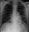
Bilateral pneumothoraces following a right subclavian catheter insertion after thymectomy for a patient with a myasthenic crisis
- PMID: 23789013
- PMCID: PMC3684336
Bilateral pneumothoraces following a right subclavian catheter insertion after thymectomy for a patient with a myasthenic crisis
Abstract
Background: Myasthenia gravis (MG) is an autoimmune disease involving the formation of antibodies against the nicotinic acetylcholine receptors. Thymectomy is the treatment in MG patients with thymoma. We report a case of an MG patient who developed postthymectomy bilateral pneumothoraces after the placement of a subclavian central venous catheter.
Case report: The 21-year-old patient with MG underwent a thymectomy and, in a later admission, complained of myasthenic crisis symptoms. He was scheduled to receive plasma exchange therapy and electromyography the following day. Plasmapheresis was initiated after the placement of a right subclavian dialysis catheter. Postinsertion chest x-ray revealed bilateral pneumothoraces after a single unilateral attempt to cannulate the right subclavian vein. A right thoracotomy tube was placed with interval resolution of the bilateral pneumothoraces.
Conclusion: The development of bilateral pneumothoraces in this case was attributed to the possible accidental communication between the 2 pleural spaces, which rarely happens during thymectomy surgery.
Keywords: Central venous catheters; chest tubes; myasthenia gravis; pneumothorax.
Conflict of interest statement
Figures
Similar articles
-
[A case of radiation-related pneumonia and bilateral tension pneumothorax after extended thymectomy and adjuvant radiation for thymoma with myasthenia gravis].Nihon Kokyuki Gakkai Zasshi. 2010 Aug;48(8):584-8. Nihon Kokyuki Gakkai Zasshi. 2010. PMID: 20803975 Japanese.
-
Bilateral pneumothoraces following central venous cannulation.Case Rep Med. 2009;2009:745713. doi: 10.1155/2009/745713. Epub 2009 Nov 5. Case Rep Med. 2009. PMID: 19901997 Free PMC article.
-
Chemotherapy-induced myasthenic crisis in thymoma treated with primary chemotherapy with curative intent on mechanical ventilation: a case report and review of the literature.J Med Case Rep. 2021 Feb 2;15(1):32. doi: 10.1186/s13256-020-02601-8. J Med Case Rep. 2021. PMID: 33526108 Free PMC article. Review.
-
Thymectomy in myasthenia gravis: proposal for a predictive score of postoperative myasthenic crisis.Eur J Cardiothorac Surg. 2014 Apr;45(4):e76-88; discussion e88. doi: 10.1093/ejcts/ezt641. Epub 2014 Feb 12. Eur J Cardiothorac Surg. 2014. PMID: 24525106
-
Treatment of myasthenia gravis with dropped head: a report of 2 cases and review of the literature.Neuromuscul Disord. 2015 May;25(5):429-31. doi: 10.1016/j.nmd.2015.01.014. Epub 2015 Feb 7. Neuromuscul Disord. 2015. PMID: 25747003 Review.
Cited by
-
The Legend of the Buffalo Chest.Chest. 2021 Dec;160(6):2275-2282. doi: 10.1016/j.chest.2021.06.043. Epub 2021 Jun 30. Chest. 2021. PMID: 34216606 Free PMC article.
-
Bilateral pneumothorax after pacemaker placement "Buffalo chest".Respir Med Case Rep. 2019 Jan 29;26:227-228. doi: 10.1016/j.rmcr.2019.01.022. eCollection 2019. Respir Med Case Rep. 2019. PMID: 30740301 Free PMC article.
-
A case of bilateral pneumothorax following computer-tomography guided transthoracic biopsy in a woman with suspected pulmonary cancer.Respirol Case Rep. 2023 Jul 17;11(8):e01157. doi: 10.1002/rcr2.1157. eCollection 2023 Aug. Respirol Case Rep. 2023. PMID: 37469569 Free PMC article.
References
-
- Palace J, Vincent A, Beeson D. Myasthenia gravis: diagnostic and management dilemmas. Curr Opin Neurol. 2001 Oct;14(5):583–589. - PubMed
-
- Kondo K. Optimal therapy for thymoma. J Med Invest. 2008 Feb;55((1-2)):17–28. - PubMed
-
- National Institute for Health and Clinical Excellence. Technology Appraisal: The Clinical Effectiveness and Cost Effectiveness of Ultrasonic Locating Devices for the Placement of Central Venous Lines. September 2002. http://www.nice.nhs.uk/guidance/index.jsp?action=byID&o=11474. Accessed February 14, 2013.
-
- Richman DP, Agius MA. Treatment of autoimmune myasthenia gravis. Neurology. 2003 Dec 23;61(12):1652–1661. - PubMed
-
- Jaretzki A, Steinglass KM, Sonett JR. Thymectomy in the management of myasthenia gravis. Semin Neurol. 2004 Mar;24(1):49–62. - PubMed
Publication types
LinkOut - more resources
Full Text Sources

