Review
. 2013 Jul;201(1):W29-39.
doi: 10.2214/AJR.12.9956.

Groove pancreatitis: spectrum of imaging findings and radiology-pathology correlation

Affiliations
Review

Groove pancreatitis: spectrum of imaging findings and radiology-pathology correlation

Siva P Raman et al. AJR Am J Roentgenol. 2013 Jul.

Abstract

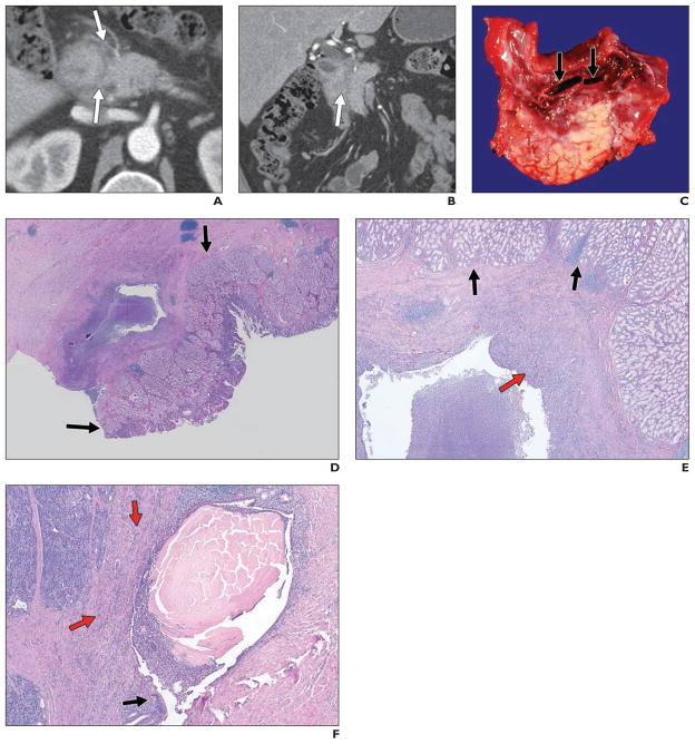
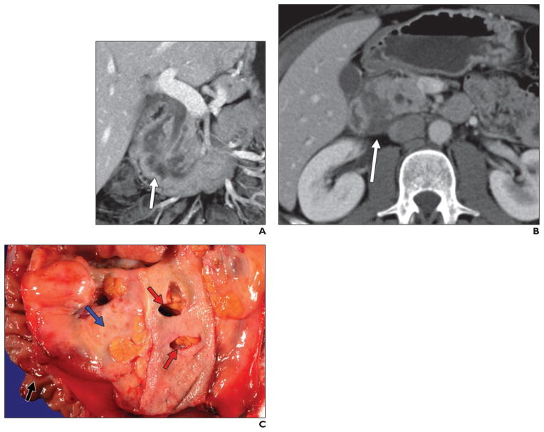
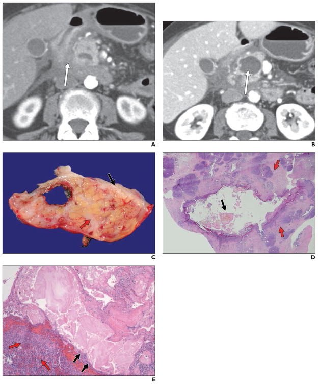
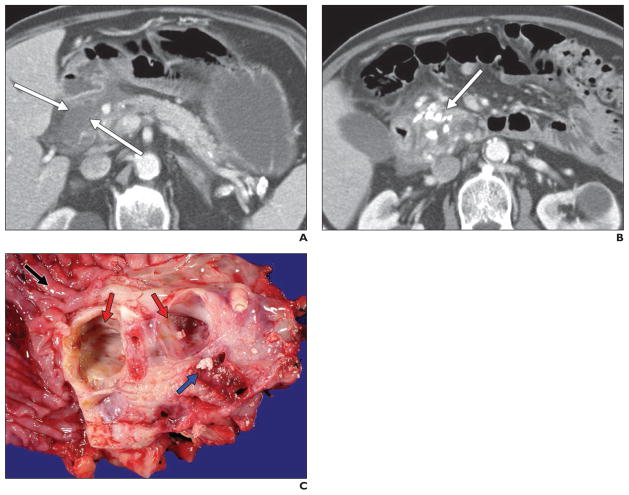
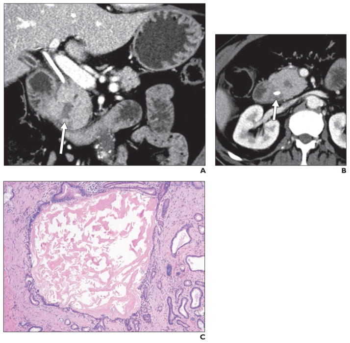
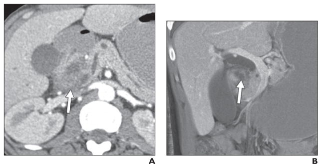
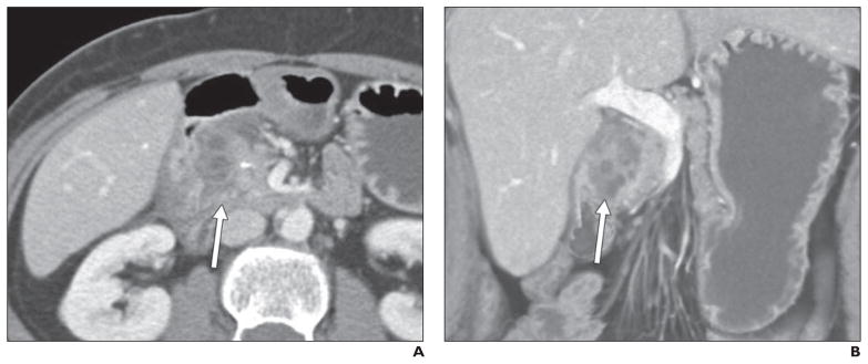
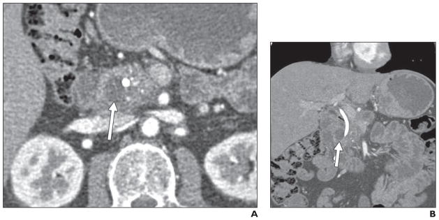
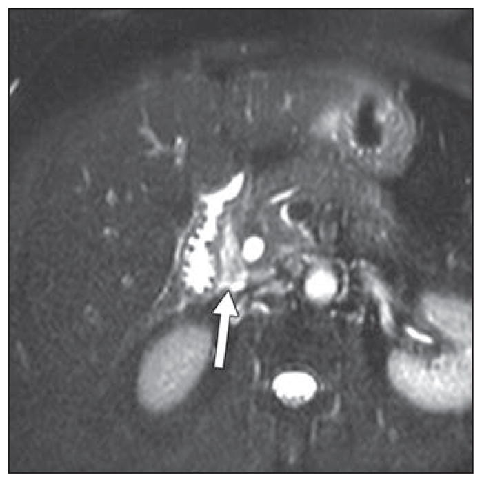
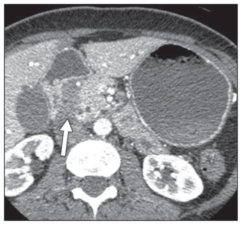
Objective: Groove pancreatitis is a rare form of chronic pancreatitis affecting the "groove" between the pancreatic head, duodenum, and common bile duct. The exact cause is unknown, although there are strong associations with long-term alcohol abuse, functional obstruction of the duct of Santorini, and Brunner gland hyperplasia.

Conclusion: Unfortunately, differentiating groove pancreatitis from malignancy on the basis of imaging features, clinical presentation, or laboratory markers can be extraordinarily difficult, and the vast majority of these patients ultimately undergo a pancreaticoduodenectomy (Whipple procedure) because of an inability to completely exclude malignancy. In certain cases, however, the imaging features on CT and MRI can allow the radiologist to prospectively suggest the correct diagnosis.


Figures

Fig. 1
Fig. 1. 43-year-old man who initially presented with epigastric pain
A and B, Contrast-enhanced CT revealed subtle infiltrating soft tissue (arrows) in pancreaticoduodenal groove. He underwent ERCP and endoscopic ultrasound, both of which suggested duodenal wall thickening and discrete mass, although biopsy results were negative. On basis of these findings, patient was thought to have primary duodenal malignancy. However, this process was found to represent groove pancreatitis after pancreaticoduodenectomy. C, Gross specimen shows paraampullary duodenal wall cysts (arrows) with surrounding erythema and edema. Duodenal wall (top) and underlying pancreas (lower half) are thickened and fibrotic. D, Histologic specimen, low-power view (H and E, ×1), shows Brunner gland hyperplasia (arrows) in duodenum, overlying inflamed cyst in groove region. E, Histologic specimen (H and E, ×10) shows duodenal Brunner gland hyperplasia (black arrows) overlying inflamed cyst. Red arrow shows inflamed fibrous tissue in cyst wall. Cyst wall is composed of markedly inflamed fibrous tissue. F, Histologic specimen (H and E, ×20) shows partially denuded duct with luminal concretions. Cyst is partially lined by ductal epithelium (black arrow) and contains thick proteinaceous secretions. Background pancreatic parenchyma (red arrows) is inflamed and fibrotic.
Fig. 2
Fig. 2. 39-year-old man who presented with abdominal pain
A and B, Contrast-enhanced coronal (A) and axial (B) images show infiltrating soft tissue (arrows) in pancreaticoduodenal groove. This “sheetlike” soft tissue is crescentic in appearance and is associated with thickening of medial duodenal wall and several cysts in wall of duodenum. Although possibility of groove pancreatitis was entertained on basis of CT appearance and negative endoscopic ultrasound biopsy results, patient underwent Whipple procedure because of inability to completely exclude duodenal malignancy. Postsurgical pathology results confirmed diagnosis of groove pancreatitis. C, Gross specimen shows paraampullary duodenal wall cyst. Cyst is present between duodenal wall (black arrow) and adjacent pancreas (blue arrow), which show marked edema. Common bile duct has been opened and has few extraneous knife marks from dissection (red arrows); it does not show luminal narrowing or stricture formation.
Fig. 3
Fig. 3. 61-year-old woman who presented with 4-month history of abdominal pain, nausea, and vomiting
A and B, Axial contrast-enhanced CT images show induration between duodenum and pancreatic head (arrow, A), some fluid in retroperitoneum or pararenal spaces, and cystic lesion in pancreatic head (arrow, B). Endoscopic ultrasound–guided fine-needle aspiration was negative for malignancy, although appearance was concerning for infiltrating neoplasm. Diagnosis of groove pancreatitis was confirmed on postsurgical pathology. C, Gross specimen shows pancreatic cyst located subjacent to duodenal wall (black arrow). Pancreatic parenchyma (red arrow) is fibrotic. D, Histologic specimen (H and E, ×1) shows dilated pancreatic duct. Duct lumen contains characteristic thick eosinophilic inspissated secretions (black arrow). Background pancreas is expanded by dense fibrosis (red arrows) and other stigmata of obstructive chronic pancreatitis. E, Histologic specimen (H and E, ×40) shows dilated pancreatic duct. At this higher magnification, luminal secretions are associated with denudation (black arrows) of ductal epithelium. Spindle cells and mixed fibroinflammatory infiltrate (red arrows) of plasma cells, eosinophils, neutrophils, and lymphocytes are present in cyst wall.
Fig. 4
Fig. 4. 72-year-old man who presented with abdominal pain and weight loss
A and B, Axial CT images with contrast agent show low-density soft tissue or fluid in pancreaticoduodenal groove (arrows, A), with multiple calcifications in pancreatic parenchyma (arrow, B). Given calcifications, it was thought that this pancreaticoduodenal groove low-attenuation material could represent sequelae of pancreatitis, but patient underwent surgery because of inability to completely exclude malignancy. Postsurgical pathology confirmed diagnosis of groove pancreatitis, with findings of chronic pancreatitis in remainder of gland. C, Gross pathologic specimen (duodenum, black arrow) shows cysts (red arrows) in duodenal wall and subjacent pancreas, which are unilocular and contain multiple calculi (blue arrow).
Fig. 5
Fig. 5. 62-year-old man who presented with painless jaundice and underwent placement of biliary drainage catheter
A and B, Coronal (A) and axial (B) contrast-enhanced CT images show masslike enlargement of pancreatic head with central cystic focus (arrow, A), which is relatively isodense to surrounding pancreas, as well as diffuse pancreatic ductal dilatation. There is subtle soft tissue in pancreaticoduodenal groove (arrow, B), which is best seen on axial image. Endoscopic ultrasound suggested presence of discrete mass in this location (although biopsy results were negative), and patient underwent Whipple procedure under assumption that mass represented pancreatic adenocarcinoma. However, postsurgical pathology revealed it to be segmental form of groove pancreatitis with involvement of pancreatic head. C, Histologic specimen (H and E, ×40) shows dilated pancreatic duct with thick proteinaceous secretions. Multiple smaller dilated ducts are present. Ducts are lined by low cuboidal-to-columnar mucinous ductal epithelium.
Fig. 6
Fig. 6. 45-year-old man with history of alcoholism who presented with acute renal failure and abdominal pain
A and B, Axial (A) and coronal maximum-intensity-projection (B) contrast-enhanced CT images with contrast agent show hypodense soft tissue (arrow, A) in pancreaticoduodenal groove, with some areas of lower density (arrow, B), which are possibly cystic changes. Possibility of groove pancreatitis was raised on basis of imaging findings. Endoscopic ultrasound was performed and raised possibility of distinct mass in this location (although biopsy results were negative for tumor). Given inability to completely exclude tumor, patient underwent Whipple procedure, where diagnosis of groove pancreatitis was confirmed.
Fig. 7
Fig. 7. 45-year-old woman with long history of multiple bouts of abdominal pain refractory to conservative management
A and B, Axial (A) and coronal (B) contrast-enhanced CT images show infiltrating crescentic soft tissue (arrows) in pancreaticoduodenal groove with multiple cystic spaces. Given her long history, this was prospectively thought to represent groove or chronic pancreatitis, and patient underwent Whipple procedure because of continued inability to control her pain symptoms. Diagnosis of groove pancreatitis was confirmed on postsurgical pathology.
Fig. 8
Fig. 8. 46-year-old man who presented with pruritus. He was found to have biliary and pancreatic ductal dilatation and underwent biliary stent placement
A and B, Axial (A) and coronal (B) contrast-enhanced CT images show subtle thickening between duodenum and pancreas, with cystic focus (arrows) in pancreaticoduodenal groove. ERCP showed irregular stricture of distal common bile duct, which was thought to be concerning for malignancy (although brush biopsy results were negative). Diagnosis of groove pancreatitis was confirmed on postsurgical pathology.
Fig. 9
Fig. 9
66-year-old woman with history of recurrent abdominal pain and presumptive groove pancreatitis. MRI shows T2-hyperintense sheetlike soft tissue in pancreaticoduodenal groove (arrow). She underwent endoscopic ultrasound, which revealed soft tissue in pancreaticoduodenal groove. However, because of imaging features and negative endoscopic ultrasound biopsy results, “lesion” was followed with surveillance scans and remained stable over time.
Fig. 10
Fig. 10
49-year-old woman with 1-month history of nausea and vomiting. CT image shows homogeneous hypodense thickening in pancreaticoduodenal groove, without evidence of vascular encasement, as well as smooth tapering of distal common bile duct and pancreatic duct (not shown). This was thought preoperatively to represent groove pancreatitis but was found on postoperative pathology to represent groove pancreatic adenocarcinoma.

References

    1. Becker V. Bauchspeicheldruse (Inselapperat ausgenommen) In: Doerr W, editor. Spezielle pathologische anatomie. Berlin, Germany: Springer-Verlag; 1973.
    1. Blasbalg R, Baroni RH, Costa DN, et al. MRI features of groove pancreatitis. AJR. 2007;189:73–80. - PubMed
    1. Balakrishnan V, Chatni S, Radhakirshnan L, et al. Groove pancreatitis: a case report and review of the literature. JOP. 2007;8:592–597. - PubMed
    1. Triantopoulou C, Dervenis C, Giannakou N, et al. Groove pancreatitis: a diagnostic challenge. Eur Radiol. 2009;19:1736–1743. - PubMed
    1. Tezuka K, Makino TY, Hirai I, Kimura W. Groove pancreatitis. Dig Surg. 2010;27:149–152. - PubMed

LinkOut - more resources