. 2014 Mar;20(3):188-97.
doi: 10.1089/ten.TEC.2013.0148. Epub 2013 Aug 16.

Pulp cell tracking by radionuclide imaging for dental tissue engineering

Affiliations

Pulp cell tracking by radionuclide imaging for dental tissue engineering

Jean-Baptiste Souron et al. Tissue Eng Part C Methods. 2014 Mar.

Abstract

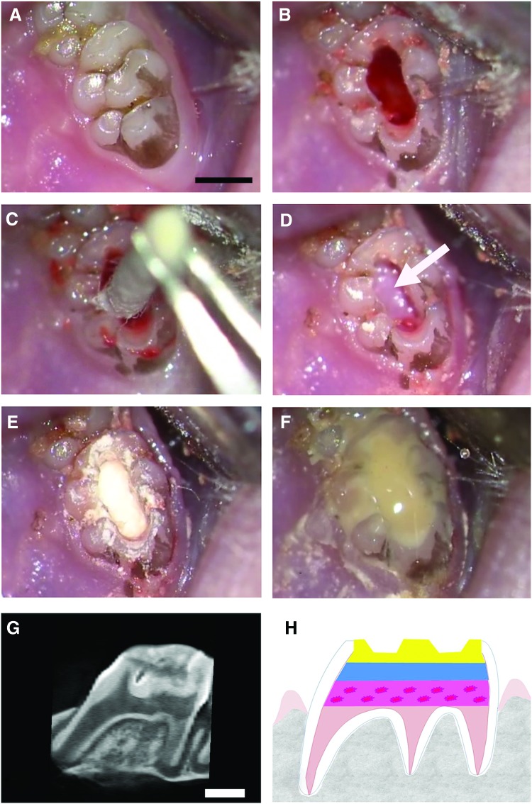
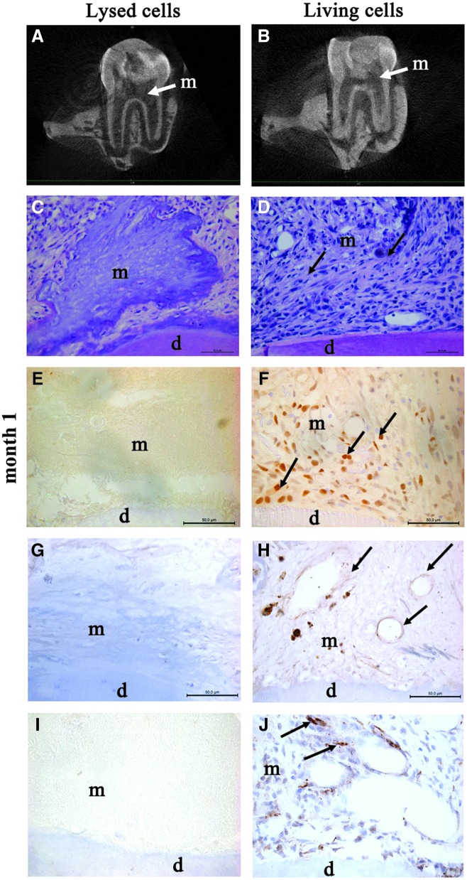
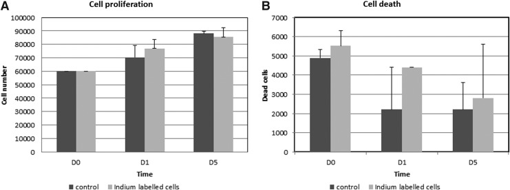
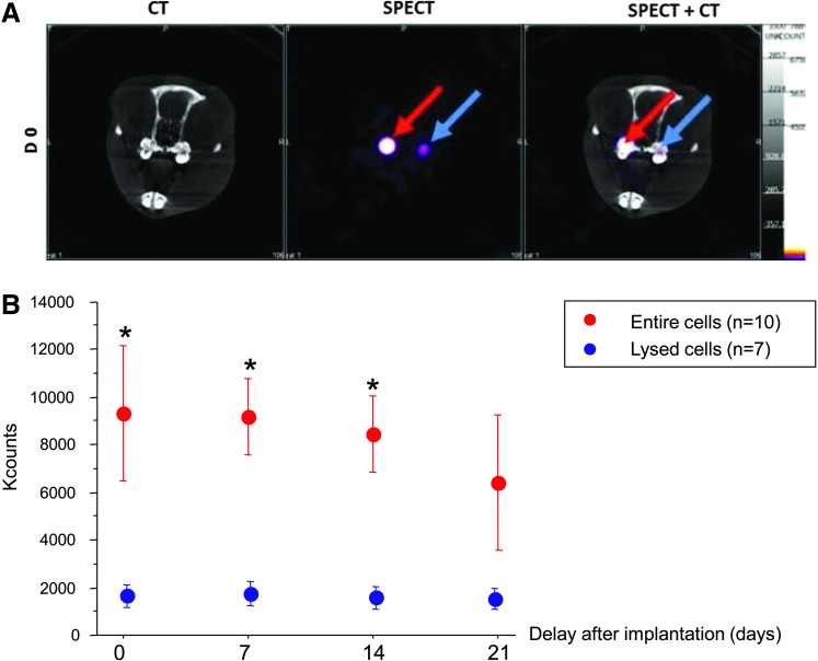
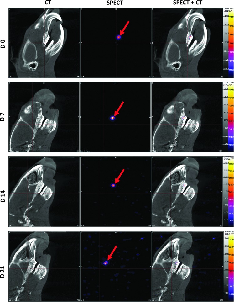
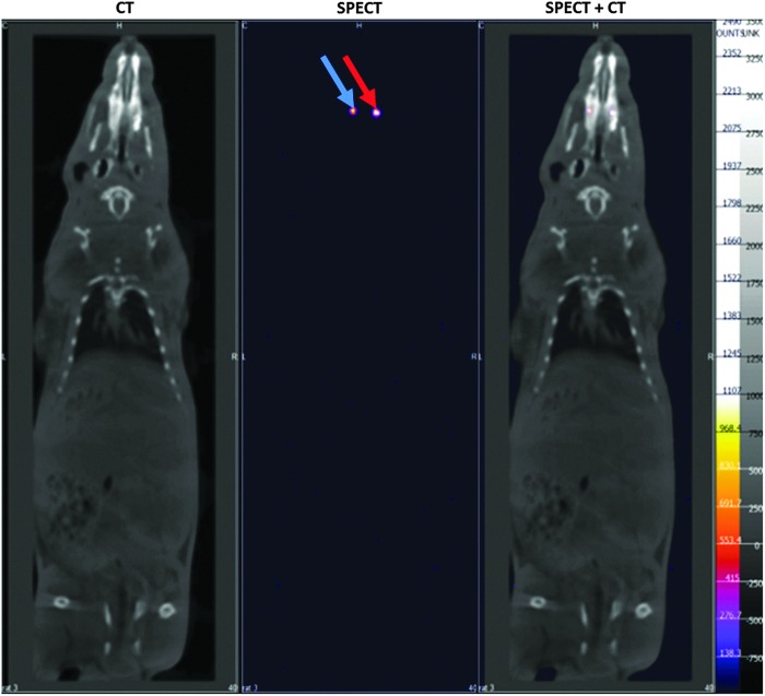
Pulp engineering with dental mesenchymal stem cells is a promising therapy for injured teeth. An important point is to determine the fate of implanted cells in the pulp over time and particularly during the early phase following implantation. Indeed, the potential engraftment of the implanted cells in other organs has to be assessed, in particular, to evaluate the risk of inducing ectopic mineralization. In this study, our aim was to follow by nuclear imaging the radiolabeled pulp cells after implantation in the rat emptied pulp chamber. For that purpose, indium-111-oxine (¹¹¹In-oxine)-labeled rat pulp cells were added to polymerizing type I collagen hydrogel to obtain a pulp equivalent. This scaffold was implanted in the emptied pulp chamber space in the upper first rat molar. Labeled cells were then tracked during 3 weeks by helical single-photon emission computed tomography (SPECT)/computed tomography performed on a dual modality dedicated small animal camera. Negative controls were performed using lysed radiolabeled cells obtained in a hypotonic solution. In vitro data indicated that ¹¹¹In-oxine labeling did not affect cell viability and proliferation. In vivo experiments allowed a noninvasive longitudinal follow-up of implanted living cells for at least 3 weeks and indicated that SPECT signal intensity was related to implanted cell integrity. Notably, there was no detectable systemic release of implanted cells from the tooth. In addition, histological analysis of the samples showed mitotically active fibroblastic cells as well as neoangiogenesis and nervous fibers in pulp equivalents seeded with entire cells, whereas pulp equivalents prepared from lysed cells were devoid of cell colonization. In conclusion, our study demonstrates that efficient labeling of pulp cells can be achieved and, for the first time, that these cells can be followed up after implantation in the tooth by nuclear imaging. Furthermore, it appears that grafted cells retained the label and are viable to follow the repair process. This technique is expected to be of major interest for monitoring implanted cells in innovative therapies for injured teeth.


Figures

<b>FIG. 1.</b>
FIG. 1.
Description of the rat pulpotomy model. The pulpotomy model based on the implantation of pulp equivalent (pulp cells seeded in a 3D collagen hydrogel) in the rat pulp chamber (scale bar, 1 mm). After anesthesia, drilling of an occlusal cavity of the first upper molar under an endodontic microscope (A). Elimination of the cameral pulp parenchyma with a rotary instrument (B). After hemostasis by compression (C), placement of radioactive pulp equivalent (with either entire cells or lysed cells) in the pulp chamber space (white arrow shows the pulp equivalent) (D). Sealing of the cavity with a calcium silicate-based cement (Biodentine™; Septodont) (E). Covering with light-cured flow composite (FloRestore; Denmat) (F). Microcomputed tomography (micro-CT) imaging of the tooth immediately after implantation showing the different layers (G), which are illustrated on a schema (dotted-pink layer: pulp equivalent seeded with cells; blue layer: calcium silicate-based cement; yellow layer: composite restoration) (H). Color images available online at www.liebertpub.com/tec
<b>FIG. 2.</b>
FIG. 2.
Repair process at 1-month postpulp cell implantation. Micro-CT imaging showed no pulp or root canal obliteration 4 weeks after implantation of lysed cells (A) or living cells (B) (white arrows show the implanted matrix). At month 1, hematoxylin and eosin staining showed the absence of cells in the collagen matrix implanted with lysed cells (C). In contrast, numerous cells were observed in the matrix (arrows) implanted with living cells (D). Immunohistochemistry for proliferating cell nuclear antigen showed proliferating cells (arrows) in the matrix seeded with living cells (F), but not in controls (E). Vascularization (arrows) was observed by immunostaining for von Willebrand factor in the matrix seeded with living cells (H), whereas no vessel was observed in controls (G). Innervation was evidenced in the matrix seeded with living cells by immunostaining for the calcitonin gene related peptide, which labels sensory nervous fibers (arrows) (J). In contrast, no labeling was observed in the control matrix (I). d, dentin; m, implanted matrix.
<b>FIG. 3.</b>
FIG. 3.
Effect of indium-111-oxine (111In-oxine) labeling on pulp cell proliferation and viability. The absence of detrimental effect of 111In-oxine incorporation on the proliferation rate of labeled cells at early time points (0, 1, and 5 days). Mean results of three individual experiments are presented with 95% confidence intervals for cultures of 111In-oxine-labeled cells and controls. There was no significant difference between the groups (A). The absence of detrimental effect of 111In-oxine incorporation on the viability of labeled cells at same time points (no significant difference) (B).
<b>FIG. 4.</b>
FIG. 4.
In vivo follow-up of implanted pulp equivalents by single-photon emission computed tomography (SPECT)—comparison of entire and lysed cells. 111In-oxine-labeled cells, entire (red arrow) and lysed (blue arrow), were followed up after implantation with SPECT/CT. The signal displays a greater intensity in tooth implanted with living cells when compared to samples implanted with lysed cells (A). Quantification of 111In-oxine activity was performed during 3 weeks. Data are presented as mean±SEM after radioactive decay correction. Although a similar 111In activity was embedded in both pulp equivalents, detected counts are 5-fold lower in the preparation with lysed cells in all time points, including immediately postimplantation. This suggests a release of cell fragments from the collagen network during implantation. There is no significant count decrease according to time in both pulp equivalents (Kruskal–Wallis p=0.6 for entire cells and p=1 for lysed cells) (B). *p<0.004 (Mann–Whitney) between entire and lysed cells.
<b>FIG. 5.</b>
FIG. 5.
Craniofacial imaging. SPECT/CT imaging of 111In-oxine-labeled cell implantation. Implanted living cells were successfully tracked during 3 weeks (red arrows). Localization of implanted cells was possible with merging SPECT and CT images (right column). No signal was detectable out of the teeth and, in particular, no cell spreading was observed in the craniofacial area.
<b>FIG. 6.</b>
FIG. 6.
Whole-body imaging. Whole-body imaging at D14 did not show spreading of labeled cells, either in the ectopic sites or in the reticuloendothelial system. red arrow: living cells; blue arrow: lysed cells.

References

    1. Parahitiyawa N.B., Jin L.J., Leung W.K., Yam W.C., and Samaranayake L.P.Microbiology of odontogenic bacteremia: beyond endocarditis. Clin Microbiol Rev 22, 46, Table of Contents, 2009 - PMC - PubMed
    1. Somma F., Castagnola R., Bollino D., and Marigo L.Oral inflammatory process and general health. Part 2: how does the periapical inflammatory process compromise general health? Eur Rev Med Pharmacol Sci 15,35, 2011 - PubMed
    1. Sun H.H., Jin T., Yu Q., and Chen F.M.Biological approaches toward dental pulp regeneration by tissue engineering. J Tissue Eng Regen Med 5,e1, 2011 - PubMed
    1. Huang A.H., Chen Y.K., Chan A.W., Shieh T.Y., and Lin L.M.Isolation and characterization of human dental pulp stem/stromal cells from nonextracted crown-fractured teeth requiring root canal therapy. J Endod 35,673, 2009 - PubMed
    1. Rosa V., Della Bona A., Cavalcanti B.N., and Nor J.E.Tissue engineering: from research to dental clinics. Dent Mater 28,341, 2012 - PMC - PubMed

Publication types

LinkOut - more resources