. 2013 Dec;19(23-24):2544-52.
doi: 10.1089/ten.TEA.2012.0686. Epub 2013 Aug 9.

Proangiogenic hydrogels within macroporous scaffolds enhance islet engraftment in an extrahepatic site

Affiliations

Proangiogenic hydrogels within macroporous scaffolds enhance islet engraftment in an extrahepatic site

Ann-Christina Brady et al. Tissue Eng Part A. 2013 Dec.

Abstract

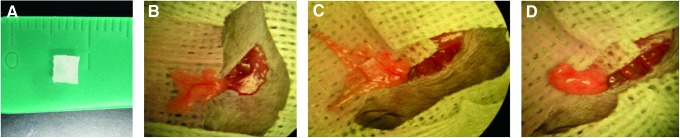
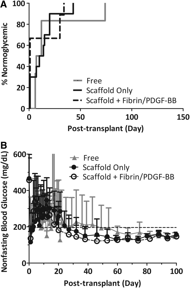
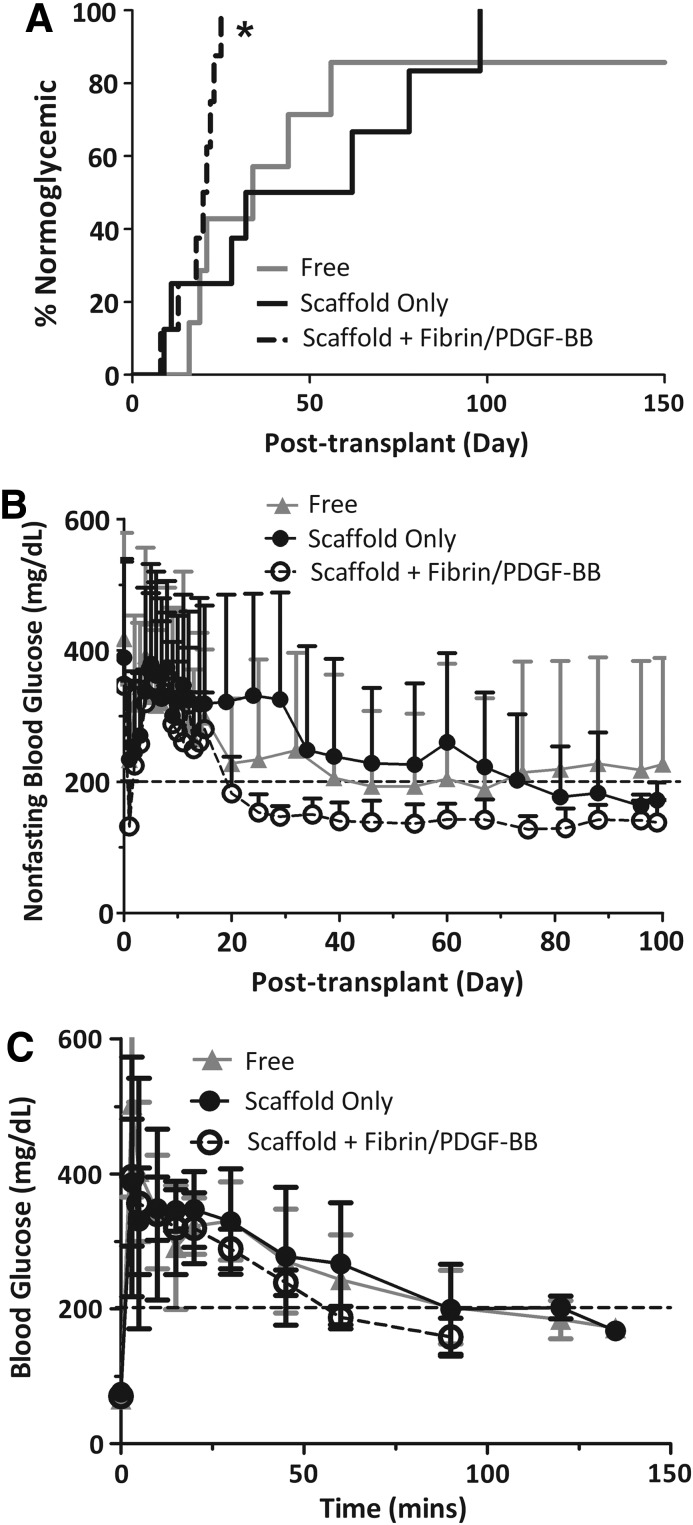
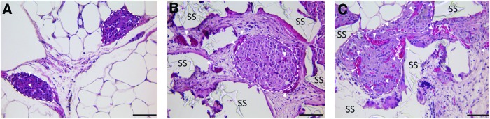
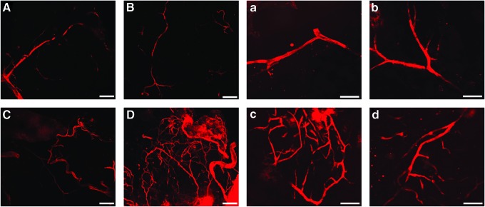
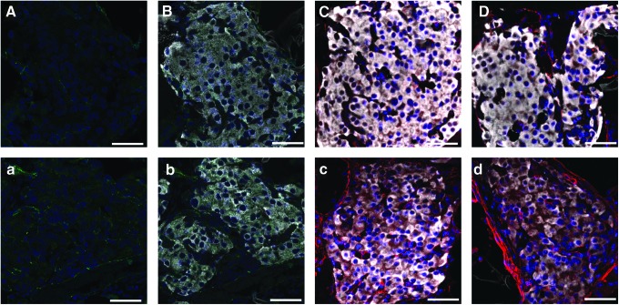
The transplantation of allogeneic islets in recent clinical trials has shown substantial promise as a therapy for type 1 diabetes; however, long-term insulin independence remains inadequate. This has been largely attributed to the current intravascular, hepatic transplant site, which exposes islets to mechanical and inflammatory stresses. A highly macroporous scaffold, housed within an alternative transplant site, can support an ideal environment for islet transplantation by providing three-dimensional distribution of islets, while permitting the infiltration of host vasculature. In the present study, we sought to evaluate the synergistic effect of a proangiogenic hydrogel loaded within the void space of a macroporous poly(dimethylsiloxane) (PDMS) scaffold on islet engraftment. The fibrin-based proangiogenic hydrogel tested presents platelet derived growth factor (PDGF-BB), via a fibronectin (FN) fragment containing growth factor and major integrin binding sites in close proximity. The combination of the proangiogenic hydrogel with PDMS scaffolds resulted in a significant decrease in the time to normoglycemia for syngeneic mouse islet transplants. This benefit was associated with an observed increase in competent vessel branching, as well as mature intraislet vessels. Overall, the addition of the proangiogenic factor PDGF-BB, delivered via the FN fragment-functionalized hydrogel, positively influenced the efficiency of engraftment. These characteristics, along with its ease of retrieval, make this combination of a biostable macroporous scaffold and a degradable proangiogenic hydrogel a supportive structure for insulin-producing cells implanted in extrahepatic sites.


Figures

FIG. 1.
FIG. 1.
Photographs of (A) poly(dimethylsiloxane) (PDMS) scaffold used for implantation (5×5×2 mm); (B–D) PDMS scaffold implantation into the epididymal fat pad site, where the PDMS scaffold containing islets, with or without fibrin/ platelet-derived growth factor (PDGF-BB), was placed on spread fat pad, wrapped in the fat pad tissue, sealed with a plain fibrin gel (no growth factor), and placed back into the peritoneum. Color images available online at www.liebertpub.com/tea
FIG. 2.
FIG. 2.
Transplants of 500 islet equivalent (IEQ) C57BL/6J islets into diabetic C57BL/6J mouse recipients. (A) Time to reversal to normoglycemia and (B) average nonfasting blood glucose of recipients following transplantation of 500 IEQ into the epididymal fat pad either free (gray line; n=6), within a PDMS scaffold (black line; n=9), or within a PDMS scaffold coloaded with fibrin/PDGF-BB (dashed black line; n=9). Reversal is defined as two consecutive readings <200 mg/dL. Implants were explanted between 110 and 150 days, where prompt reversal to a diabetic state was observed for all animals. Error bars=standard deviation. No statistical difference in time to reversal was observed for any of the groups.
FIG. 3.
FIG. 3.
Transplants of 250 IEQ C57BL/6J islets into diabetic C57BL/6J mouse recipients. (A) Time to reversal to normoglycemia and (B) average nonfasting blood glucose of recipients following transplantation of 250 IEQ into the epididymal fat pad either free (gray line; n=7), within a PDMS scaffold (black line; n=8), or within a PDMS scaffold coloaded with fibrin/PDGF-BB (dashed black line; n=8). Reversal is defined as two consecutive readings <200 mg/dL. (C) Intravenous glucose tolerance test performed on functional graft recipients at 95 days post-transplant. Blood glucose measurements were collected at time points indicated, following injection of bolus glucose, for islets in either free (gray triangle; n=2), within a PDMS scaffold (black circle; n=3), or within a PDMS scaffold coloaded with fibrin/PDGF-BB (dashed black open circle; n=3). Implants were explanted between 110 and 150 days, where prompt reversal to a diabetic state was observed for all animals. Error bars=standard deviation. *Statistically significant difference in time to reversal from both scaffold only (p=0.009) and free (p=0.045). No difference was observed between free and scaffold-only groups (p=0.90).
FIG. 4.
FIG. 4.
Representative histopathological images of hematoxylin/eosin-stained islets explanted from the epididymal fat pad. (A) Free islets (day 115); (B) islets within a PDMS scaffold (day 110); and (C) islets within PDMS scaffold with fibrin/PDGF-BB (day 110); dashed lines encircle islets; white arrows highlight functional vasculature with red blood cells; SS=PDMS scaffold; scale bar=50 μm. Color images available online at www.liebertpub.com/tea
FIG. 5.
FIG. 5.
Multislice confocal microscopy projections of lipophilic carbocyanine dye DiI within scaffold cross sections. Scaffolds contained either none (top row, A, B & a, b) or fibrin/PDGF-BB (bottom row, C, D & c, d) and were perfused with lipophilic carbocyanine dye DiI before explantation on day 10. Scale bars: 200 μm for A–D, 100 μm for a–d. Color images available online at www.liebertpub.com/tea
FIG. 6.
FIG. 6.
Histopathological evaluation of islet-loaded PDMS scaffolds, removed from the epididymal fat pad site on day 10 (A–C; a–c) or day 30 (D & d), which contained either no (A–D) or fibrin/PDGF-BB (a–d). Representative images of immunoflourescence staining of explants for CD31 (green) (A & a), CD31 plus insulin (white) (B & b), and α-smooth muscle actin (red) plus insulin (white) (C–D & c–d). All sections were counterstained with DAPI (blue); scale bar=50 μm. Color images available online at www.liebertpub.com/tea

References

    1. The effect of intensive treatment of diabetes on the development and progression of long-term complications in insulin-dependent diabetes mellitus. The Diabetes Control and Complications Trial Research Group. N Engl J Med. 1993;329:977. - PubMed
    1. Pileggi A. Cobianchi L. Inverardi L. Ricordi C. Overcoming the challenges now limiting islet transplantation: a sequential, integrated approach. Ann N Y Acad Sci. 2006;1079:383. - PubMed
    1. Shapiro A.M. Ricordi C. Hering B.J. Auchincloss H. Lindblad R. Robertson R.P., et al. International trial of the Edmonton protocol for islet transplantation. N Engl J Med. 2006;355:1318. - PubMed
    1. Hardy M.A. Witkowski P. Sondermeijer H. Harris P. The long road to pancreatic islet transplantation. World J Surg. 2010;34:625. - PMC - PubMed
    1. Cattan P. Berney T. Schena S. Molano R.D. Pileggi A. Vizzardelli C., et al. Early assessment of apoptosis in isolated islets of Langerhans. Transplantation. 2001;71:857. - PubMed

Publication types

LinkOut - more resources