

Comment
doi: 10.1016/j.cell.2013.05.054.
Inside-out connections: the ER meets the plasma membrane
Affiliations
- PMID: 23791170
- PMCID: PMC6783275
- DOI: 10.1016/j.cell.2013.05.054
Item in Clipboard
Comment
Inside-out connections: the ER meets the plasma membrane
Cell.
.
Abstract
Junctions that connect the endoplasmic reticulum (ER) and the plasma membrane (PM) are unique yet ubiquitous subcellular compartments. Giordano et al. now report that extended synaptotagmins (E-Syts) promote their formation, providing fundamental insight into the molecular machinery controlling ER and plasma membrane crosstalk.
Copyright © 2013 Elsevier Inc. All rights reserved.
Figures
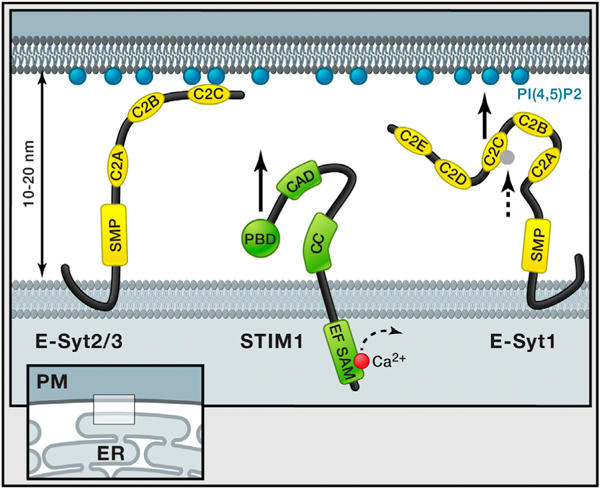
ER-PM junctions are ubiquitous cell compartments In which the two membranes are stably kept at a distance of 10 to 20 nm. Giordano et al. (2013) show that three ER-localized proteins, E-Syts, play an important role in tethering the ER to the plasma membrane. Overexpression of E-Syt2 and E-Syt3 connects large regions of the ER to the plasma membrane mediated by E-Syt C2C domains that bind plasma membrane PI(4,5)P2 independent of Ca2+. In contrast, E-Syt1 is at basal Ca2+ levels not associated with the plasma membrane, but, as the intracellular Ca2+ level increases, the C2C domain will bind Ca2+ (dashed arrow), resulting in binding to PIP2 in the plasma membrane (black arrow) and trapping of E-Syt1 at the ER-PM junction. This is very different from the regulation of STIM1, for which previous studies showed that STIM1 translocation to these same ER-PM junctions is mediated by lowering of lumenal ER Ca2+ levels (illustrated with dashed arrow), which triggers its oligomerization and the exposure of a PI(4,5)P2 binding and an Orai interaction region (black arrow). Giordano et al. (2013) further show that the previously predicted transmembrane region at the N terminus of E-Syts forms a hairpin insertion in the ER membrane, making both N and C termini accessible to the cytosol. All E-Syts have a cytosolic SMP domain, whereas E-Syt1 has five C2 domains compared to three for E-Syt2 and E-Syt3. Domains depicted on STIM1; EF, EF hand; SAM, sterile α motif; CC, coiled-coil; CAD, CRAC activation domain; and PBD, polybasic domain.
Comment on
-
PI(4,5)P(2)-dependent and Ca(2+)-regulated ER-PM interactions mediated by the extended synaptotagmins.Cell. 2013 Jun 20;153(7):1494-509. doi: 10.1016/j.cell.2013.05.026. Cell. 2013. PMID: 23791178 Free PMC article.
References
-
- Manford AG, Stefan CJ, Yuan HL, Macgurn JA, and Emr SD (2012). Dev. Cell 23, 1129–1140. - PubMed
Publication types
MeSH terms
Substances
Grants and funding
LinkOut - more resources
Full Text Sources
Other Literature Sources
Miscellaneous
