. 2013 Jun 20;153(7):1510-25.
doi: 10.1016/j.cell.2013.05.021.

Terminal axon branching is regulated by the LKB1-NUAK1 kinase pathway via presynaptic mitochondrial capture

Affiliations

Terminal axon branching is regulated by the LKB1-NUAK1 kinase pathway via presynaptic mitochondrial capture

Julien Courchet et al. Cell. .

Abstract

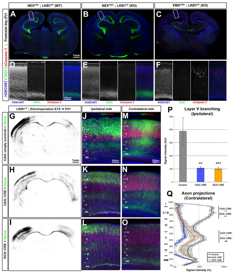
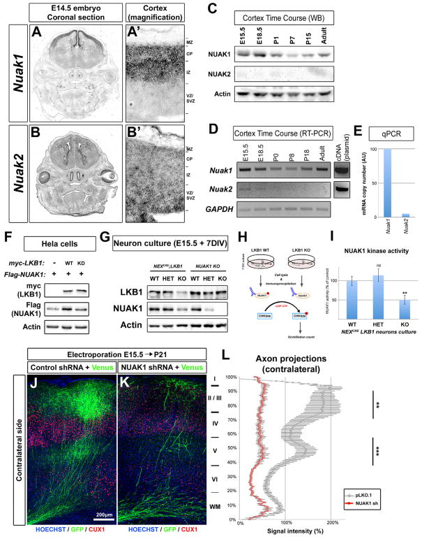
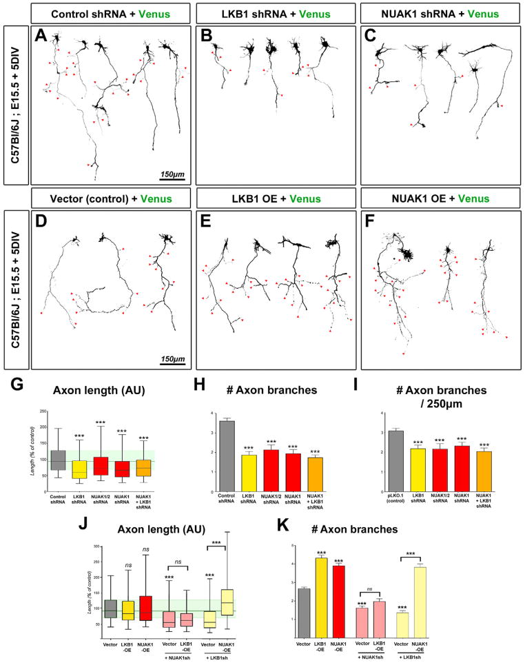
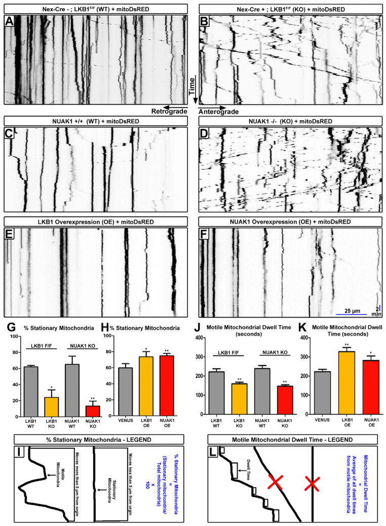
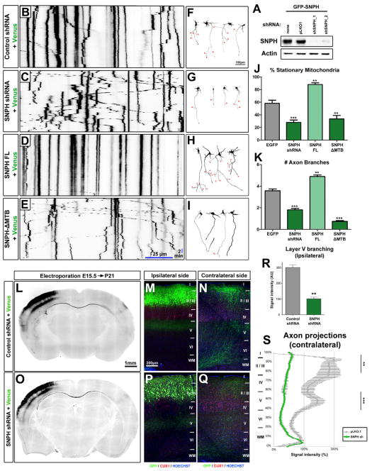
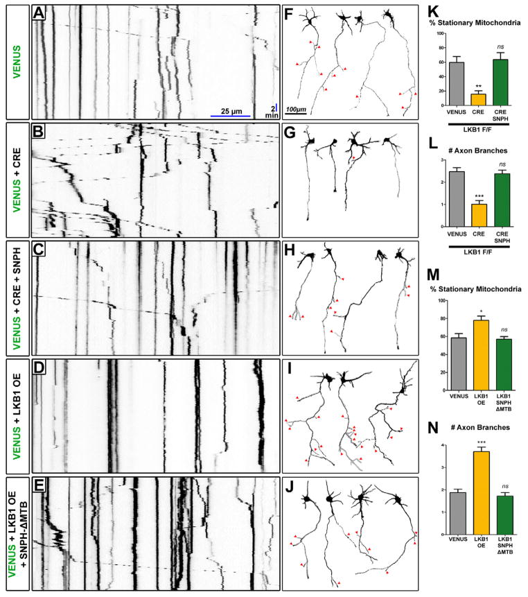
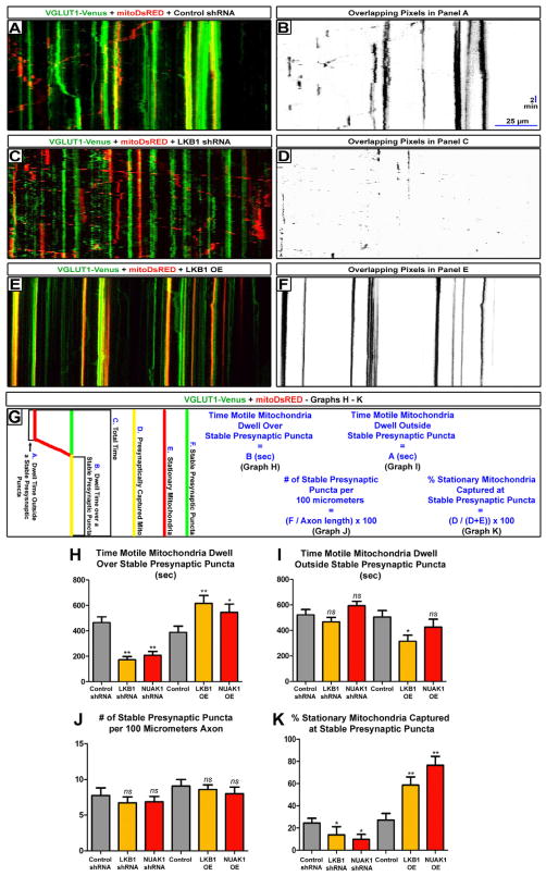
The molecular mechanisms underlying the axon arborization of mammalian neurons are poorly understood but are critical for the establishment of functional neural circuits. We identified a pathway defined by two kinases, LKB1 and NUAK1, required for cortical axon branching in vivo. Conditional deletion of LKB1 after axon specification or knockdown of NUAK1 drastically reduced axon branching in vivo, whereas their overexpression was sufficient to increase axon branching. The LKB1-NUAK1 pathway controls mitochondria immobilization in axons. Using manipulation of Syntaphilin, a protein necessary and sufficient to arrest mitochondrial transport specifically in the axon, we demonstrate that the LKB1-NUAK1 kinase pathway regulates axon branching by promoting mitochondria immobilization. Finally, we show that LKB1 and NUAK1 are necessary and sufficient to immobilize mitochondria specifically at nascent presynaptic sites. Our results unravel a link between presynaptic mitochondrial capture and axon branching.


Figures

Figure 1
Figure 1. LKB1 deletion after axon initiation does not impair axon maintenance but reduces axon branching in vivo
(A–C) Coronal section of newborn LKB1F/F mouse brains not expressing Cre recombinase (Wildtype WT, A) or expressing Cre under the NEX (KO) (B) or EMX (KO) (C) promoters. (D–F) Higher-magnification images of the cortex region boxed in A, B and C respectively. (G–I) Low magnification images of coronal brain sections of P21 Lkb1F/F mice electroporated at E15 with plasmids expressing mVenus alone (G) or co-expressing Cre recombinase (under CAG promoter, H or Doublecortin promoter, I) and mVenus. (J–O) Higher magnification of the ipsilateral (J–L) or the contralateral side (M–O) showing reduced axon branching in both Cre-electroporated neurons (K–O) compared to control (J, M). (P–Q) Quantification of normalized Venus fluorescence in layer 5 of the ipsilateral cortex (P, ±SEM) and along the radial axis of the cortical wall in the contralateral cortex (Q, ±SEM). Statistical analysis: Mann-Whitney (P) or two-way ANOVA (Q). See also Fig. S1 and S2.
Figure 2
Figure 2. AMPK-related kinase NUAK1 is required for axon branching in vivo
(A–B) Expression of Nuak1 and Nuak2 genes in the head of Balb/C mouse by in situ hybridization. Magnification (A′–B′) shows that Nuak1 and Nuak2 have complementary expression patterns in the cerebral cortex. (C) Time-course of NUAK1 and NUAK2 protein expression in Balb/C mouse cortex by Western-blot. (D) Time-course of Nuak1 and Nuak2 mRNA expression in Balb/C mouse cortex by RT and PCR. (E) Quantitative PCR after RT (qRT-PCR) in E15.5 mouse cortex revealed that Nuak1 transcript is 20 times more abundant than Nuak2 transcript. (F) Overexpression of catalytically-active LKB1 increases NUAK1 protein expression. (G) Endogenous expression of LKB1 and NUAK1 proteins in LKB1 and NUAK1 knockout neurons. Western-blot analysis was performed with the indicated antibodies (H) Schema of NUAK1 kinase assay. (I) NUAK1 kinase activity is reduced in LKB1 KO neurons (±SEM). (J–K) In utero cortical electroporation of the indicated constructs revealed that loss of NUAK1 decreased axonal branching in the contralateral hemisphere similar to the loss of LKB1. (L) Quantification of EGFP normalized fluorescence along the radial axis of the cortical wall in the contralateral cortex. Statistical analysis: Mann-Whitney test (I) or two-way ANOVA (L).
Figure 3
Figure 3. LKB1 regulates axon growth and branching in vitro through NUAK1
(A–C) Representative neurons imaged after 5 day of culture in vitro (DIV) following inhibition of LKB1 (B) or NUAK1 (C) expression. (D–F) Overexpression of LKB1 (E) or NUAK1 (F) in 5 DIV cortical neurons induced the formation of supernumerary axonal branches. Red arrowheads in A–F point to axon branches. (G–I) Quantification of axon morphology shows that LKB1 or NUAK1 inhibition results in a shortened axon (G) and decreased collateral branch formation (HI). (J–K) Quantitation of axon length (J) or number of collateral branches at 5DIV (K) after overexpression of the indicated constructs. Data represent 25th, 50th and 75th percentile (G, J) or average value ±SEM (H–I, K). Statistical analysis: Mann-Whitney test. See also Fig. S3–S5.
Figure 4
Figure 4. LKB1 and NUAK1 are necessary and sufficient for mitochondrial immobilization in cortical axons
(A–F) Representative kymographs of axonal mitochondria in dissociated neurons electroporated with the indicated constructs. (G) Loss of LKB1 or NUAK1 caused a significant decrease in the number of stationary mitochondria, (H) while overexpression of LKB1 or NUAK1 produced more stationary mitochondria (±SEM). (J) Loss of LKB1 or NUAK1 lead to a decrease in motile mitochondrial dwell time, (K) while overexpression of LKB1 or NUAK1 increased the dwell time of motile mitochondria (±SEM). (I) Kymograph illustration of the parameters used to determine motile and stationary mitochondria. (L) Schema illustrating the parameters used to determine mitochondrial dwell time. Motile mitochondria which did not pause for at least 60 seconds were excluded as were stationary mitochondria. All kymographs throughout the paper are oriented as shown in A–B. Statistical analysis: Mann-Whitney test. See also Fig. S6.
Figure 5
Figure 5. Syntaphilin-dependent mitochondria immobilization is necessary and sufficient for proper axon branching
(A) Validation of shRNA targeting mouse Snph in HEK293T cells. Western-blot analysis was performed with the indicated antibodies. (B–E) Representative kymographs of axonal mitochondria in dissociated neurons electroporated with the indicated constructs. (F–I) Representative neurons at 5 DIV electroporated with the constructs indicated in B–E. Red arrowheads point to axon branches. (J–K) Loss or disruption of SNPH decreased, while overexpression of SNPH increased the number of stationary mitochondria (J) and of axonal branches (K). (L–Q) Low magnification images of coronal brain sections at P21 (L & O) and high magnification for the ipsilateral (M & P) or contralateral (N & Q) side. (R) Loss of SNPH caused a reduction in axonal branching on layer 5 of the ipsilateral hemisphere. (S) Loss of SNPH reduced axonal branching in both layers 2/3 and 5 of the contralateral hemisphere. Data represent average value ±SEM (J–K, R–S). Statistical analysis: Mann-Whitney test or two-way ANOVA (S).
Figure 6
Figure 6. LKB1 mediates axonal branching through mitochondria immobilization along the axon
(A–E) Representative kymographs of axonal mitochondria in dissociated neurons electroporated with the indicated constructs. (F–J) Representative neurons at 5 DIV electroporated with the constructs indicated in A–E. Red arrowheads point to axon branches. (K) Loss of LKB1 caused a decrease in the percentage of stationary mitochondria, while overexpression of wildtype human SNPH upon loss of LKB1 returned mitochondrial transport to normal levels. (L) Loss of LKB1 decreased the number of axonal branches, while overexpression of wildtype human SNPH upon loss of LKB1 returned axonal branching to normal levels. (M) Overexpression of LKB1 increased the percentage of stationary mitochondria, while overexpression of dominate negative human SNPH-ΔMTB at the same time as LKB1 returned mitochondrial transport to normal levels. (N) Overexpression of LKB1 also increased the number of axonal branches, while overexpression of dominate negative human SNPH-ΔMTB at the same time as LKB1 returned axonal branch number to normal levels. Data represent average value ±SEM (K–N). Statistical analysis: ANOVA non-parametric test.
Figure 7
Figure 7. LKB1 and NUAK1 are necessary and sufficient for mitochondrial immobilization at nascent presynaptic sites
(A, C, E) Representative dual color kymographs of axonal VGLUT1-Venus puncta and mitochondria dynamics in cortical neurons electroporated with the indicated constructs. (B, D, F) Overlapping pixels maps of A, C, E were created in Fuji/ImageJ using the Colocalization Threshold program. (G) Schematic illustration of the parameters used to quantify the results shown in H–K. (H) Knockdown of LKB1 or NUAK1 decreased the dwell time of mitochondria over nascent presynaptic sites, while overexpression of LKB1 or NUAK1 increased it. (I) Knockdown of LKB1 or NUAK1 did not affect the dwell time of mitochondria outside nascent presynaptic sites, while overexpression of LKB1 or NUAK1 slightly decreased it. (J) Knockdown or overexpression of LKB1 and NUAK1 do not affect the linear density of stable (for 30 minutes) VGLUT1-Venus nascent presynaptic sites. (K) Loss of LKB1 or NUAK1 decreased, while overexpression of LKB1 or NUAK1 increased the percentage of mitochondria stably captured (for 30 minutes) at presynaptic sites. Data represent average value ±SEM (K–N). Statistical analysis: Mann-Whitney test. See also Fig. S7.

Comment in

References

    1. Alessi DR, Sakamoto K, Bayascas JR. LKB1-dependent signaling pathways. Annu Rev Biochem. 2006;75:137–163. - PubMed
    1. Amato S, Liu X, Zheng B, Cantley L, Rakic P, Man HY. AMP-activated protein kinase regulates neuronal polarization by interfering with PI 3-kinase localization. Science. 2011;332:247–251. - PMC - PubMed
    1. Baas AF, Kuipers J, van der Wel NN, Batlle E, Koerten HK, Peters PJ, Clevers HC. Complete polarization of single intestinal epithelial cells upon activation of LKB1 by STRAD. Cell. 2004;116:457–466. - PubMed
    1. Bardeesy N, Sinha M, Hezel AF, Signoretti S, Hathaway NA, Sharpless NE, Loda M, Carrasco DR, DePinho RA. Loss of the Lkb1 tumour suppressor provokes intestinal polyposis but resistance to transformation. Nature. 2002;419:162–167. - PubMed
    1. Barnes AP, Lilley BN, Pan YA, Plummer LJ, Powell AW, Raines AN, Sanes JR, Polleux F. LKB1 and SAD kinases define a pathway required for the polarization of cortical neurons. Cell. 2007;129:549–563. - PubMed

Publication types

MeSH terms