. 2013 Jun 27;3(6):1874-84.
doi: 10.1016/j.celrep.2013.05.037. Epub 2013 Jun 20.

Cryptopatches are essential for the development of human GALT

Affiliations

Cryptopatches are essential for the development of human GALT

Tomonori Nochi et al. Cell Rep. .

Abstract

Abnormal gut-associated lymphoid tissue (GALT) in humans is associated with infectious and autoimmune diseases, which cause dysfunction of the gastrointestinal (GI) tract immune system. To aid in investigating GALT pathologies in vivo, we bioengineered a human-mouse chimeric model characterized by the development of human GALT structures originating in mouse cryptopatches. This observation expands our mechanistic understanding of the role of cryptopatches in human GALT genesis and emphasizes the evolutionary conservation of this developmental process. Immunoglobulin class switching to IgA occurs in these GALT structures, leading to numerous human IgA-producing plasma cells throughout the intestinal lamina propria. CD4+ T cell depletion within GALT structures results from HIV infection, as it does in humans. This human-mouse chimeric model represents the most comprehensive experimental platform currently available for the study and for the preclinical testing of therapeutics designed to repair disease-damaged GALT.


Figures

Figure 1
Figure 1. Hematopoietic cells and structures within the small intestine of N/S, NSG and BALB/c mice
H&E (A) and IFA (B) images of SI from N/S (top), NSG (center) and BALB/c (bottom) mice are shown. Corner images in (A): high magnification of the cryptopatches in N/S and BALB/c mice. N/S mouse cryptopatches can become enlarged with defined follicle-associated epithelium giving rise to structures that are morphologically similar to BALB/c mouse GALT (i.e., ILFs and PPs). (B) IFA revealed the expected absence of mouse T and B cells in the cryptopatches found in N/S mice. Corner images: higher magnification minus DAPI. Scale bars =100 μm. (C-D) Flow cytometry analyses of the intestinal lamina propria from N/S and NSG mice confirmed the absence of mCD3+ T cells and mCD19+ B cells in these immunodeficient mouse strains and their presence in BALB/c mice. Representative flow plots (C) and cumulative data (D) (n=3 per strain). All cell isolations were performed under the same conditions and equal numbers of events were collected per sample. (E-F) mCD45+mLinnegmIL-7Rα+mc-kit innate lymphoid cells are present in the SI lamina propria of both N/S and BALB/c mice, but not NSG mice. (E) Innate lymphoid cells in the SI lamina propria were identified via flow cytometry (n=3, each strain). Far left panels: isotype controls lineage cocktail. Pairs of middle panels: mCD45+mLinneg cells examined for mIL-7Rα and mc-kit expression. Far right panels: isotype control for mIL-7Rα and mc-kit. Representative plots from 1 mouse of 3 analyzed per strain. (F) Quantification of the absolute numbers of innate lymphoid cells. Student’s t-test was used to confirm the statistical significance of the difference in the number of mLinnegmIL-7Rα+mc-kit+ cells present in N/S, NSG and BALB/c mice (n=3 per strain). See also Figure S1.
Figure 2
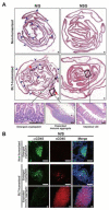
Figure 2. Endogenous cryptopatches, devoid of mouse lymphocytes, mature into expanded immune aggregates in the SI of N/S mice following BLT humanization
(A) Representative low magnification images of H&E-stained lower SI (ileum, ~6 cm long when embedded) of non-humanized and BLT-humanized N/S and NSG mice. Cryptopatches (blue arrowheads) are present in the SI of both non-humanized and BLT-humanized N/S mice. In addition, expanded immune aggregates (red arrows) are observed in N/S-BLT mice. High magnification images illustrate a representative enlarged cryptopatch and an expanded immune aggregate in N/S-BLT mice. Also shown are intestinal villi from NSG-BLT mice. Scale bar=250 μm. (B) IFA show that the enlarged cryptopatches in non-humanized N/S mice harbor only mCD45+ cells (green, left panel) (top row), while in N/S-BLT mice there is an accumulation of hCD45+ cells (red, center panel) alongside the mCD45+ cells (middle row). The relative proportion of hCD45+ cells to mCD45+ cells shifts dramatically as cryptopatches expand to become immune aggregates (bottom row). Merged plus DAPI (right). Scale bar=50 μm. See also Figure S1.
Figure 3
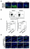
Figure 3. The distribution of hCD19+ B cells in the SI of N/S-BLT mice is restricted to GALT structures
(A) Expanded immune aggregates of N/S-BLT mice were defined as human GALT structures by the presence of hCD3+ T cells, hCD19+ B cells, hCD11c+ dendritic cells and hCD68+ macrophages. (B and C) Flow cytometry analyses show equivalent numbers of hCD3+ T cells in the SI of N/S-BLT (n=4) and NSG-BLT (n=4) mice. In contrast, the absolute number of hCD19+ B cells in the SI of N/S-BLT mice is higher versus NSG-BLT mice (Student’s t-test; N.S.=not significant). Gating: lymphoid→hCD45 positive →hCD3 or hCD19+. (D) The lack of human GALT structures in NSG-BLT mice limits direct comparison to the intestinal villi of the 2 types of BLT mice. Scale bars=50 μm.
Figure 4
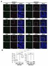
Figure 4. Small intestinal lamina propria of N/S-BLT mice exhibits a large number of human immunoglobulin-producing cells
(A) SI of both N/S-BLT (n=4) and NSG-BLT (n=4) mice were stained with anti-hIgM, hIgG or hIgA (green, left column) and anti-hCD45 (red, center column). The merged images with a DAPI-counterstained image are shown on the right panel. Higher magnification images are presented under the original low magnification images. Scale bar=50 μm. (B) Images similar to those in (A) from 4 mice per group were digitally converted using Definiens Tissue Studio® to quantitate the numbers of hCD45+ cells expressing either hIgM or hIgG or hIgA. (Student’s t-test; lines connect compared data sets). See also Figures S2 and S3.
Figure 5
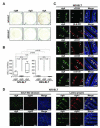
Figure 5. hIgA-secreting cells are the most common plasma cells found in the SI of N/S-BLT mice
(A-B) Mononuclear cells isolated from the SI of N/S-BLT (n=4) and NSG-BLT (n=4) mice were used for ELISPOT analyses to quantitate antibody-secreting plasma cells (ASCs). Images from a representative mouse per group (A). Cells secreting each Ig from each type of BLT mouse were compared by Student’s t-test (lines connect compared data sets) (B). (C) Fully differentiated hIgA+ plasma cells in the SI lamina propria of N/S-BLT mice express hCD138, hCD27 and hCD38 but not hCD20 or HLA-DR. Images are hIgA (green, left), hCD138, hCD20, hCD27, hCD38 or HLA-DR (red, center) and merged plus DAPI (right). (D) hIgA1- and hIgA2-producing plasma cells are found in both the human GALT-structures and the lamina propria of N/S-BLT mice. Images include: polyclonal anti-hIgA with binding specificities for both hIgA1 and hIgA2 (green, left), monoclonal antibodies specific for either hIgA1 or hIgA2 (red, center) and the merged plus DAPI (right). Scale bars=50 μm. See also Figure S4.

References

    1. Benckert J, Schmolka N, Kreschel C, Zoller MJ, Sturm A, Wiedenmann B, Wardemann H. The majority of intestinal IgA+ and IgG+ plasmablasts in the human gut are antigen-specific. J. Clin. Invest. 2011;121:1946–1955. - PMC - PubMed
    1. Brenchley JM, Douek DC. HIV infection and the gastrointestinal immune system. Mucosal Immunol. 2008;1:23–30. - PMC - PubMed
    1. Brenchley JM, Price DA, Schacker TW, Asher TE, Silvestri G, Rao S, Kazzaz Z, Bornstein E, Lambotte O, Altmann D, et al. Microbial translocation is a cause of systemic immune activation in chronic HIV infection. Nat. Med. 2006;12:1365–1371. - PubMed
    1. Cao X, Shores EW, Hu-Li J, Anver MR, Kelsall BL, Russell SM, Drago J, Noguchi M, Grinberg A, Bloom ET, et al. Defective lymphoid development in mice lacking expression of the common cytokine receptor γ chain. Immunity. 1995;2:223–238. - PubMed
    1. Denton PW, Estes JD, Sun Z, Othieno FA, Wei BL, Wege AK, Powell DA, Payne D, Haase AT, Garcia JV. Antiretroviral pre-exposure prophylaxis prevents vaginal transmission of HIV-1 in humanized BLT mice. PLoS Med. 2008;5:e16. - PMC - PubMed

Publication types

MeSH terms