Review
. 2014 Jan 25;382(1):673-682.
doi: 10.1016/j.mce.2013.06.003. Epub 2013 Jun 19.

The immune system and inflammation in breast cancer

Affiliations
Review

The immune system and inflammation in breast cancer

Xinguo Jiang et al. Mol Cell Endocrinol. .

Abstract

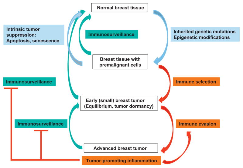
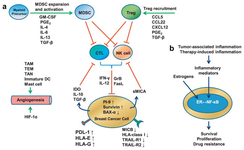
During different stages of tumor development the immune system can either identify and destroy tumors, or promote their growth. Therapies targeting the immune system have emerged as a promising treatment modality for breast cancer, and immunotherapeutic strategies are being examined in preclinical and clinical models. However, our understanding of the complex interplay between cells of the immune system and breast cancer cells is incomplete. In this article, we review recent findings showing how the immune system plays dual host-protective and tumor-promoting roles in breast cancer initiation and progression. We then discuss estrogen receptor α (ERα)-dependent and ERα-independent mechanisms that shield breast cancers from immunosurveillance and enable breast cancer cells to evade immune cell induced apoptosis and produce an immunosuppressive tumor microenvironment. Finally, we discuss protumorigenic inflammation that is induced during tumor progression and therapy, and how inflammation promotes more aggressive phenotypes in ERα positive breast cancers.

Keywords: Breast cancer; ER-alpha; Immunity; Immunosurveillance; Inflammation.


Figures

Fig. 1
Fig. 1
Immunosurveillance and inflammation in breast cancer. Inherited genetic mutation and epigenetic modifications cause premalignant transformation of mammary cells. Transformed cells can be eliminated by intrinsic or extrinsic tumor suppression mechanisms. Immune selection and immune evasion result in the development of advanced breast tumor. Immunosurveillance inhibits or reverses tumor development through killing the tumor cells. Protumorigenic inflammation accompanied advanced breast tumor promotes immune evasion and suppresses effective immunosurveillance.
Fig. 2
Fig. 2
RNAi knockdown of PI-9 blocks estrogen protection against NK cell-mediated cytotoxicity. (A and B) MCF-7, human breast cancer cells were transfected with the control pGL3 luciferase siRNA, or with the PI-9 siRNA. After 24 h, ethanol vehicle or E2 was added and the cells were maintained for an additional 24 h and either harvested for Western blotting using antibodies to PI-9 and the internal standard calnexin (A) or incubated with the indicated ratios of effector NK92 cells to MCF-7 target cells and assayed for cytoxicity using the time-resolved fluorescence assay (B) (filled and open bars, pGL3 siRNA − and + E2, respectively; hatched bars, PI-9 siRNA + E2). For example, at a ratio of 8 NK cells to 1 MCF-7 cell, in MCF-7 cells in which PI-9 was not induced with E2 (E/T ratio 8, black bar), and in MCF-7 cells treated with E2 and PI-9 was knocked down with PI-9 siRNA (E/T ratio 8, hatched bar) there was substantial NK cell induced cytotoxicity (~18% and 27%, respectively). In contrast, in MCF-7 cells in which PI-9 was induced with E2 and the cells were transfected with the control siRNA (open bar), the cells were protected and NK cell induced cytotoxicity was minimal (<5%) (From Jiang et al., 2007, reprinted with permission).
Fig. 3
Fig. 3
Dietary genistein induces PI-9 in MCF-7 tumors in mice. (A) Growth rates of MCF-7 tumors in ovariectomized athymic mice. At week 0, the estrogen pellets were removed, and the mice were divided into three treatment groups: PC (positive control, E2), GEN 500 (genistein 500 ppm), and NC (negative control, regressing tumor) that were fed AIN-93G diet alone. Tumor size was then measured weekly for 23 week. Data are expressed as means ± sem cross-sectional tumor area for all tumors in each group. (B) Dietary genistein and soy induce PI-9 in MCF-7 solid tumors. Mice were exposed to high (2 mg) E2 in implanted cholesterol pellets for ~11 weeks and tumors harvested (black bar) and frozen. Mice were fed diets containing genistein (500 ppm) or soy flour (diet containing 20% soy protein, ~400 ppm genistein) (black bars). Tumors were harvested at about 23 week when they reached the same size as the E2 tumors. Control MCF-7 cells were maintained in medium lacking hormones, or in 10 nm E2, or 10 nM genistein for 24 h and harvested (open bars). RNA was extracted and analyzed for PI-9 mRNA by quantitative RT-PCR. Data represent the mean ± sem for at least three samples (From Jiang et al., 2008; reprinted with permission).
Fig. 4
Fig. 4
Model showing how the immune system and inflammation eliminate or promote breast cancer. (A) Scheme showing the mechanisms by which breast cancers evade immunosurveillance. Major immunosuppressive cells including Treg and MDSCs are recruited or expanded and activated by proinflammatory mediators produced in the breast tumor microenvironment. Activated Treg and MDSCs suppress CTL and NK cells which are potent antitumor effector cells. Breast cancer cells also produce soluble factors such as IDO, IL-10, TGF-β and sMICA to suppress the activity of CTL and NK cells. Breast cancer cells evade immunosurveillance by changing the expression levels of apoptosis-associated intracellular proteins (PI-9, Survivin and BAX-α) and immune recognition- or activation-associated membranous proteins (MICB, HLA-class I, TRAIL-R1, TRAIL-R2, PDL-1, HLA-E, HLA-G). HIF-1α and proinflammatory cells such as TAM, TEM, TAN, immature DC and mast cells promote tumor angiogenesis. (B) Inflammation promotes an aggressive phenotype in ERα+ breast cancers. ERα and NF-κB activated by estrogens and proinflammatory mediators promote breast cancer cell survival, proliferation, and drug resistance. Crosstalk between ERα and NF-κB is context-dependent and can result in synergistic activation or mutual suppression. Abbreviations: Treg, regulatory T cells; MDSC; myeloid-derived suppressor cell; GM-CSF, granulocyte–macrophage colony-stimulating factor; PG, prostaglandin; CCL, CC chemokine ligand; CXCL, chemokine (C-X-C motif) ligand; CTL, cytotoxic T lymphocyte; NK, natural killer; IDO, indoleamine-pyrrole 2,3-dioxygenase; GrB, granzyme B; FasL, Fas ligand; IFN, interferon; IL, interleukin; TGF, transforming growth factor; sMICA, soluble Major Histocompatibility Complex class I related chain A; TRAIL, TNF-related apoptosis-inducing ligand; PI-9, proteinase inhibitor 9; PDL-1, programmed death ligand 1; HLA, human leukocyte antigen; TAM, tumor associated macrophage; TEM, Tie2 expressing monocyte; TAN, tumor associated neutrophil; DC, dendritic cell; HIF, hypoxia-inducible factor.

References

    1. Acharyya S, Oskarsson T, Vanharanta S, Malladi S, Kim J, Morris PG, Manova-Todorova K, Leversha M, Hogg N, Seshan VE, Norton L, Brogi E, Massague J. A CXCL1 paracrine network links cancer chemoresistance and metastasis. Cell. 2012;150:165–178. - PMC - PubMed
    1. Araki S, Eitel JA, Batuello CN, Bijangi-Vishehsaraei K, Xie XJ, Danielpour D, Pollok KE, Boothman DA, Mayo LD. TGF-beta1-induced expression of human Mdm2 correlates with late-stage metastatic breast cancer. J Clin Invest. 2010;120:290–302. - PMC - PubMed
    1. Bargou RC, Wagener C, Bommert K, Mapara MY, Daniel PT, Arnold W, Dietel M, Guski H, Feller A, Royer HD, Dorken B. Overexpression of the death-promoting gene bax-alpha which is downregulated in breast cancer restores sensitivity to different apoptotic stimuli and reduces tumor growth in SCID mice. J Clin Invest. 1996;97:2651–2659. - PMC - PubMed
    1. Basu S, Nachat-Kappes R, Caldefie-Chezet F, Vasson MP. Eicosanoids and adipokines in breast cancer: from molecular mechanisms to clinical considerations. Antioxid Redox Signal 2012 - PubMed
    1. Bates GJ, Fox SB, Han C, Leek RD, Garcia JF, Harris AL, Banham AH. Quantification of regulatory T cells enables the identification of high-risk breast cancer patients and those at risk of late relapse. J Clin Oncol. 2006;24:5373–5380. - PubMed

Publication types